
1
Fifth stage
Surgery-Ortho
Lec-7
.د
مثنى
28/4/2016
Foot and Ankle orthopedics
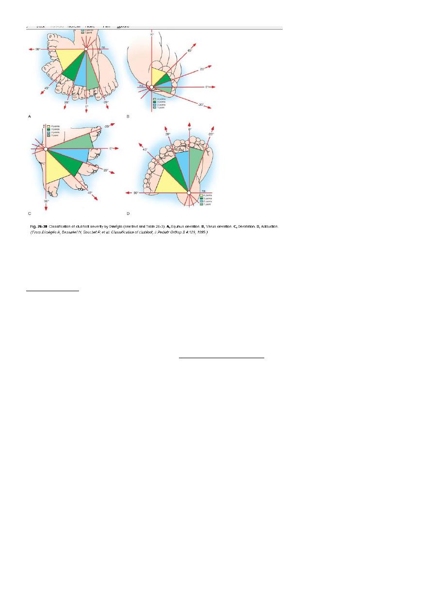
Talipes Equinovarus (Idiopathic clubfoot)
talus= L anke bone, pes= L foot
Equinus= heal tilted backward
varus= heel deviated medially
Incidence: 1-2/thousand births.
Male/female = 2/1.
Bilateral in 1/3rd of cases.
Etiology:
Idiopathic in most of cases, but may be associated with tight uterus as in primegravida
uterus.
Associated with myelomeningocele, congenital tibial deficiency and arthrogriposis.
Clinical features:
Components:
Equinus of heel.
Varus of hind foot.
Adduction and supination of forefoot.
Small heel and calf muscle.
Exam for associated:
DDH.
Spina bifida.
Arthrogriposis (absent creases in legs).
2
Treatment:
Conservative
Begin in the first few days of life.
Ponseti Method: Weakly manipulation and POP casting.
Adhesive strapping
If conservative treatment fails ; then Operative treatment is considered.
Pes Planus (flat-foot)
The medial border (arch of the foot) is in contact with the ground.
Common among children.
Types:
o Flexible flat-foot: normal variation usually disappears spontaneously.
o Rigid flat-foot: cannot be corrected passively.
o Compensatory flat-foot e g. short tendo achillis, or external rotation of lower
limbs (Charlie Chaplin look) .
High Arched feet (Pes Cavus)
Caused by muscle imbalance usually due to neuromuscular disorder eg. Poliomyelitis.
Associated with claw toes.

3
Both feet affected.
Often NO treatment required apart from well moulded shoes.
Halux Valgus
Lateral angulation of the big toe due to varus angulation of the first metatarsal.
Prominent head of first metatarsal (Bunion).
In people who wear shoes.
Loss of muscle tone in forefoot in elderly.
In rheumatoid arthritis.
Family history is common.
Clinical features:
In elderly women 50-70 years or adolescents with positive family history.
Usually bilateral.
Painless deformity.
Pain on inflammed buninion or OA of 1
st
MPJ.
Difficulty in purchasing comfortable shoes.
Treatment:
Asymptomatic, mild and non-progressive deformity; conservative treatment:
comfortable footwear with low heels.
Severe, progressive and painful deformity requires surgical treatment.
Claw Toes
Interphalangeal joints are flexed and metatarsophalangeal joint is extended.
Seen in poliomyelitis, Rheumatoid arthritis but usually idiopathic.
Associated with pes cavus.
Hammer Toes
Proximal IPJ flexed, distal IPJ and MPJ extended.
Cause is obscure.
Shoe pressure produce painful corns and callosities.

4
Rupture of Tendo Achillis
Age >40.
After running or jumping feels a struck above the heel.
Cannot tip toe.
Gap is felt 5cm above heel.
Simmond’s test: squeezing the calf causes planterflexion of foot normally. Absent
planterflexion means rupture of the tendon.
Treatment Operative repair then POP in equinus for 8 weeks.
Traction apophysitis (Sever’s disease)
Pain and tenderness over Achillis tendon insertion.
In boys around 10years old.
Treat by raising the heel and abandon strenuous exercise.
Plantar Fasciitis
Painful Heel
Common in Males 30-60yrs.
Pain with first steps after inactivity (rising from bed).
Obesity, prolonged standing, and stiff shoe soles are predisposing factors.
Local tenderness.
X-ray: boney spur.
Treatment: NSAID, pad under heel, local C/S injection.
Köhler’s disease
Osteochondritis of the navicular
Painful limp and tenderness in midfoot in children <5yrs.
X-ray: dense, fragmented navicular bone.
Usually resolve spontanuously.
Freiberb’s disease
Crushing type of osteochondritis.
Affects head of 2nd metatarsal.
Usually seen in young adult women.

5
Fractures of the talus
Rare, resulted from high energy trauma (fall from height or road traffic accident). The
fractures might occur in head, neck, body, or lateral process. Might associated with
dislocations of midtarsal, subtalar, ankle joints, or complete talar dislocation. There are
pain, sever swelling, deformity and skin tenting which may lead to skin sloughing, and skin
laceration sometimes.
Imaging: X ray shows the type and severity of fractures and associated injuries , CT scan is
of important value, MRI and radioactive isotope might be used.
Treatment:
Undisplaced fractures treated by below knee P.O.P in planter flexion for 8-12 weeks
Displaced fractures: closed reduction , if failed open reduction and internal fixation
followed by POP for 8-12 weeks. Non weight bearing continue for other 8-10 weeks.
Complications:
1- Skin damage.
2- Avascular necrosis of the body of the talus.
3- Nonunion.
4- Osteoarthritis.
Fractures of calcaneum
In most cases the patient falls from a height, often from a ladder, onto one or both heels.
Over 20 per cent of these patients suffer associated injuries of the spine, pelvis or hip.
The fractures might be extra-articular fractures or intra-articular fractures (runs into
superior articular surface involving subtalar joint.
Clinical features: There is usually a history of a fall from height or road traffic accident .
The foot is painful and swollen and a large bruise appears on the lateral aspect of the heel,
after1-2 days ecchymosis spreads into the sole of the foot . The heel may look broad and
squat.
Always check for signs of a compartment syndrome of the foot (intense pain, very extensive
bruising and diminished sensation, with pain on passive toe movement).
X- ray in lateral and axial view may shows chip, split, or crush fractures, CT sometimes used
to assess the fracture details.
Treatment:
Elevation and ice packing till the swelling subsides.

6
Displaced avulsion or intra-articular fractures needs open reduction and internal fixation
followed by elevation and non-weight bearing mobilization for 8 weeks.
Undisplaced fractures need p.o.p immobilization for 4 – 6 weeks followed by pressure
bandage with analgesic .
Comolications:
1- Stiffness of subtalar joint and midtarsal joint causing difficulty in walking especially on
uneven surfaces.
2- Osteoarthritis.
3- Broadening of the heel: problems in shoe fitting.
Fracture base of 5
th
metatarsal
This injury is very common, caused by foot torsion. It is nearly always caused by a twisting
injury in which the foot is forced into inversion and equines (planter flexion), the styloid
process at the base of the 5
th
metatarsal being pulled off by the tendon of the peroneus
brevis muscle, which is inserted into it . There is pain and tenderness over lateral side of
foot. It regarded as muscle injury. Treated by pressure bandage and analgesia, if pain sever
a below-knee walking plaster for 3 weeks.
Metatarsal stress fracture ( March fracture)
Young adult (a military recruit or a nurse) or osteoporotic women affected usually, the foot
may become painful and slightly swollen after overuse. A tender lump is palpable just distal
to the midshaft of a metatarsal bone (usually the second metatarsal) .
X-ray appearance may at first be normal but a radioisotope scan will show an area of
intense activity in the bone. Later a hairline crack may be visible and later on a callus. No
displacement occurs and neither reduction nor splintage is necessary. The forefoot may be
supported with an elastic bandage and normal walking is encouraged.