1
Forth stage
Surgery
Lec-1
د.هيثم النجفي
1/1/2014
Appendicitis
Incidence:
It usually occurs at 20 - 30 years of age
Before puberty M:F are equal, at and after puberty M < F.
Rare in old age due to atrophy of the lymphoid tissue & fibrosis of the appendix with
complete obliteration of the lumen.
lf occurs in an old patient, you must suspect cancer caecum.
Rare in children > 5 years of age due to short wide lumen of the appendix obstruction
& stasis does not occur.
Etiology:
Till now, there is no single microorganism to be the soul factor; there is mixture of
microorganisms which are claimed to be the affecting factor (E.coli, staph, Strept. Fecalis,
Pseudomonas
).
Precipitating Factors:
a) Obstruction:
It is the commonest & most important precipitating factor, it may be due to causes in the:
Lumen: hard feces, oxyuriasis, ascaris.
Wall:
- Rarely tumors of the appendix or ceacum
- Swelling of lymphoid tissue in response to viral infection.
Outside: Cancer cecum (in old age) & adhesion.
b) Anatomical Factor:
The appendix has a narrow lumen (Stricture).
N.B: due to obstruction there will be accumulation of secretion, with stasis will lead to
infection. (The cascade of the process will be started)
Pathology:
A. Acute Non Obstructive Appendicitis (Catarrhal):
It is claimed to be caused by viral inflammation edema and swelling of
lymphatic colical which is present in the submucosa obstruction and ulceration
invading of the submucosa by microorganism cascade.
N.B: actually this process also considered as obstruction, but there is no intraluminal agent
which occludes the obstruction.
B. Acute Obstructive Appendicitis (Cascade):
Accumulation of secretion super added infection secreting of
microorganism through mucosa then submucosa then muscularis mucosa then into
serosa, once it reaches the serosa when the appendicular artery lies in endemic conduct

2
with the appendix, inflammation of this artery will lead to thrombosis ischemia
necrosis, sluffing, perforation and either localized or generalized peritonitis.
Complications:
1. If the immunity of the patient is excellent, there will be raping of the appendix by the
greater omentum, small bowel, and cecum this will lead to formation of appendicular
mass.
2. If the patient is immunocompromised (e.g. Diabetic Pt.)
If the microorganism is very virulent there will be no raping of the appendix and if the
appendix is pelvic so the appendix will perforate and due to this perforation without
localization will lead to generalized peritonitis.
3. Appendicular Abscess: Perforation of the appendix within an appendicular mass forms
an abscess.
4. Fecal Fistula: After drainage of abscess & appendectomy in presence of mass.
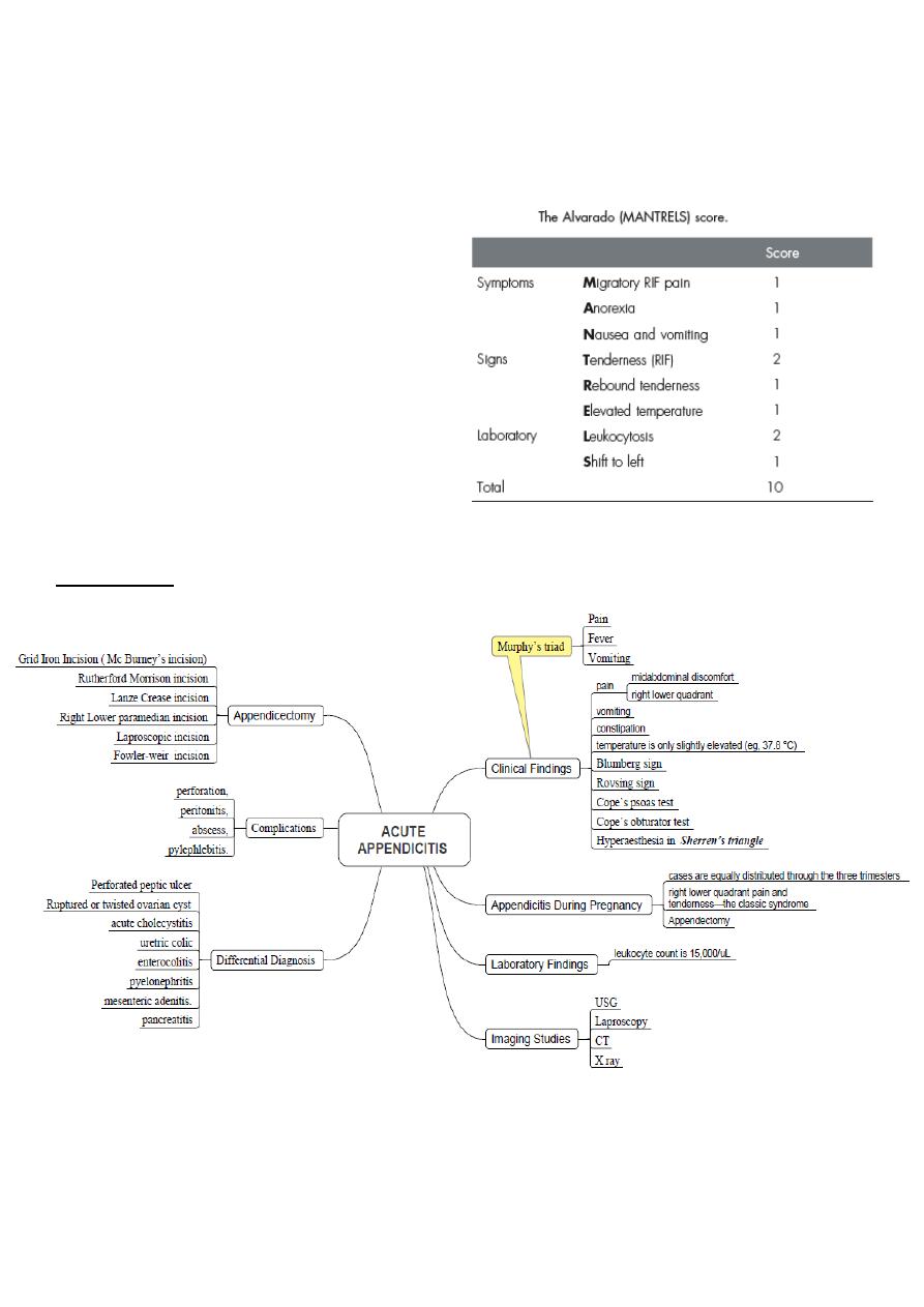
Clinical Presentation:
Svmptoms
1. Acute Pain that shifts (presenting symptom):
It starts periumbilical and is ill defined dull (visceral pain), because of the appendix is
present (embryologically) in the mid and hind gut, this pain is due to visceral
peritoneal irritation.
Then the pain is shifted to the right iliac fossa, now the pain becomes sharper
(somatic).
It is aggravated by movement (jumping) and cough.
2. Anorexia & Nausea: it is an important symptom.
3. Vomiting: once or twice
4. Constipation: Usually present.
5. Mild" fever < 38'C (lf higher indicates complications or suggest other causes of acute
abdomen), it is late symptom.
Signs:
I. Inspection
Abdomen is not move freely with inspiration.
Pointing sign (ask the patient to cough), pain at McBurney’s point.
II. Palpation
Localized tenderness in right iliac fossa.
Rebound tenderness on the right iliac fossa.
Guarding on the right iliac fossa (voluntary muscle spasm).
Rigidity on the right iliac fossa (involuntary muscle spasm)
: if the appendicitis is due to obstruction the patient comes from the
.1
N.B
start with colicky abdominal pain.
if the patient is asked to cough, the pain becomes sharper and
N.B.2:
localized to the site of appendix (pointing sign).

3
Special Signs
Rovesing sign pressure on Lt iliac fossa pain on Rt iliac fossa due
to shifting of gases (air) from the pelvic colon to the cecum.
Obturator test (Zachary cope)(in pelvic appendicitis) the tip of the appendix is in
contact obturator internus muscle, internal rotation of hip joint and extension of the
knee pain in Rt. Iliac fossa (due to stretching of the obturator internus muscle).
Psoas test (in retroceacal appendicitis): appendix in contact with psoas
muscle, when the patient flexes right hip pain in Rt. Iliac fossa (due
to stretching of psoas muscle).
Hyperaesthesia of triangle of Sheren (in sub-hepatic type)
III. Percussion
Tenderness in the Rt. Iliac fossa.
IV. Auscultation:
There may be diminished intestinal sounds.
V. PV& PR.
To detect pelvic appendix & exclude gynecological causes
Investigations:
1. GUE: for oxalate, pus cells and RBCs
2. KUB: to see if there is stone, calcification or obstruction the UT.
3. WBC count: to see if there is leucocytosis.
4. CT scan: to confirm the diagnosis
Differential Diagnosis:
Children
1. Gastroenteritis: the child comes with high fever and dehydration, sever deep pain
during PR examination indicates appendicitis.
2. Mesenteric lymphadenitis: (the most important) caused by viral infection, the patient
comes with pain, tenderness and rebound thenderness in the Rt. Iliac fossa (similar to
appendicitis). To distinguish between them, the patient with mesenteric lymphadenitis
has high fever and enlargement in the tonsils, there may be circum-oral pallor, and
also there is shifting tenderness.
3. Intussusception: Appendicitis is uncommon before the age of two years, whereas the
median age for intussusception is 18 months. A mass may be palpable in the right
lower quadrant, intermittent severe abdominal pain and
"red currant jelly" stool (stool
mixed with blood and mucus)
the preferred treatment of intussusception is reduction
by careful barium enema.
4. Meckel’s diverticulitis It may be impossible to clinically distinguish Meckel’s
diverticulitis from acute appendicitis.
Adults
1. Ureteric stone: pain from the loin to the groin, in males it descends to the scrotum
and referred tenderness in the renal angle. X-ray and US will confirm the diagnosis.

4
2. Pyelonephrosis: hyperpyrexia with sever dull pain in the loin, frequency, and
urgency. GUE show the field is full with pus cells.
N.B: in GUE there will be pus cells, see if the bacteria is positive this means infection, if it is
negative this means pyuria (T.B or Malignancy)
3. Testicular torsion: Pain can be referred to the right iliac fossa, suspect appendicitis
unless the scrotum is examined in all cases.
4. Crohn’s disease or Yersinia infection (Yersinia enterocolitica): An antecedent
history of abdominal cramping, weight loss and diarrhoea suggests Yersinia
enterocolitis rather than appendicitis.
5. Perforated duodenal ulcer (DU): As a rule, there is a history of dyspepsia and a
very sudden onset of pain that starts in the epigastrium and passes down the right iliac
fossa. Rigidity and tenderness and rebound tenderness in the right iliac fossa are
present. X-ray will show gas under the diaphragm.
6. Pancreatitis: sever pain, tenderness and rebound tenderness in the mid-
hypochondirum, the patient looks ill, serum amylase and serum lipase are the corner
stone in diagnosis.
7. Intestinal obstruction: high intestinal obstruction (vomiting, distention and
constipation), low intestinal obstruction (constipation, distention and vomiting). With
present of colicky abdominal pain. X-ray will show multiple air fluid level.
Females
1. Mittelschmerz: midcycle rupture of a follicular cyst with bleeding produces lower
abdominal and pelvic pain, typically midcycle, and symptoms usually subside within
hours.
2. Primary peritonitis: pain, tendeness and rebound tenderness, usually in virgin
female, due to pneumococcal microorganism.
3. Acute salpingitis: pain, fever, vaginal discharge, tenderness often bilateral. X-ray to
confirm Dx.
4. Ectopic pregnancy: the pain commences on the right side and stays there. The pain is
severe and continues unabated until operation. Usually, there is a history of a missed
menstrual period, and a urinary pregnancy test may be positive. Severe pain is felt
when the cervix is moved on vaginal examination. Pelvic ultrasonography should be
carried out in all cases in which an ectopic pregnancy is a possible diagnosis
.
5. Torsion/haemorrhage of an ovarian cyst: This can prove a difficult differential
diagnosis. When suspected, pelvic ultrasound and a gynaecological opinion should be
sought.
6. UTI: common in females, advanced UTI lead to peyonephrosis, treat both male and
female.
N.B. if the field of GUE shows more than 4 pus cells send to culture.
Elderly
1. Carcinoma of the caecum: when obstructed or locally perforated, carcinoma of the
caecum may mimic or cause obstructive appendicitis in adults. A mass may be
palpable.

5
2. Diverticulitis (Cecal diverticulum): Right-sided diverticulitis may be clinically
indistinguishable from appendicitis. Abdominal CT scanning is particularly useful in
making the distinction. As with left-sided diverticulitis, treatment should be
conservative with intravenous antibiotics with recourse to laparoscopy or laparotomy
in the face of deterioration
.
3. UTI
4. T.B
5. Typhoid: lead to perforation
Summary
