APPENDIX
Dr. ZAID MUWAFAQ AL-HAMIDMRCS England(UK), FJMC Jordan, HSM SURGERY Jordan, MBChB Mosul
Specialist General & Laparoscopic Surgeon
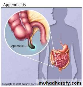

ACUTE APPENDICITIS
Incidence:rare in infants, increasingly common in childhood and early adult life, peak incidence in the teens and early 20s.
before puberty males = females.
In teenagers and young adults the male–female ratio 3:2
Thereafter, the greater incidence in males declines.
Aetiology
No definite cause1-Decreased dietary fibre and increased consumption of refined carbohydrates (low fiber diet)
2-Bacterial proliferation within the appendix,
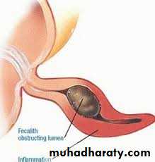
no single organism is responsible(mixed growth of aerobic and anaerobic organisms ).3- Obstruction of the appendix lumen
- Lymphoid hyperplasia
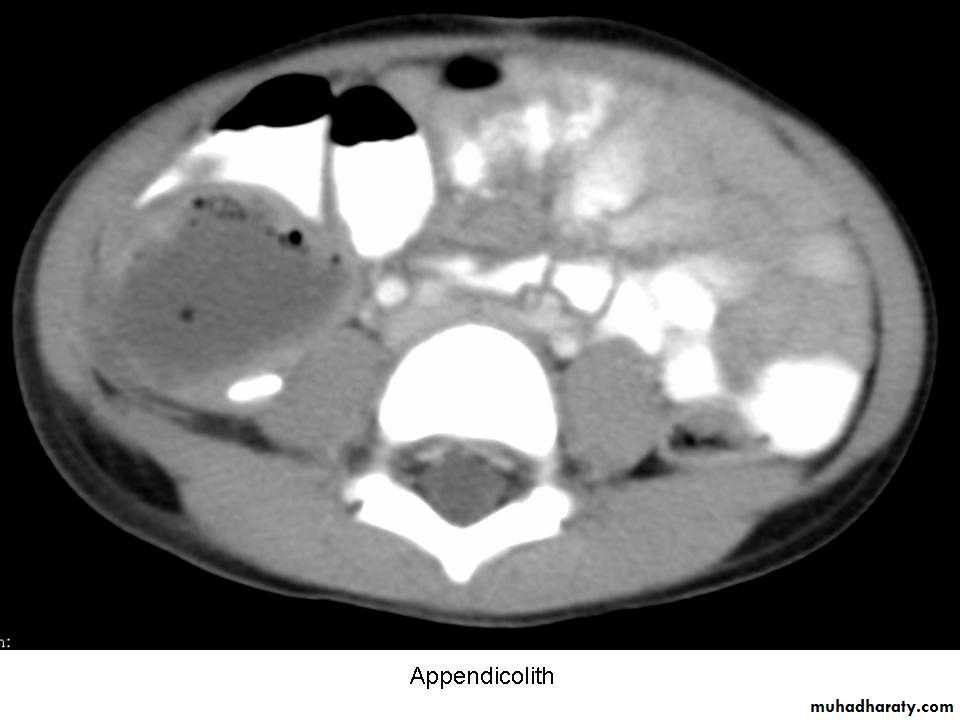
- faecolith (composed of inspissated faecal material,
calcium phosphates, bacteria and epithelial debris
- stricture
- foreign body(rare)
- tumour, particularly carcinoma of the caecum(middle age and elderly)
- Intestinal parasites, particularly Oxyuris vermicularis
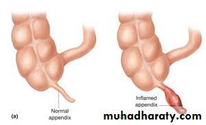
Pathology
Lymphoid hyperplasia narrows the lumen luminal obstruction.Continued mucus secretion and inflammatory exudation increase intraluminal pressure obstructing lymphatic drainageOedema
- Mucosal ulceration
- Bacterial translocation to the submucosa.
Resolution may occur (spontaneously/antibiotic therapy).
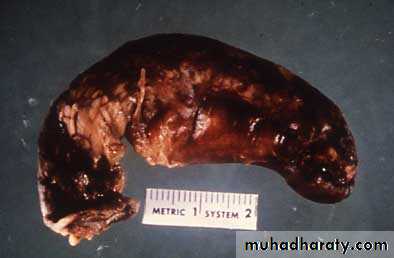
orCondition progresses,more distension venous obstruction & wall ischaemia bacterial invasion through the muscularis propria and submucosa, producing acute appendicitis.
Ischaemic necrosis gangrenous appendicitisfree bacterial contamination of the peritoneal cavity(peritonitis).
Alternatively, the greater omentum and loops of small bowel become adherent to the inflamed appendix, walling off the spread of peritoneal contamination, and resulting in a phlegmonous mass or paracaecal abscess.
Rarely, appendiceal inflammation resolves, leaving a distended mucus-filled organ termed a ‘mucocoele’ of the appendix .
Diffuse peritonitis is the great threat of acute appendicitis, it ocuurs as a result of:
free migration of bacteria through an ischaemic appendicular wallfrank perforation of a gangrenous appendix
delayed perforation of an appendix abscess.
Factors that promote this process include:
- extremes of age
- immunosuppression
- diabetes mellitus
- faecolith obstruction of the appendix lumen
- free-lying pelvic appendix
- previous abdominal surgery
In these situations, a rapidly deteriorating clinical course is accompanied by signs of diffuse peritonitis and systemic
sepsis syndrome.
History
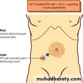
PAIN-poorly localised, colicky abdominal pain(midgut visceral discomfort in response to appendiceal inflammation and obstruction).
-The pain in periumbilical region.
- Central abdominal pain + anorexia, nausea and usually one or two episodes of vomiting that follow the onset of pain.
Clinical diagnosis
With progressive inflammation of the appendix:
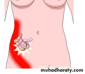
Shifting of pain: from central abdominal pain to right iliac fossa(irritation of RIF parietal peritoneum) visceral to somatic pain which is :- more intense
- constant
- localised to right iliac fossa
Typically, coughing or sudden movement exacerbates the right iliac fossa pain.
ANOREXIA
-constant clinical feature, particularly in children.Family history
1/3 of children with appendicitis have a first-degree relative with a similar history .
One half of acute appendicitis classic visceral–somatic sequence of pain .
Signs
Temperature and pulse rate:
- After 6 hours, slight pyrexia (37.2–37.7°C) with a corresponding increase in the pulse rate to 80 or 90 is usual.
-Changes of greater magnitude may indicate complications.
- Unwell patient with low-grade pyrexia
- Inspection of the abdomen limitation of respiratory movement in the lower abdomen.
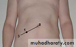
- localised abdominal tenderness maximum at McBurney’s point.
- Muscle guarding
Rebound tenderness
Asking the patient to cough or gentle percussion over the site of maximum tenderness will elicit rebound tenderness .
- Pointing sign: The patient is asked to point to where the pain began and where it moved .
- Rovsing’s sign: Deep palpation of the left iliac fossa may cause pain in the right iliac fossa.
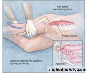
- Psoas sign : passive extension of hip or active flexion of hip against resistance pain. The patient will lie with the right hip flexed for pain relief .
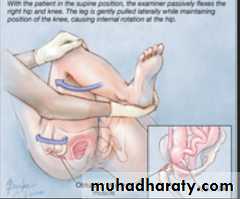
- Obturator sign : hip flexion and internal rotation cause pain in the hypogastrium (the obturator test).
-Cutaneous hyperaesthesia may be demonstrable
in the right iliac fossa

Two clinical syndromes of acute appendicitis:
1- acute catarrhal (non-obstructive) appendicitis2- acute obstructive appendicitis:
-more acute course(abrupt)
-generalised abdominal pain from the start.
- Temperature normal
- vomiting common.
Special features, according to position of the appendix
1-Retrocaecal appendicitis- Rigidity is often absent
- Application of deep pressure may fail to elicit tenderness (silent appendix)
Deep tenderness is often present in the loin, and rigidity of the quadratus lumborum.
Psoas spasm leading to flexion of the hip joint. Hyperextension of the hip joint may induce abdominal pain (Psoas sign +ve).
2-Pelvic appendicitis
- More common in children
- Early diarrhoea (inflamed appendix in contact with the rectum).
- Complete absence of abdominal rigidity
- Tenderness over McBurney’s point is also lacking.
- Deep tenderness can be made out just above and to the right of the symphysis pubis.
- Rectal examination reveals tenderness in the rectovesical pouch or the pouch of Douglas, especially on the right
side.
- +ve psoas and obturator signs.
- Frequency of micturition (inflamed appendix in contact with the bladder).
3-Postileal appendicitis
The inflamed appendix lies behind the terminalIleum(difficult to diagnose)
- Pain may not shift
- Diarrhea is a feature
- Marked retching
- Tenderness, if any, is ill defined, although it may be
present immediately to the right of the umbilicus.
Infants
- Rare in infants under 36 months of age.- The patient is unable to give a history. Diagnosis is often delayed.
- Higher incidence of perforation and postoperative morbidity than older children.
- Diffuse peritonitis can develop rapidly because of the underdeveloped greater omentum.
Children
- Vomiting is more common.
- complete aversion to food.
Special features, according to age
The elderly
- Gangrene and perforation occur much more frequently .- The abdominal clinical picture is not obvious even in the presence of gangrenous appendicitis (lax abdominal walls or obesity)
- Clinical picture may simulate subacute intestinal obstruction.
- Higher mortality
(coincident medical conditions plus the previous factors)
The obese
- Obesity can obscure and diminish all the local signs of acute
appendicitis.
- Midline abdominal incision
(Delay in diagnosis + technical difficulty of operating in the obese)
- Laparoscopy is particularly useful in the obese.
Pregnancy
Appendicitis is the most common extrauterine acute abdominal condition in pregnancy.
- Delay in presentation (early non-specific symptoms are often attributed to the pregnancy).
- The physiologic leukocytosis of pregnancy (high as 16,000 cells/mm3).
Obstetric teaching has been that the caecum and appendix are progressively pushed to the right upper quadrant of the abdomen as pregnancy develops during the second and
third trimesters.
- Pain in the right lower quadrant of the abdomen remains the cardinal feature of appendicitis in pregnancy.
- Fetal loss occurs in 3–5 per cent of cases, increasing to 20 per cent if perforation is found at operation.
Children
1- Acute gastroenteritis and mesenteric lymphadenitis:the pain is diffuse, and tenderness is not as sharply localized and cervical lymph nodes may be enlarged.
2- Meckel’s diverticulitis:
signs may be central or left sided.
history of antecedent abdominal pain or intermittent lower gastrointestinal bleeding.
3- Intussusception:
Appendicitis is uncommon before the age of two years, whereas the median age for intussusception is 18 months.
A mass may be palpable in the right lower quadrant,
Differential diagnosis
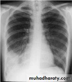
5-Lobar pneumonia and pleurisy, especially at the right base
Abdominal tenderness is minimal,pyrexia is marked
+ve pleural friction rub or altered breath sounds on auscultation.
A chest radiograph is diagnostic.

4- Henoch–Schönlein purpura
preceded by a sore throat or respiratory infection.Abdominal pain can be severe.
ecchymotic rash, affecting the extensor surfaces of the limbs and on the buttocks.
The face is usually spared.
Microscopic haematuria is common.
Adults
1- Terminal ileitis- a doughy mass of inflamed ileum may be felt.
- history of abdominal cramping, weight loss and diarrhoea.
The ileitis may be non-specific, due to Crohn’s disease or Yersinia infection.
- serum antibody titres are diagnostic, and treatment with intravenous tetracycline is appropriate.
2- Ureteric colic:
Urinalysis should always be performed, and the presence of red cells supine abdominal radiograph. Renal ultrasound or intravenous urogram is diagnostic.
3- Right-sided acute pyelonephritis
increased frequency of micturition.
tenderness confined to the loin, fever (temperature 39°C) and possibly rigors and pyuria.
4- perforated peptic ulcer
the duodenal contents pass along the paracolic gutter to the right iliac fossa.
- history of dyspepsia
- very sudden onset of pain that starts in the epigastrium and passes down the right paracolic gutter.
- rigidity is usually greater in the right hypochondrium.
- rigidity and tenderness in the right iliac fossa
- erect chest radiograph gas under the diaphragm in 70 %.
- abdominal (CT) examination in difficult cases.
5- Testicular torsion (teenage or young adult male)
Pain can be referred to the right iliac fossa, and shyness on the part of the patient may lead the unwary to suspect appendicitis unless the scrotum is examined in all cases.
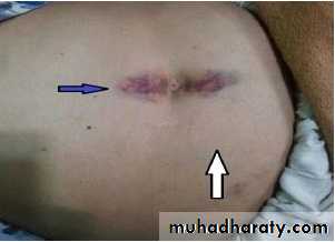
7- Rectus sheath haematoma
relatively rareacute pain and localised tenderness in the RIF
after an episode of strenuous physical exercise.
Localised pain without gastrointestinal upset is the rule. Occasionally, in an elderly patient, particularly one taking anticoagulant therapy, a rectus sheath haematoma may present as a mass and tenderness in the right iliac fossa after minor trauma.
6-Acute pancreatitis
should be considered in the differential diagnosis of all adults suspected of having acute appendicitis and, when appropriate, should be excluded by serum or urinary amylase measurement.Adult female
pelvic disease in women of childbearing age most often mimics acute appendicitis.
A careful gynaecological history should be taken in all women with suspected appendicitis, concentrating on:
- menstrual cycle
- vaginal discharge
- possible pregnancy .
The most common diagnostic mimics are
pelvic inflammatory disease (PID), Mittelschmerz, torsion or haemorrhage of an ovarian cyst and ectopic pregnancy.
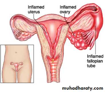
1-Pelvic inflammatory disease
(salpingitis, endometritis and tubo-ovarian sepsis)- The pain is lower than in appendicitis and is bilateral.
- history of vaginal discharge, dysmenorrhoea and dysurea .
- vaginal examination adnexal and cervical tenderness .
- High vaginal swab & culture Chlamydia trachomatis and Neisseria gonorrhoeae.
- gynaecologist opinion should be obtained.
- Transvaginal ultrasound
- diagnostic laparoscopy
2- Mittelschmerz
Midcycle rupture of a follicular cyst with bleeding produces lower abdominal and pelvic pain, typically midcycle.
- No systemic upset
Pregnancy test is negative
Symptoms usually subside within hours.
Occasionally, diagnostic laparoscopy is required. Retrograde menstruation may cause similar symptoms.
3- Torsion/haemorrhage of an ovarian cyst
(difficult differential diagnosis)pelvic ultrasound and a gynaecological opinion should be sought.
If encountered at operation untwisting of the involved adnexa and ovarian cystectomy should be performed.
Right-sided unruptured tubal pregnancy :
- pain commences on the right side and stays there.- pain is severe.
- history of a missed menstrual period
- Signs of intraperitoneal bleeding with referred pain in the shoulder.
- cervical excitation test positive
- urinary pregnancy test may be positive.
- Pelvic ultrasonography.
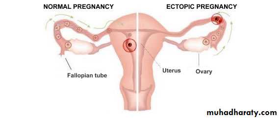
4- Ectopic pregnancy
- Ectopic preg. signs of haemoperitoneum
right-sided tubal abortion or
right-sided unruptured tubal pregnancy.
Elderly
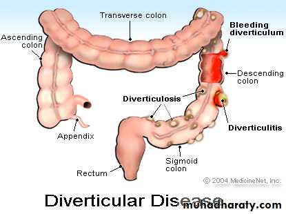
1- DiverticulitisAbdominal CT scanning is particularly useful and should be considered in the management of all patients over the age of 60 years.
Right-sided diverticulitis is unusual and may be clinically indistinguishable from appendicitis.
2-Intestinal obstruction
In elderly, occasionally, it may be difficult to differentiate IO from acute appendicitis
3-Carcinoma of the caecum
-Carcinoma of the caecum when obstructed or locally perforated, may mimic or cause obstructive appendicitis in adults.-history of antecedent discomfort, altered bowel habit or unexplained anaemia.
- mass may be palpable and an abdominal CT scan
diagnostic.
Rare differential diagnoses
1-Preherpetic pain of the right 10th and 11th dorsal nerves.- no shift of pain
- marked hyperaesthesia.
- no intestinal upset, no rigidity.
The herpetic eruption may be delayed for 3–8 hours.
2-Tabetic crises.
3-Spinal conditions include tuberculosis of the spine, metastatic carcinoma, osteoporotic vertebral collapse and multiple myeloma.4-porphyria and diabetes mellitus
5-Typhlitis or leukaemic ileocaecal syndrome
Investigation
The diagnosis of acute appendicitis is essentially clinicalRoutine
Full blood count WBC > 10,000
Urinalysis hematuria/ pyuria due to irritation of nearby ureter/ urinary bladder
Selective
Pregnancy test
Urea and electrolytes
Supine abdominal radiograph
Ultrasound of the abdomen/pelvis
Contrast-enhanced abdomen and pelvic computed tomography scan
Alvarado score
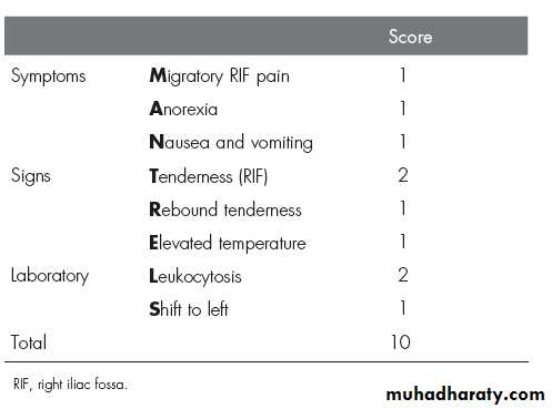
Alvarado score:
A score of 7 or more is strongly predictive of acute appendicitisEquivocal score (5–6)abdominal ultrasound or contrast-enhanced CT examination .
Abdominal ultrasound is useful in:
- children and thin adults
- if gynaecological pathology is suspected, with a diagnostic accuracy >90 %.
Contrast-enhanced CT scan is most useful in:
- diagnostic uncertainty+older patients
- acute diverticulitis, intestinal obstruction and neoplasm suspected.
Treatment
The traditional treatment for acute appendicitis is appendicectomy.Some research the conservative management in non-obstructive appendicitis including :
1- bowel rest(NPO)2- intravenous antibiotics, usually metranidazole and third-generation cephalosporin
conservative management 80-90% successful, 15 recurrence within one year.
patients over the age of 40 should be followed up to ensure there is no underlying malignancy
Appendicectomy:
no unnecessary delay
short period of intensive preoperative preparation:
1- NPO
2- Intravenous fluids
(catheterisation is needed only in the very ill)
3- IV antibiotics
In the absence of purulant peritonitis give single preoperative dose of antibiotics .
When peritonitis is suspected, therapeutic intravenous antibiotics to cover Gramnegative bacilli, as well as anaerobic cocci, should be given.
Appendicectomy
- general anaestheticpatient supine on the operating table.
When a laparoscopic technique is to be used, the bladder must be empty
(ensure that the patient has voided before leaving the ward).
palpated for a mass prior to preparing the entire abdomen with an antiseptic solution.(mass conservative)
Draping of the abdomen
Incisions used:
1- The gridiron incision(most common)
2- Rutherford Morison incision(muscle cutting)
3- Transverse skin crease (Lanz) incision
4- lower midline abdominal incision( doubtful diagnosis)
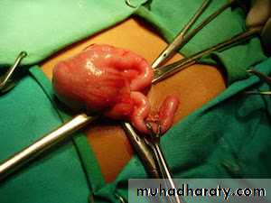
1-The caecum is identified by the presence of taeniae coli and is withdrawn.
2- Appendix may be felt at the base
of the caecum.
3- Inflammatory adhesions must be gently broken with a finger, the appendix delivered into the wound.
4- Mesoappendix devided between ligature, the base of appendix ligated and divided.
5- Purse-string to invaginates the stump of the appendix(?????)
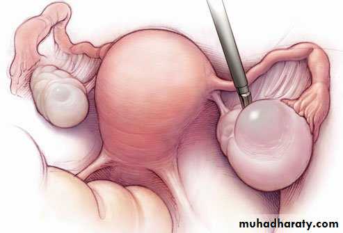
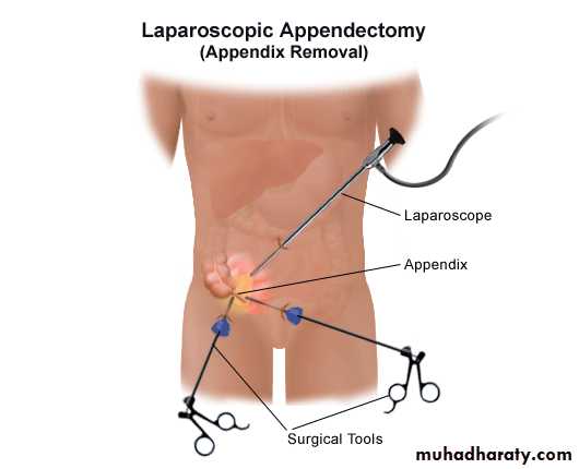
Laparoscopic appendicectomy
Laparoscopy is diagnostic tool(esp. in female) and therapeutic.Used more in:
Female (child bearing age)
in obese patients
early pregnancy
Advantages:
less postoperative pain
Early discharge from hospital
Early return to daily activities
Natural Orifice Transluminal Endoscopic Surgery
NOTES1-A normal appendix is found
- exclude other possible diagnoses, particularly terminal ileitis, Meckel’s diverticulitis and tubal or ovarian causes in women.- remove the appendix (avoid future diagnostic difficulties)
2-The appendix cannot be found.
The caecum should be mobilised, and the taeniae coli should be traced to their confluence on the caecum before the diagnosis of ‘absent appendix’ is made.
Problems encountered during appendicectomy
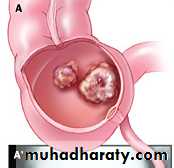
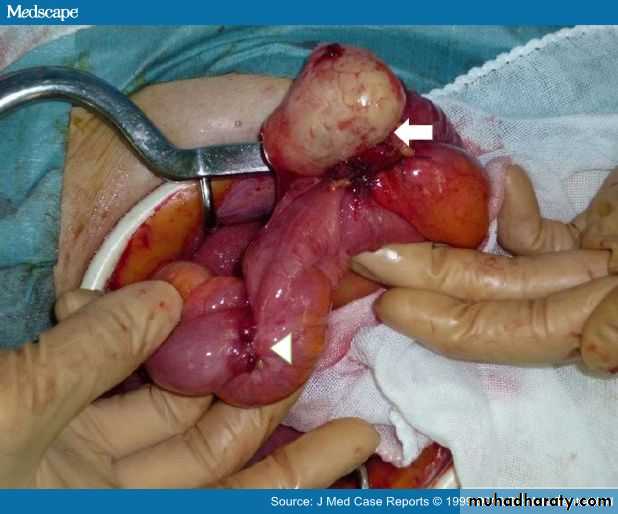
3-An appendicular tumour is found.
Small tumours (under 2.0 cm in diameter) can be removed by appendicectomy.Larger: right hemicolectomy
4- An appendix abscess is found and the appendix cannot be removed easily.
Preoperatively diagnosed abscessPercutaneous drainage of the abscess and intravenous antibiotic .
At operationdraine the abscess +intravenous antibiotics.
Frankly necrotic appendix(very rarely) caecectomy or partial right hemicolectomy .
Appendix abscess
Failure of resolution of an appendix mass or continued spiking pyrexia usually indicates that there is pus within the phlegmonous appendix mass.Treatment :
Ultrasound or abdominal CT scan percutaneous drain.
If unsuccessful laparotomy through a midline incision is indicated.
Pelvic abscess
- an occasional complication of acute appendicitis.Presented with spiking pyrexia several days after appendicitis
Pelvic pressure or discomfort
loose stool or tenesmus is common.Rectal examination mass in the pelvis.
Pelvic ultrasound or CT scan confirm.
Treatment:- Radiologically guided percutaneous drainage
- Transrectal drainage under general anaesthetic.
1- Wound infection
most common postoperative complicationusually presents with pain and erythema of the wound on
the 4th or 5th postoperative day.
Treatment: wound drainage + antibiotics.
Postoperative complications2- Intra-abdominal abscess
presented with spiking fever, malaise and anorexia developing 5–7 days after operation .
Sites of collection:
Interloop, paracolic, pelvic and subphrenic
Abdominal ultrasonography and CT scanning diagnostic and allow percutaneous drainage.
Laparotomy in intra-abdominal sepsis without localised collection.3- slipped ligature
internal bleeding, hypotension, tacchycardia reoperate4- Ileus
Ileus persisting for more than 4 or 5 days + fevercontinuing intra-abdominal sepsis investigation.
5- Respiratory
- rare
- Adequate postoperative analgesia and physiotherapy reduce the incidence.
6- Venous thrombosis and embolism
- rare after appendicectomy
- more in elderly and in women taking the oral contraceptive pill and prophylactic measures should be considered.
7- Portal pyaemia (pylephlebitis)
rarecomplication of gangrenous appendicitis
high fever, rigors and jaundice.
caused by septicaemia in the portal venous system intrahepatic abscesses (multiple).
Treatment: systemic antibiotics + percutaneous drainage of hepatic abscesses.
screen for underlying thrombophilia
8- Faecal fistula
- Rare
- leakage from appendicular stump.
- More in appendicectomy in Crohn’s disease.
Treatment: Conservative management with low-residue enteral nutrition.
9- Adhesive intestinal obstruction
This is the most common late complication of appendicectomy.
Conservative Ochsner–Sherren regimen;
A nonoperative programme but to be prepared to operate should clinical deterioration occur . This includes:Careful recording of the patient’s condition
1- Temperature and pulse rate should be recorded 4-hourly
2- Fluid balance record
3- The abdomen regularly reexamined.
4- The extent of the mass should be made (mark the limits of the mass on the abdominal wall using a skin pencil)
5- A contrast-enhanced CT examination of the abdomen
6- Antibiotic
7- An abscess, if present, should be drained radiologically.
Management of an appendix mass
Criteria for stopping conservative treatment of an appendix mass
_ A rising pulse rate
_ Increasing or spreading abdominal pain
_ Increasing size of the mass
Clinical deterioration or evidence of peritonitis is an indication for early laparotomy.
If the mass resolve, Patients over the age of 40 should have colonoscopy and follow-up imaging to ensure resolution and exclude appendicular or colonic malignancy.
Recurrent acute appendicitis
- not uncommon- attacks vary in intensity and may occur every few months.
majority of cases ends in severe acute appendicitis.
The appendix in these cases shows fibrosis indicative of previous inflammation.
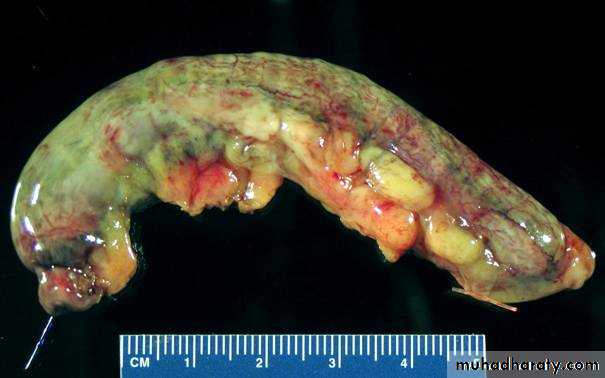
Neoplasms of the appendix
1-Carcinoid tumours- arise in argentaffin tissue (Kulchitsky cells of the crypts of Lieberkühn)
most common in the vermiform appendix.
- appendix removed because of symptoms of subacute or recurrent appendicitis.
frequently in the distal third of the appendix.
moderately hard , yellow tumour
the intact mucosa .
Microscopicallycharacteristic pattern using immunohistochemical stain for chromogranin B .
rarely gives rise to metastases.
Treatment:
- Appendectomy .- Right hemicolectomy is indicated if:
caecal wall involvement .
2 or more in size.
Involved lymph nodes.
2- Goblet cell carcinoid tumour
3- Primary adenocarcinoma of the appendixextremely rare, presented as appendicitis
treated by right hemicolectomy
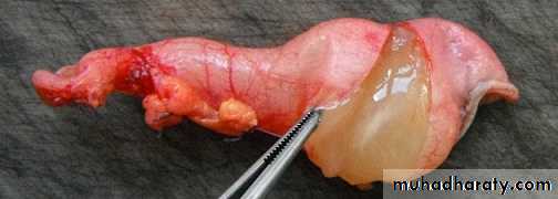
Mucinous cystadenoma
- A mucin-secreting adenoma of the appendixrupture into the peritoneal cavity , seeding it with mucussecreting cells.
delayed presentation with gross abdominal distension as a result of pseudomyxoma peritoneii, which may mimic ascites .
Treatment
radical resection of all involved parietal peritoneal
surfaces and aggressive intraperitoneal chemotherapy.
Thank you