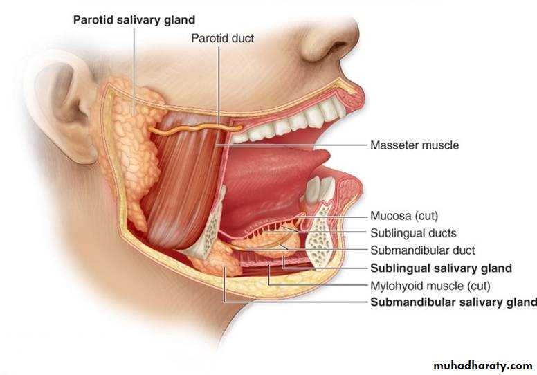
Salivary Glands
Dr. ZAID MUWAFAQ AL-HAMIDMRCS England(UK), FJMC Jordan, HSM SURGERY Jordan, MBChB Mosul
Specialist General & Laparoscopic SurgeonTwo submandibular glands
Two parotid glandsTwo sublingual glands
Approximately 450 minor salivary glands
Anatomy of salivary glands
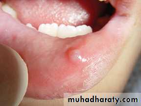
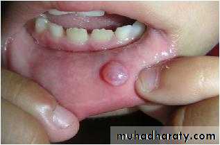
1- Extravasation Cysts
Commontrauma to the overlying mucosa.
affect lower lip producing painless swelling and usually translucent .
resolve spontaneously
most require formal surgical excision +overlying mucosa +underlying minor salivary gland.
Recurrence is rare.
Common disorders of minor salivary glands
90 % malignant.
anywhere in the upper aerodigestive tractcommon sites : upper lip, palate and retromolar regions.
Less common sites nasal and pharyngeal cavities.
Benign minor salivary gland tumors present as painless, firm, slow-growing swellings. Overlying ulceration is extremely rare.
Treatment: excision of the tumor +overlying mucosa+ primary closure
Tumours of minor salivary glands
rare.
Firmdiscoloration overlying mucosa (pink to blue or black) .
late necrotic with ulceration.
Treatmentwide excision +/- partial or total maxillectomy + reconstruction.
Malignant minor salivary gland tumours
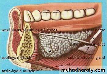
Sublingual Salivary Gland
1- Cysts(ranula)
mucous extravasation cyst that arises from a sublingualgland.
- translucent swelling ‘frog’s belly’ .
resolve spontaneously.
many require formal surgical excision of the cyst and the affected sublingual gland.
Common disorders of the sublingual glands
rare mucous retention cyst
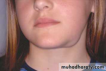
arise from sublingual and submandibular s g.Mucus collects within the cyst, which perforates through the mylohyoid muscle diaphragm to enter the neck.
dumb-bell-shaped swelling
Soft
fluctuant
Painless
in the submandibular or submental region of the neck .
Diagnosis : ultrasound or magnetic resonance imaging (MRI).Treatment:
- Excision transcervical ( removing the cyst+ submandibular+ sublingual glands).
- Smaller plunging ranulas transoral sublingual gland excision+/- marsupialisation.
2- Plunging ranula
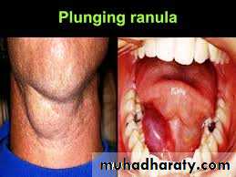
- extremely rare
85 per malignant.hard or firm
painless swelling
in the floor of the mouth.
Treatment
wide excision + overlying mucosa+ neck dissection. +reconstruction .
Tumors of sublingual
Ectopic/aberrant salivary gland tissue
Stafne bone cyst
- most common ectopic salivary tissue .
Asymptomatic
clearly demarcated radiolucency of the angle of the mandible.
below the inferior dental neurovascular bundle.
No treatment is required.
THE SUBMANDIBULAR GLANDS
Inflammatory disorders of thesubmandibular gland(sialadenitis)
acute, chronic or acute on chronic.Common causes are:
Acute submandibular sialadenitis:
1- Bacterial sialadenitis
more common than viral sialadenitis
- secondary to obstruction.
- antibiotics, if chronically inflamed formal excision.
2- Viral. The paramyxovirus (mumps)
Usually parotitis.occasionally submandibular glands
painful tender swollen glands.
Other viral infections rare.
Chronic submandibular sialadenitis.
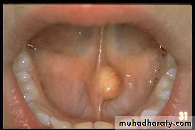
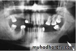
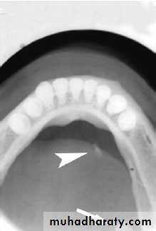
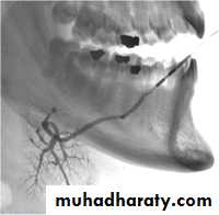
Stone formation (sialothiasis): most common cause of obstruction within the submandibular gland is within the gland and duct system.
80%of all salivary stones occur in the submandibular glands because highly viscous secretions .
80% submandibular stones are radio-opaque
Obstruction and trauma to submandibular gland
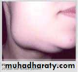
acute painful swelling in the region of the submandibular gland
precipitated by eatingcompletely obstruct(less common) opening of the submandibular ductswelling develop rapidly 1–2 hours after the meal resolves spontaneously
partial obstruction(more common) (hilum of the gland or within duct in the floor of the mouth)
- infrequent symptoms
- minimal discomfort and swelling
- not confined to mealtimes.
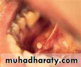
- examination enlarged firm submandibular gland, tender on bimanual examination.
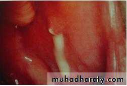
Pus from the sublingual papilla .
Clinical symptoms
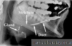
sialogram
Management
stone within the submandibular duct in the floor of the mouth anterior to the point at which the duct crosses the lingual nerve (second molar region) incising over the duct+stone delivered+ leave the wall of the ductstone is proximal to the lingual nervesubmandibular gland excision a removal of the stone + ligation of the submandibular duct
endoscopic retrieval of stone, lithotripsy(sialadenoscope)
Indication:sialadenitis
Salivary tumours.
Complications:
• haematoma
• wound infection
• marginal mandibular nerve injury
• lingual nerve injury
• hypoglossal nerve injury
• transection of the nerve to the mylohyoid muscle producing submental skin anaesthesia.
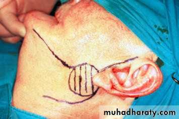
Submandibular gland excision
- uncommon
- (benign and malignant)present as a slow-growing, painless swelling within the submandibular triangle.50 % of submandibular gland tumours are benign.
pain is not a reliable indication of malignancy as benign tumours often present with pain in the affected gland, presumably due to capsular distension or outflow obstruction.
Tumours of the submandibular gland
• facial nerve weakness
• rapid enlargement of the swelling• induration and/or ulceration of the overlying skin
• cervical node enlargement.
Clinical features of malignant salivary tumours
1-Computed tomography (CT) and MRI scanning
the extension, circumscribed (benign, or diffuse, invasive and probably malignant).2- Biopsy
Open surgical biopsy is contraindicated as this may seed the tumour into surrounding tissues, making it impossible to eradicate
microscopic deposits of tumour cells.
Fine-needle aspiration biopsy
no risk of seeding viable
Investigation
surgical excision with a cuff of normal tissue is the goal.
- Small tumor+ localised ( entirely within the submandibular gland parenchyma) intracapsular submandibular gland excision is
- Benign tumors (large and beyond the submandibular gland) suprahyoid neck dissection
(preserving the marginal mandibular branch of the facial nerve, lingual nerve and hypoglossal nerves).- Overt malignancymodified neck dissection /radical neck dissection .
(may sacrifice of the lingual and hypoglossal nerves )Management of submandibular gland tumours
Developmental disorders
Rareagenesis, duct atresia and congenital fistula
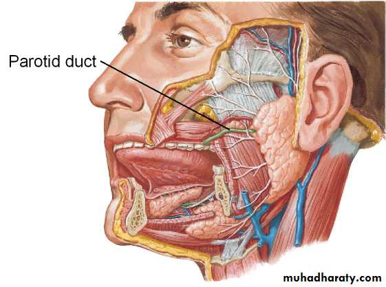
THE PAROTID GLAND
Viral infections
Mumps (most common)acute painful parotid swelling
Mostly affects children.
spread by airborne droplets .
1–2 days fever, nausea and headache
pain and swelling in parotid glands.
pain very severe and exacerbated by eating and drinking.
Symptoms resolve within 5–10 days.
Treatment : regular paracetamol + adequate oral fluid intake.
Complications
orchitis, oophoritis, pancreatitis, sensorineural deafness and meningoencephalitis are rare.
Other viral agents that produce parotitis include Coxsackie A and B, parainfluenza 1 and 3, Echo and lymphocytic choriomeningitis.
Inflammatory disorders

Acute ascending bacterial sialadenitis
dehydrated elderly patients following major surgery Reduced salivary flow ascending infection.
Staphylococcus aureus /Streptococcus viridans
Can occur with no obvious precipitating factors.
presentation tender, painful parotid swelling that arises over several hours .
generalised malaise, pyrexia and occasional cervical lymphadenopathy.
The pain is exacerbated by eating or drinking.
The parotid swelling may be diffuse/localises (lower pole of the gland)
Pus may exuding from the parotid gland papilla
Bacterial infections

Treatment: - intravenous antibiotics. abscess drainage (large bore needle aspiration / drainage under general anaesthesia. Chronic bacterial sialadenitis is rare in the parotid gland.
Papillary obstruction
less common than obstructive submandibular sialadenitiscaused by trauma to the parotid papilla
overextended upper denture flange or a fractured upper molar tooth.
inflammation and oedema obstructs salivary flow(mealtimes)
rapid onset pain and swelling at mealtimes.
untreated progressive scarring and fibrosis in and around the parotid duct papilla will produce a permanent stenosis.
Treatment: papillotomy(under either local or general anaesthesia).
- Recurrent parotitis of childhood- Obstructive parotitis
Stone formation(Sialolithiasis)
- less common in the parotid gland (20 %)
(submandibular gland (80 %).
- Parotid duct stones radiolucent.
- Stone either proximal in the collecting duct or distal near the papilla.
- Diagnosis: Parotid gland sialography.
- Treatment: stone located in the collecting duct or within the gland endoscopic retrieval, lithotripsy or rarely parotidectomy.
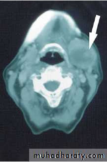
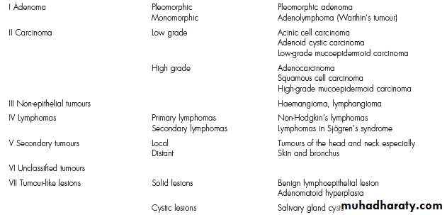
- The parotid gland is the most common site for salivary tumors.
Most tumors arise in the superficial lobe80–90 % of tumours of the parotid gland are benign
the most common is pleomorphic
Slow growing, painless swellings below the ear, in front of the ear or in the upper aspect of the neck.
- Less commonly, tumors in accessory lobe persistent swellings in the cheek.
- Rarely, tumours in deep lobe as parapharyngeal massesdifficulty in swallowing and snoring.
Tumours of the parotid gland
Malignant salivary gland tumours are divided into twodistinct subgroups:
1- Low-grade malignant tumours, e.g. acinic cell carcinoma,
are indistinguishable on clinical examination from benign
neoplasms.
2- High-grade malignant tumours :
Rapidly growing
painless swellings
discrete mass with infiltration into the overlying skin
or diffuse + hard swelling + no discrete mass(advanced disease)
- Cervical lymph node metastases.
CT and MRI scanning
Fine-needle aspiration biopsy
(open surgical biopsy is contraindicated )
(no enucleation even if a benign lesion is suspected)
Investigations
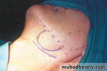
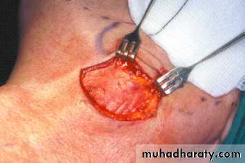
Superficial parotidectomy for Superficial lobe tumorThe aim of superficial parotidectomy is to remove the tumor with a cuff of normal surrounding tissue.
Low-grade malignant tumours superficial parotidectomy.
Radical parotidectomy
Indicated in high-grade malignant tumour(squamous cell carcinoma)Radical parotidectomy = removal of all parotid gland tissue +sectioning of the facial nerve+ ipsilateral masseter muscle +/- neck dissection(if positive LN mets.)
Treatment of parotid tumor
• haematoma formation;
• infection• temporary facial nerve weakness
• transection of the facial nerve and permanent facial weakness
• sialocoele
• facial numbness
• permanent numbness of the ear lobe associated with great auricular nerve transection;
• Frey’s syndrome.
Complications of parotid gland surgery
Frey’s syndrome (gustatory sweating)
- damage to the autonomic innervation of the salivary gland with inappropriate regeneration of parasympathetic nerve fibres that stimulate the sweat glands of the overlying skin.
sweating and erythema over the region of surgical excision of the parotid gland as a consequence of autonomic stimulation of salivation by the smell or taste of food.
Dgxstarch iodine test.
Rx antiperspirants( aluminium chloride)
denervation by tympanic neurectomy;
botulinum toxin injection into the affected skin.
Frey’s syndrome
Pleomorphic adenoma
Benign Tumor- On gross inspection : tumors is smooth and lobular and demonstrates a well defined capsule
On microscopic examination : both epithelial and mesenchymal elements are present
MOST COMMON NEOPLASM IN THE PAROTID GLAND ACCOUNTS FOR 65% OF ALL OF THE PAROTID TUMORS.
The most common salivary T.In middle aged & more in woman than in men,
Slowly growing
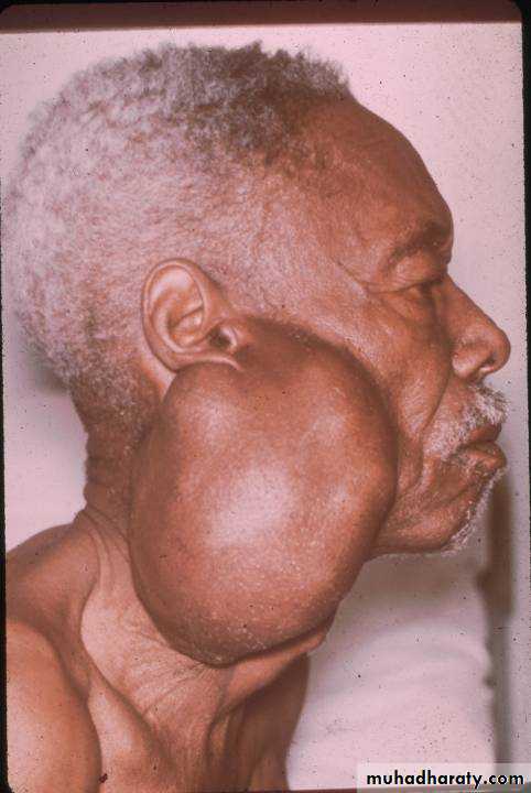
Treatment :
Superficial parotidectomyWIDE RESECTION OF THE TUMOR
AVOID SHELLING OUT THE LESION
RECURRENCE: PRIMARY DUE TO INADEQUATE RESECTIONLESIONS ARE MORE AGGRESSIVE WHEN THEY RECUR
WARTHIN’S TUMOR (ADENOLYMPHOMA)SECOND MOST COMMON PAROTID TUMOR
MALE : FEMALE 5 : 1
BILATERAL 10%
May (MULTICENTRICITY).TREATMENT: superficial parotidectomy
90%CURED WITH RESECTION
10%RECUR DUE TO MULTICENTRICITY OR INADEQUATE RESECTION
Malignant neoplasm
Mucoepidermoid carcinomaAdenoid cystic carcinoma
Acinic cell carcinoma
adeno carcinoma
Carcinoma Ex. Pleomorphic adenoma or malignant mixed tumor
Squamous cell carcinoma
Undifferentiated carcinoma
miscellaneous
A-Granulomatous sialadenitis:
Mycobacterial infectionSarcoidosis
B-Tumour-like lesions
Sialadenosis
C-Degenerative conditions
- Sjögren’s syndrome
autoimmune condition causing progressive
destruction of salivary and lacrimal glands.
- Benign lymphoepithelial lesion
Xerostomia(decrease salivary flow)
Sialorrhoea(increase salivary flow)
Other diseases of salivary glands