mesodermal condesation in the dorsal
mesogastrium
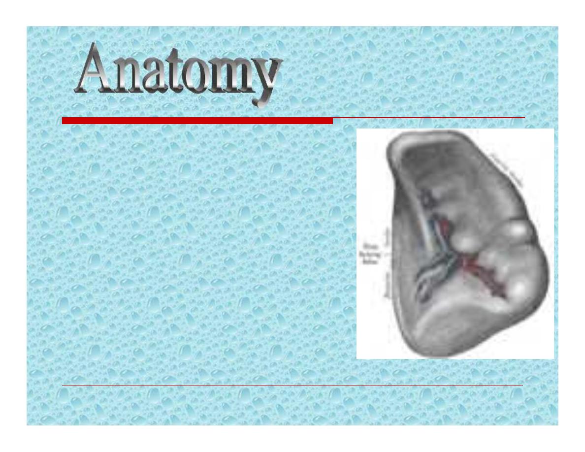
It weighs 75 – 250 g
It lies in the left
hypochondrium
Along the line of tenth rib
Its hilum lies in the angle
between the stomach and
kidney, and is in contact
with the tail of pancreas
There is a notch in its
inferolateral border.
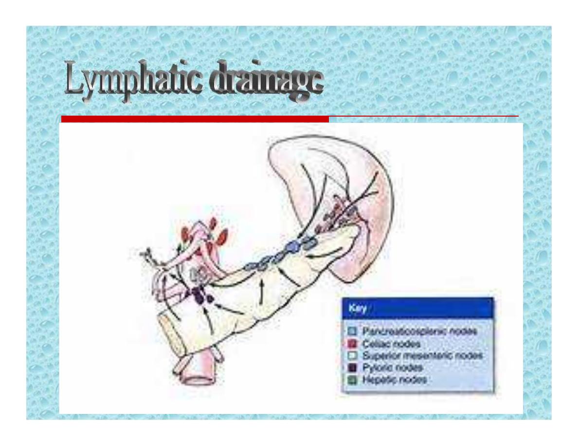
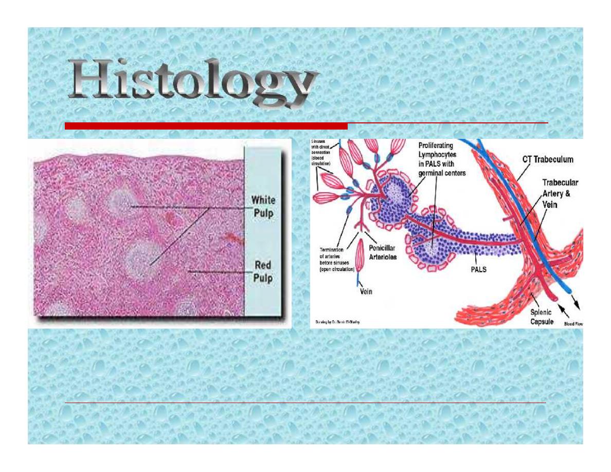
PALS;periarterial lymphatic sheath

Immune function
Major site of IgM production
Opsonins, tuftsin, and properdin
Filter function
Macrophages in the reticulum capture
cellular and non-cellular material from
the blood and plasma.
Effeet platelets & RBCs
Bacteria , pneumococci

Pitting
Removal of particulate particles from
RBCs, like the Howell-Jolly & Heinz
bodies.
Reservoir function
It is less marked but dose the spleen
contain approximately 8% of the red cell
mass.

Cytopoiesis
Haemopoiesis during fetal life.
Proliferation of T & B cells and
macrophages following antigenic
stimulation.

Imaging:
Plain radiology:
Rarely used, but incidental finding of
calcification
of the splenic artery __
splenic artery aneurysm
calcification
of spleen__an old infarct, a
benign cyst or Hydatid cyst.
multiple calcifications__tuberculosis

Ultrasonography;
Determine the size & consistency
SOL, cystic or solid
CT scan;
Better in determing the nature of
suspected splenic pathology.
MRI scan similar value to CT scan
Radioisotope:
Less commonly requested
Technetium99mm-labelled colloid is
restricted to determine whether the
spleen is a significant site of destruction
of RBCs.
Congenital abnormalities of the
spleen:
Splenic agenesis
, is rare but present in
10% of children with congenital heart
disease.
Polysplenia
, is a rare condition resulting
from failure of splenic fusion.

Splenunculi:
Are single or multiple accessory spleens
that are found in approximately 10-30% of
the population.
Located near the hilum in 50%,
Related to the splenic vessels or behind the
tail of pancreas in 30%.
Remainder are located in the mesocolon or
the splenic ligaments.
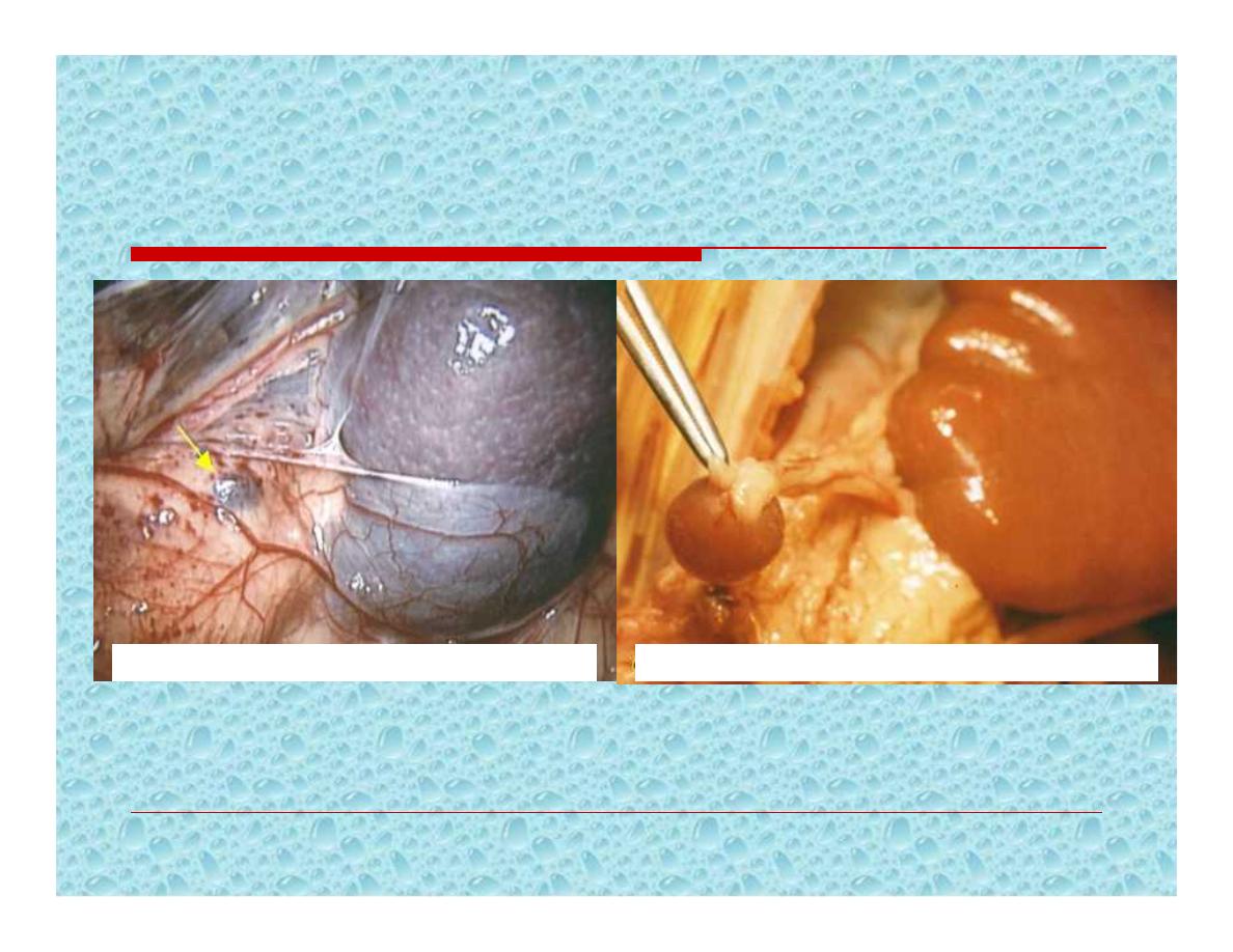
ACCESSORY SPLEENS
Accessory spleen in greater omentum
Accessory spleen
Hamartomas:
Are rarely found in life
Non-parasitic cysts:
Are rare. True cysts form from
embryonal rests.

Aetiology:
Blunt abdominal trauma
, splenic
injury should be suspected in any case
of blunt abdominal trauma,
particularly when the injury occurs to the
left upper quadrant of the abdomen.
And specially if there are fractures of the
overlying ribs ( 9
th
,10
th
,11
th
).

Penetrating abdominal trauma:
Stab and missile injury
Iatrogenic:
Splenic injury is a frequent complication of
any surgical procedure, particularly those in
the left upper quadrant.

Presentation
:
may present in three ways
The patient succumbs rapidly
Initial shock, recovery, signs of late
bleeding
The delayed case

Clinical signs of ruptured spleen:
General signs of internal hge
Left upper quadrants guarding and
tenderness.
Kehr’s sign
Shifting dullness
Fullnes in the pelvis
Diagnosis:
Abdominal ultrasonography (FAST)
CT scan

Management:
Conservative: applied in blunt trauma
Only in Haemodynamically stable patients
Minimal or no abdominal findings
CT scan *isolated injury.
* absence of hilar involvement or
massive disruption of spleen.

Immediate laparotomy:
Obvious evidence of continuous blood loss
despite adequate resuscitation.
Strong suspicion of trauma to other organs
Splenic preservation should be
considered wherever possible.
Persons with splenomegaly like in
malaria are more liable to splenic
rupture after trivial trauma.
Trend for early splenectomy
Delayed Rupture of the Spleen

May arise from:
Infected splenic embolus
In association with typhoid and paratyphoid
fever, osteomyelitis, otitis media, and
purperal sepsis.
Panreatic necrosis
In association with intraabdominal
infection.
Complications:
Rupture:
Subphrenic abscess
Peritonitis
Diagnosis:
Ultrasound
CT scan
Treatment:
Underlying cause
drainage
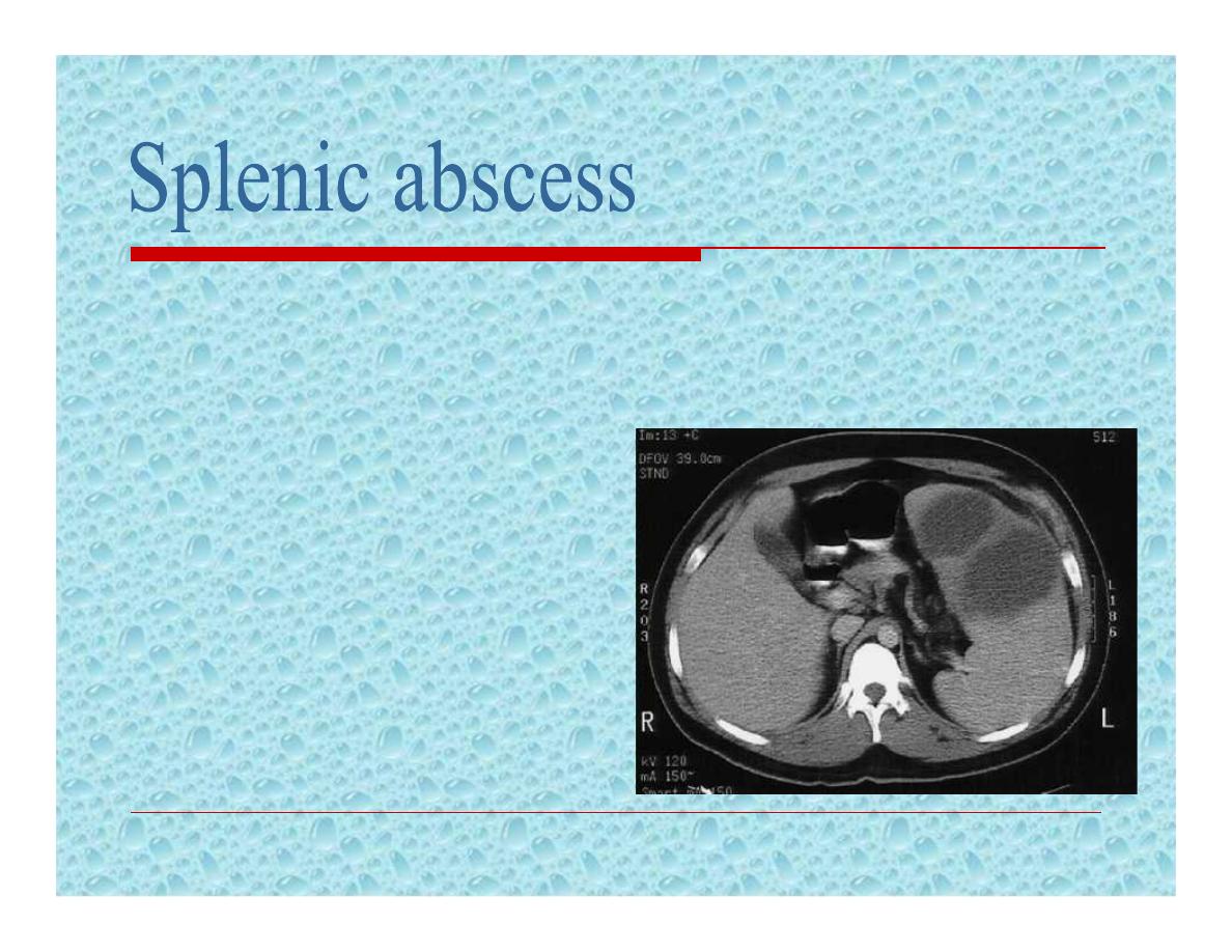
Considered in young patients with:
Splenomegaly, asthenia, loss of weight,
and fever.
Complications:
Cold abscess
Portal hypertension
Treatment:
Anti-TB drugs
Splenectomy
Splenic enlargement
, frequently
massive, found in the tropics
attributed to;
malaria
,
schistosomiasis
, and
kalaazar
.
Occasiosionally attributed to
malnutrition.
Removal of spleen is frequently
required.
Prevalent in Africa, Asia, and South
America.
S.mansoni 75%,S.haematobium 25%
Pathogenesis:
enlargement of spleen arise
either as a result of ;
Hepatic fibrosis portal hypertension
Or splenic enlargement may result from
hyperplasia induced by phagocytosis.
Diagnosis:
Exam. of urine & faeces for ova
Abnormal liver function test
Hypochromic anaemia
Treatment:
Medical not helpful for regression of
splenic enlargement
Usually removal of spleen is needed.

Pathogenesis:
ABs. That damage the patient’s own platelets
It is of two types
Acute in childhood
Chronic in adults

Acute in childhood usually follows an
acute infection and has a spontaneous
resolution within 2 months.
Chronic in adults seen in associated with
other conditions, including systemic
lupus erythematosus, chronic lymphatic
leukaemia and Hodgkin’s disease

Clinical features:
Adult type Affects females of 15 – 50
years
Purpuric patches in the skin and mucous
membrane spontanous or after trivial
trauma

Hemorrahage from the urinary tract
and GIT and haemoarthrosis are rare.
Intracranial hge is uncommon but it is
the most common cause of death.
Signs:
Ecchymosis
Tourniquet test
Palpable spleen in less than 10%

Investigations:
Prolonged bleeding time
Clotting and prothrombin time normal
Reduced platelets count (usually <
60*10^9/litre.
Bone marrow aspiration; plentiful platelet-
producing megakaryocytes.

Treatment:
Medical; steroids, good prognosis in
pediatrics with 75% incidence of
spontaneous regression
Splenectomy:
Is indicated when;
The patient develops two relapses on steroid
therapy
Low platelets in spite of steroid therapy
When ITP persisted for >than 6-9months
Splenectomy: will lead to;
cure in up to two-thirds of patients,
15% of patients show improvement
The remainder show no benefit.
Preoperative:
Fresh blood transfusion
Platelet concentrate.

Indications:
Urgent
Splenic injury, accidental or iatrogenic
Ellective
1- Oncological:
Part of en bloc resection
Diagnostic
Therapeutic

Indications:
2- Haematological
ITP
Haemolytic anaemias
1- Hereditary spherocytosis
2- Acquired autimmune haemolyic anaemia
3- Thalassaemia
4- Sickle cell anaemia
Hypersplenism
3- Portal hypertension
Variceal surgery

Preoperative preparation
I- Normalization of coagulation profile:
In the presence of bleeding tendency;
Transfusion of blood
Fresh-frozen plasma
Cryoprecipitate
platelets

Preoperative preparation
III- Vaccination; against
Pneumococcus (those over 2 years)
H. influenzae ( for all ages )
Meningococcus ( recommended in high risk
areas )
Influenza virus.
Note: in trauma victim, vaccination can be
given in the postoperative period.

Technique:
Open splenectomy
Laparoscopic splenectomy
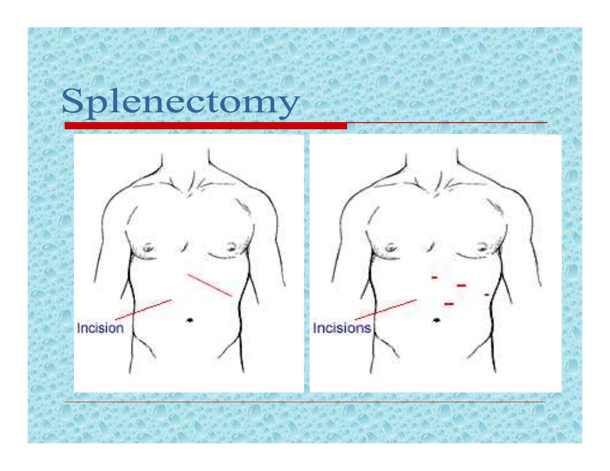
Open splenectomy
Laparoscopic splenectomy
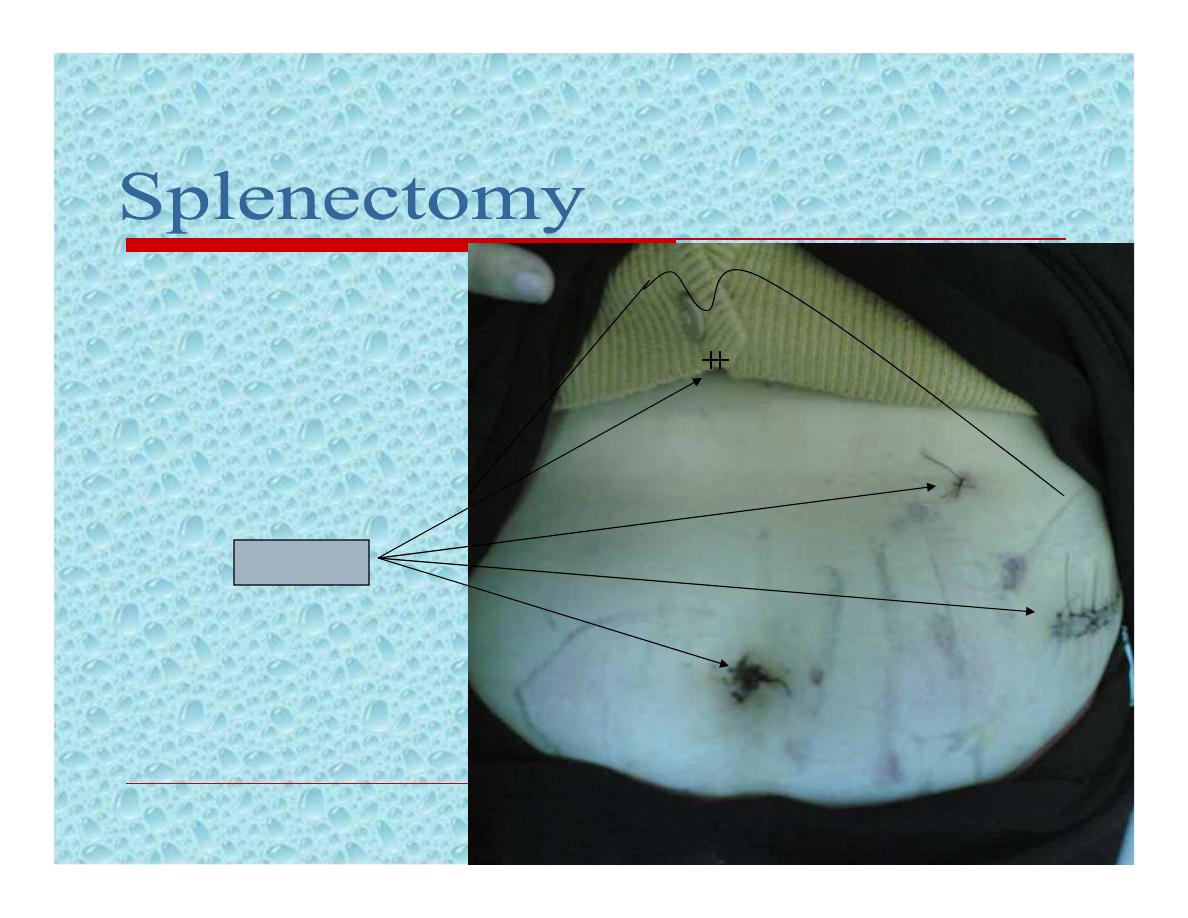
4 ports
10mm
5 mm
5 cm
10 mm

Postoperative complications:
Local:
Haemorrhage
Haematemesis
Gastric dilatation

Postoperative complications:
Local:
Iatrogenic injury to adjacent organs;
like
1-
Pancreas
; may lead to:
Pancreatitis
Local abscess
Pancreatic fistula
2-
Stomach
; a fistula may result from
damage to the greater curvature during
ligation of short gastric vessels.
3-
Colon
, splenic flexure

Postoperative complications:
Systemic:
1- Left basal atelectasis.
2- Pleural effusion.
3- Thrombocythemia;
Prophylactic aspirin is recommended if
platelet count exceeds one million per
millilitre, to prevent axillary or other venous
thrombosis.

Postoperative complications:
Systemic:
4- Post-splenectomy septicaemia.
5- Opportunist pos-splenectomy infection.
May result from S. pneumonia, N.
meningitidis, H. influenzae, and E. coli.
Is a major concern for children who
undergone splenectomy before the age of 5
years.

Postoperative complications:
Systemic:
The risk is increased in:
Young patients
Chemotherapy
Splenectomy for haematological disorders.
How do we can decrease the risk:
Vaccination
Prophylactic antibiotics
Postoperative recommendation
II- Antibiotic prophylaxis;
Daily oral penicillin, or erythromycin, or
amoxicillin, or co-amoxiclav until the age of 10
years for those children who have undergone
splenectomy before the age of 5 years.
For older children and adults is controversial;
but since the risk of overwhelming sepsis is
greatest during the first 2 – 3 years of
splenectomy it seems reasonable to give
prophylaxis antibiotics during this time.
Autosomal dominant
Defect in cell membrane; increased permeability
to Na. Increased entry of H2O inside the RBC
spherocytic RBC.
Spherocytic RBCs are fragile and need increased
energy and O2 to pump sodium outside the cell.
Spherocytic RBCs are destroyed in the spleen,
indirect bilirubin liver excretion of
bilirubin with bile bile pigments stones.

Clinical features:
Presentation generally in childhood
Intermittent jaundice, associated with
anaemia, splenomegaly and gall stones.
Haemolytic crisis; characterised by the
onset of pyrexia, abdominal pain, nausia
and vomiting and extreme pallor followed
by increased jaundice

Examination:
Pallor, mild jaundice, leg ulcers
Splenomegaly
Normal color urine

Investigation:
Increased indirect bilirubin
Anaemia, immature reticular cells
Increased urobilinogen in faeses and urine
Increased fragility test of RBCs;
Normal RBCs begin to haemolyse in 0.47%
saline
In HS aemolysis occur in 0.6% saline

Any child with gall stone disease
should be investigated for hereditary
spherocytosis and a family history
sought

Investigation:
Radioactive chromium (51Cr) labelling of
the patient’s own RBC
Decreased life span of RBCs
Increased sequestration of RBCs in spleen.
Ultrasound examination:
Splenomegaly
Gall stones

Treatment:
Splenectomy;
In young children it is preferable to delay
splenectomy until 6 years of age to
minimise the risk of post-splenectomy
sepsis.

NEOPLASMS
Haemangioma
Lymphoma is the most common
cause of neoplastic enlargement
The spleen is rarely the site of
metastatic disease

Cysts of the Spleen
Non parasitic
Embryonal cysts
Parasitic
Hydatid cyst

Cysts of the Spleen
Selected nonparasitic cyst may be
managed by aspiration
Splenectomy should be performed for all
large cyst and those with an uncertain
diagnosis