1
Forth stage
Surgery (Urology)
Lec-19
د.محمد فوزي
20/12/2015
Carcinoma of the prostate CAP
One of the most common malignant tumor affecting males over the age of 65 in western
countries.
Pathology :
95% of the tumor are adenocarcinoma and derived from acinar epithelium
75% of CAP arise from peripheral zone.
grading:
Gleason’s grade based on the degree of glandular differentiation and growth pattern.
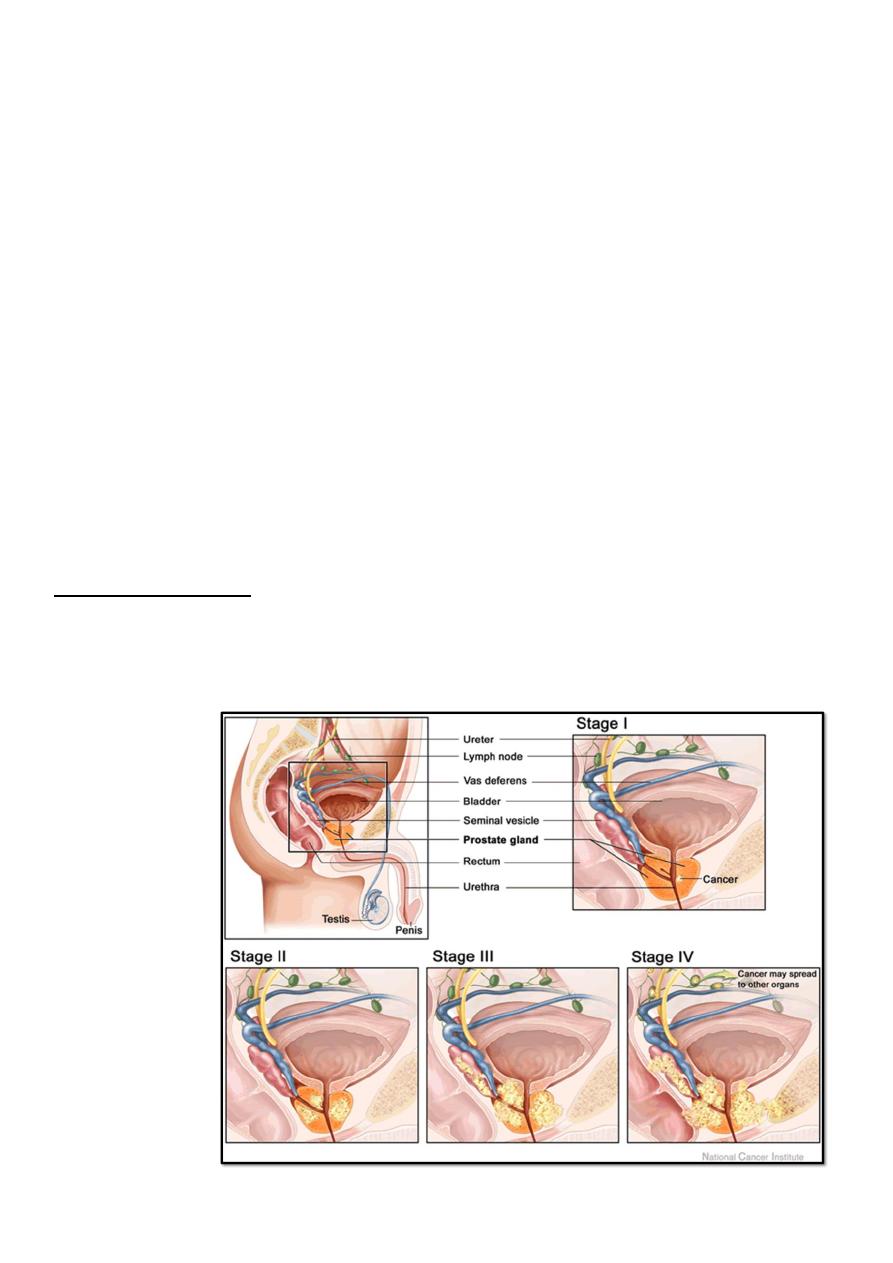
Spread :
Direct invasion: to nearby structures.
Denonvvilliar’s fascia act as barrier.
Lymphatic: internal, external & common iliac
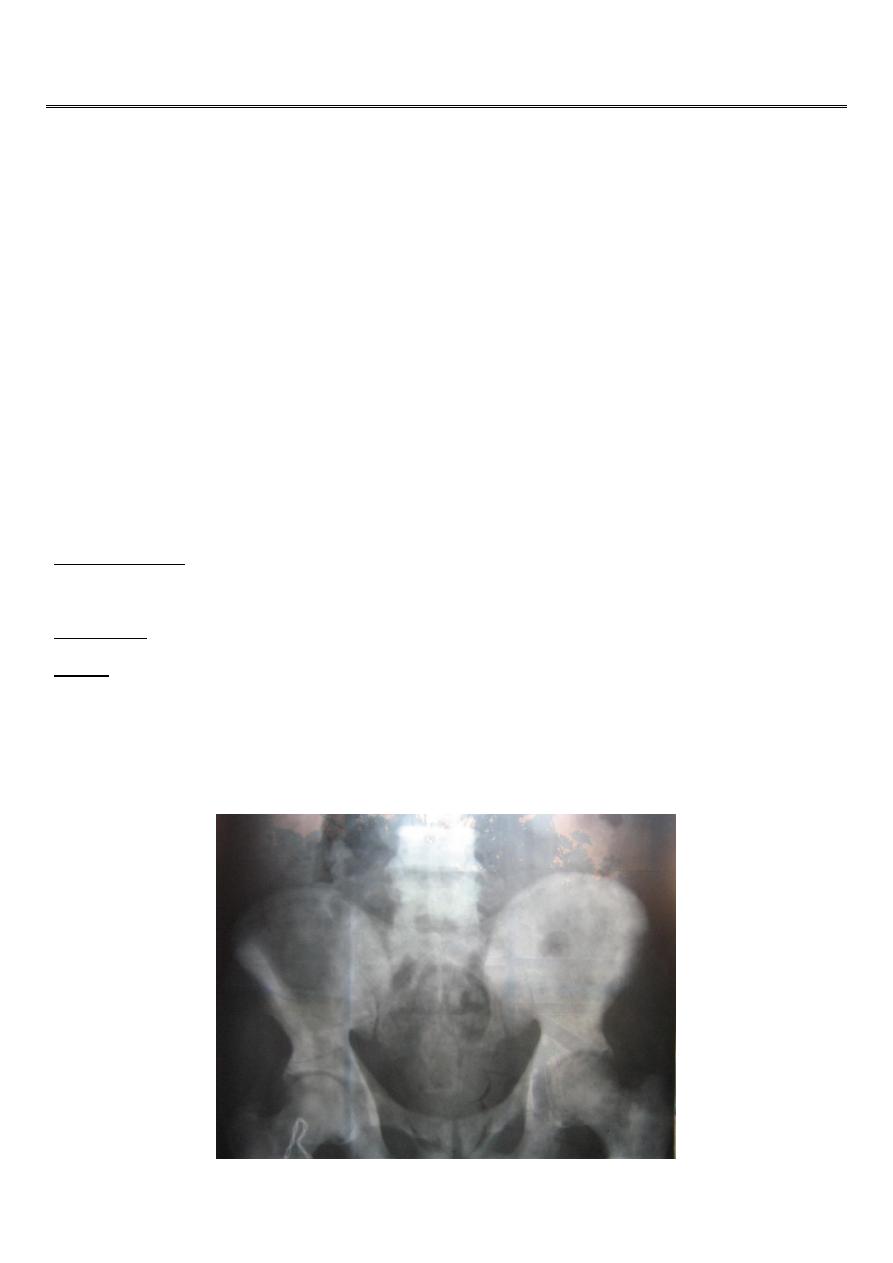
Blood: to the lower lumber vertebrae & pelvic bones due to reverse blood flow from
vesicoprostatic plexus to the emissary veins of the bones during coughing & sneezing
(OSTEOBLASTIC)
Osteoblastic lesion of secondary CAP
2
Presentation :
- Accidental during histopathological ex after prostatectomy.
- During PR ex
- High PSA
- BOO.
- Metastatic: back ache, sciatica, paraplegia or pathological fractures.
BPH CAP
Younger age older
Symptoms slowly progressive rapid progression
Usually no back or bone pain more back ache &neurological symptoms
Smooth rubbery prostate with sulcus
hard irregular prostate with obliterated
Sulcus
Rectal examination:
Stony hard irregular prostatic nodule, obliterated median sulcus, difficulty in moving the
rectal mucosa over it and fixity.
Normal PR ex does not exclude CAP.
3
Investigations :
PSA: prostatic tumor marker for diagnosis and follow up, it may also increase in prostatitis
and BPH.
10 ng/ml normal, 10-15 suspicious.
>15 is diagnostic.
Acid phosphatase: prostatic fraction.
Alkaline phosphatase: in bone metastasis.
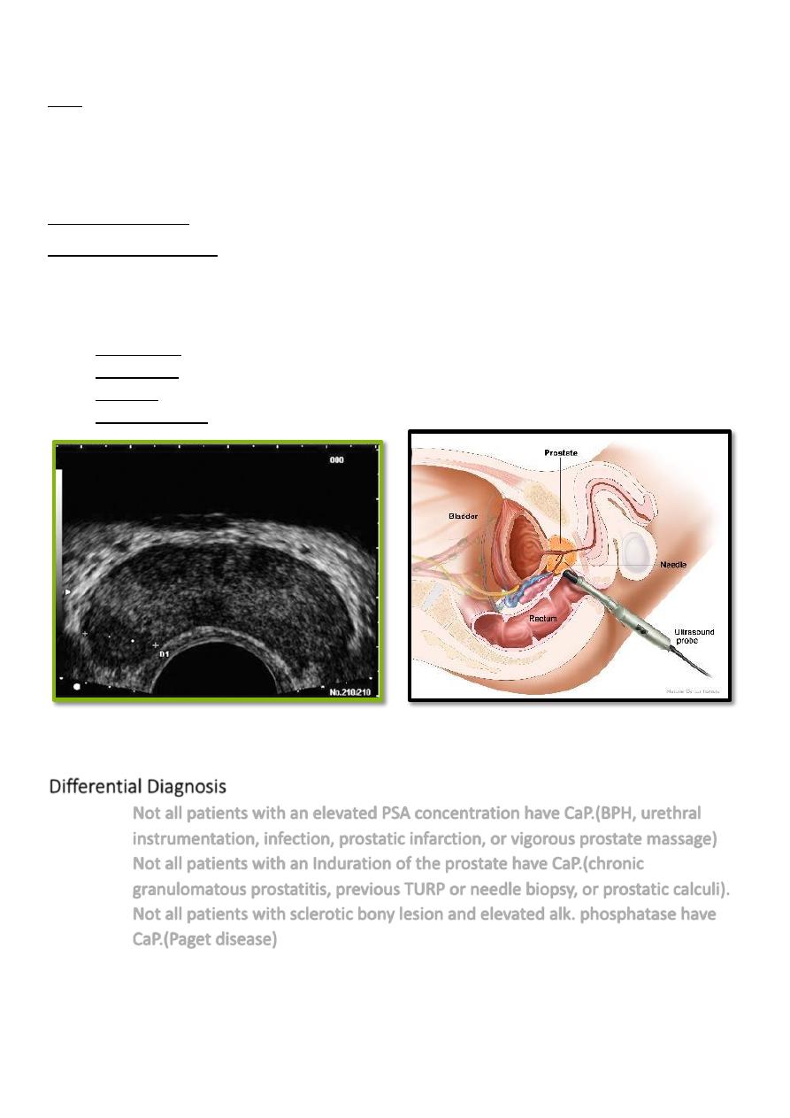
Radiological investigations :
- Plain X ray: osteoblastic lesion.
- Bone scan: hot areas (active).
- CT scan.
- TRUS & biopsy (sixtant biopsy).
Differential Diagnosis :
Not all patients with an elevated PSA concentration have CaP.(BPH, urethral
instrumentation, infection, prostatic infarction, or vigorous prostate massage)
Not all patients with an Induration of the prostate have CaP.(chronic
granulomatous prostatitis, previous TURP or needle biopsy, or prostatic calculi).
Not all patients with sclerotic bony lesion and elevated alk. phosphatase have
CaP.(Paget disease)
4
Treatment :
Watchful waiting:
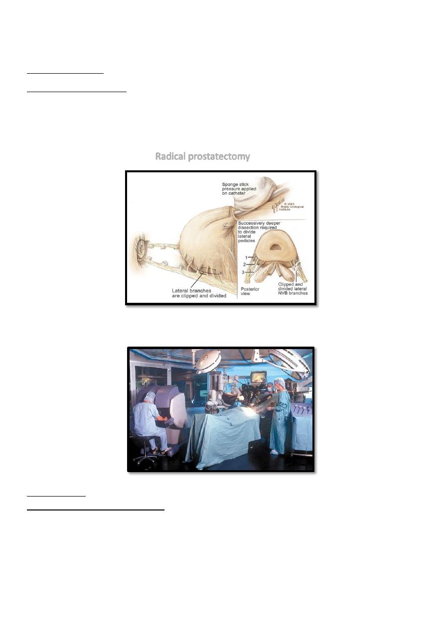
Radical prostatectomy:
Enblock surgical removal of the entire prostate, seminal vesicles and pelvic lymph nodes.
The bladder anastomosed to the urethra.
Indicated for early disease and healthy fit pt.
Radical prostatectomy
ROBOTIC RADICQL PROSTQTECTOMY
Radiotherapy
external beam & brachytherapy
Indication:
1- Locally advanced disease.
2- Unfit patient for surgery.
3-Symptomatic metastases to relieve pain.

5
Brachytherapy
external beam therapy
Hormonal therapy
Its trearment of choice for metastatic tumor
Cap is hormonal dependant (androgen), and about one third of tumors are hormone-
insensitive.
Androgen ablation may change the course of the disease
Methods of androgen ablation :
-Surgical:
Bilateral orchiectomy: complete or subcapsular.
-Medical:
LHRH agonist: (Zoladex)/28 days SC.
Anti androgen: (Nilutemide) 250 mg/6h.