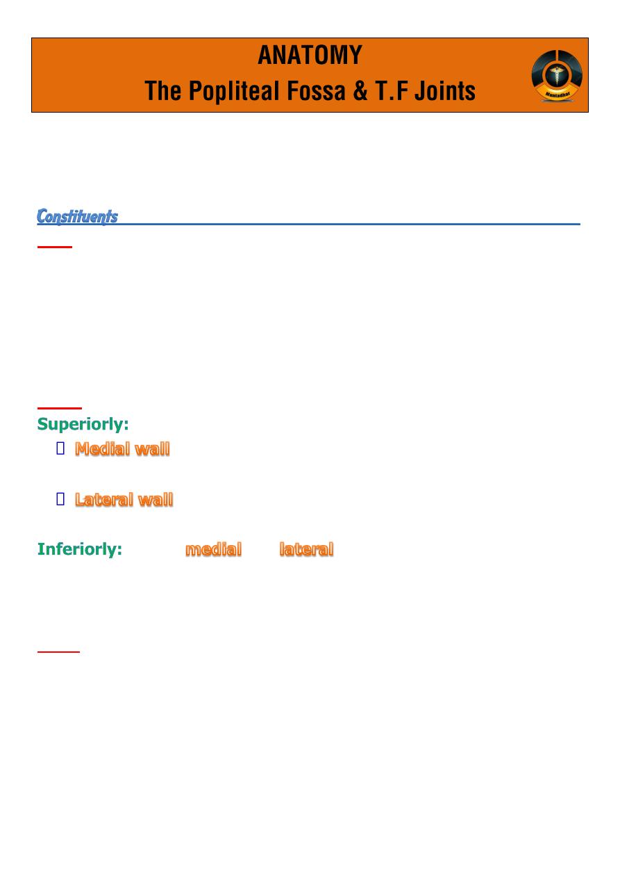
Is a diamond-shaped space behind the knee joint.
It has roof, 4 walls and floor :
Roof
is formed by
investing layer of deep fascia
.
The superficial fascia overlying the roof contain :
lateral cutaneous nerve of thigh
short saphenous vein
Walls:
is formed by diverging tendon of hamstring muscles :
is formed by tendon of semimembrenosis and
Semitendnosis muscles
is formed by tendon of biceps
are the
and
head of
gastrocnemius
which
converge at inferior angle.
Floor:
is formed from above downwards by :
popliteal surface of femur
capsule of the knee joint
reinforced by
the oblique popliteal ligament
popliteus
deep fascia covering popliteus.
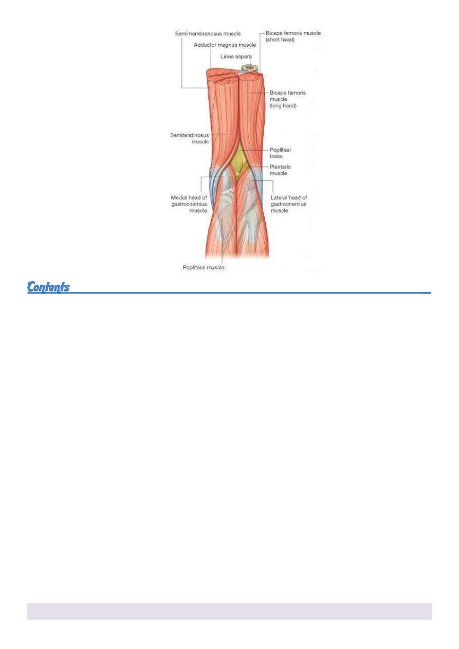
1. popliteal artery (lies deep)
it enters the fossa through the opining in
adductor magnus
and
descends vertically on the floor of the fossa.
it leaves the fossa
beneath the head of gastrocnemius muscles
.
it supplies the surrounding muscles and give anatomizing branches
around knee.
2. popliteal vein
lies superficial to the artery and is formed by the union of
venae
comitantes of tibial arteries in leg
.
3. Tibial and common peroneal nerve (branches of sciatic nerve)
entering the fossa from posterior compartment
lie just beneath the roof superficial to popliteal vessels.
4. Popliteal lymph nodes
deeply embedded, closed to popliteal artery.
5. Lower part of posterior femoral cutaneous nerve
articular branch from obturator nerve .
All these contents embedded in a fat and connective tissue.
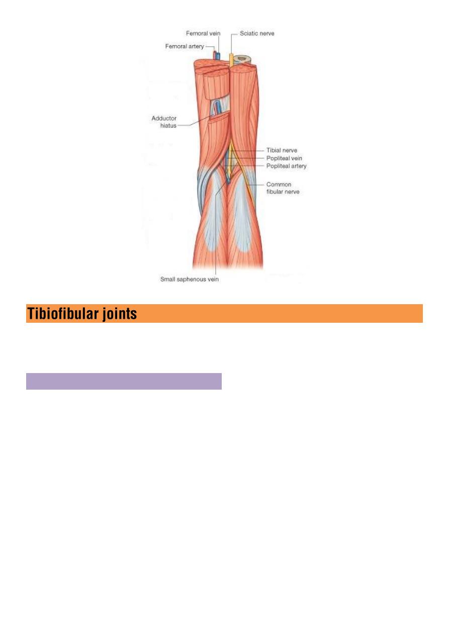
The tibia and fibula articulate at
proximal and distal
tibiofibular joint
and are also connected by
interosseous membrane
.
Proximal Tibiofibular joint
Is synovial gliding joint.
It is an articulation between the
lateral condyle of tibia
and
head of
fibula
.
The articular surfaces are flattened and covered by
hyaline cartilage
The
capsule
surrounds the joint is attached to the margins of articular
surfaces
The capsule lined by synovial membrane, and attached to articular
surfaces.
Anterior and posterior ligaments
strengthening the joint.
The joint supplied by
common peroneal nerve
.
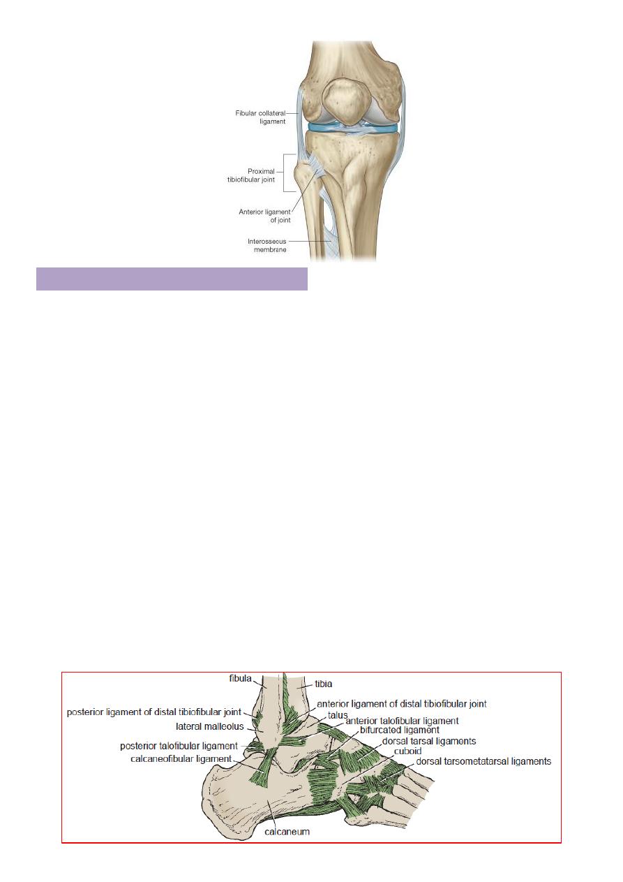
Distal Tibiofibular joint
It is fibrous joint.
It is an articulation between the
fibular notch of tibia
and
lower end of
fibula
.
There is
no capsule
.
Anterior and posterior ligaments (
band of connective tissue
)
connecting the two bones together.
Interosseous ligament (
strong thick band of fibrous tissue connecting
the two bones together
).
Inferior transvers ligament runs from
medial surface of upper part of
lateral malleolus
to
the posterior border of lower end of tibia
.
The joint supplied by
tibial and common peroneal
nerve
.
A small amount of movement takes place
during movement of ankle
joint