Fourth stage
MedicineLec-1
اسم الدكتور
1/1/2014
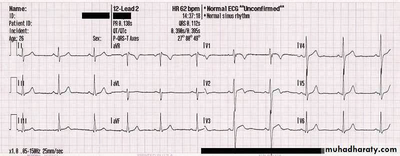
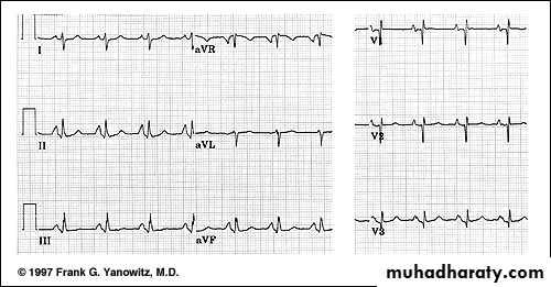
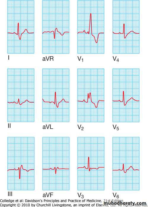
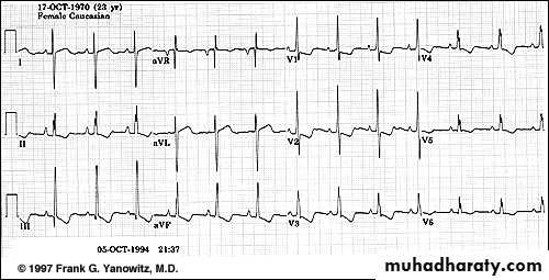
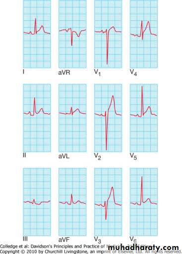
The 12 leads stndard ecgNormal ECG
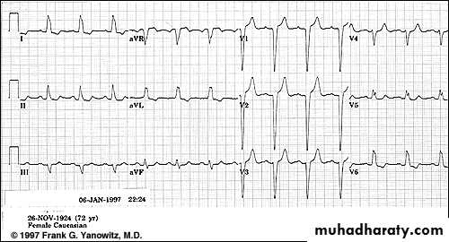
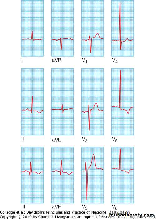
Abnormal ECG
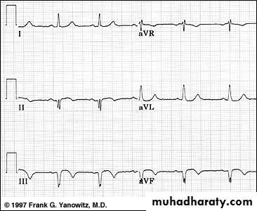
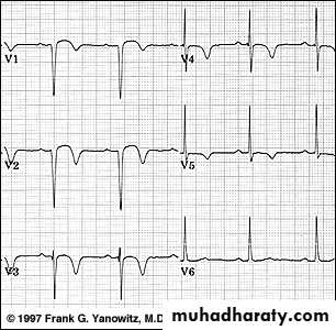
The 12 leads stndard ecg
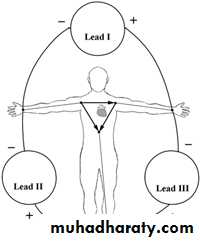
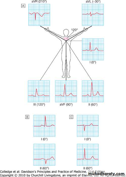
*6 standard limb leads
(orianted on the frontal plane of the heart)
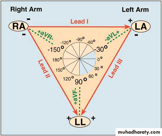
-bipolar limb leads
Lead I :RA(-)to LA(+)
Lead II :RA(-)to LF(+)
Lead III :LA(-)to LF(+)
-augmented unipolar limb leads
lead AVR : RA(+) to (LA&LF) (-)
lead AVL : LA(+) to (RA&LF) (-)
lead AVF : LF(+) to (LA&RA) (-)
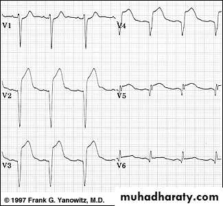
*6 unipolar (+) chest leads
Orianted on the horizental plane of the heart)
-leads v1,v2,v3,v4,v5,v6
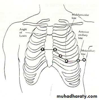
V1: right 4th intercostal space
V2: left 4th intercostal space
V3: halfway between v2 and v4
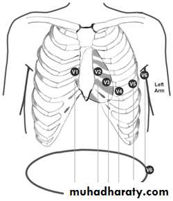
v4: left 5th intercostal space, mid-clavicular line
V5: horizontal to v4, anterior axillary line
V6: horizontal to v5, mid-axillary's line.
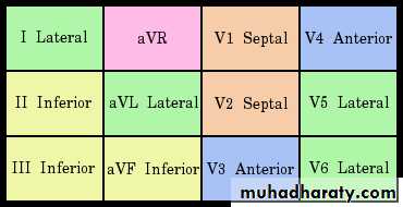
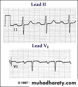
*leads II,III,AVF: record changes from inf. Border of the heart.
*v1-v2 : record changes from the RV
*v3-v4 : record changes from the intervent. Septum.
*v5-v6 -I-AVL: record changes from LV
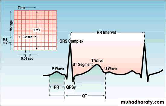
P wave: the sequential activation (depolarization) of the right and left atria.
qrs complex: right and left ventricular depolarization (normally the ventricles are activated simultaneously).st-t wave: ventricular repolarization.
u wave: origin for this wave is not clear - but probably represents "afterdepolarizations" in the ventricles
PR interval: time interval from onset of atrial depolarization (p wave) to onset of ventricular depolarization (QRS complex)
QRS duration: duration of ventricular muscle depolarization
QT interval: duration of ventricular depolarization and repolarization
RR interval: duration of ventricular cardiac cycle (an indicator of ventricular rate)
PP interval: duration of atrial cycle (an indicator of atrial rate)

measurements
Sensivity :10mm = 1mv =2 large sq vertically
Paper speed :25mm/sec
Each large 5mm sq=0,2s
Each small 1mm sq=0,04
H.R =1500/r-r interv.in 1mm ssq
=300/r-r interv. In 5mm l sq
P-r interv.=0,12-0,2 sec
Qrs duration=0,06-0,10 sec
Qt interv.=/< 0,4 sec
Reading & interpriting ecg
Methods of reading &interpreting the ecg
1.determine the cardiac rate
2.determine the cardiac rhythem
3.determine the axis of the heart
4.examine the p wave
5.asses the pr interval
6.examine the qrs complex
7.examine the st segment
8.examine the t wave
9.examine the u wave
Examination of p wave
*represent sequencial right &left atria activation
*duration >0,12 s.
*amplitude >2,5 mm
*best studied in lead ii
abnormalities of p wave
1.p mitral (l a. Hypertrophy) *broadening of p wave *m shape configeration
2. P pulmonal (r atr. Hypertrophy) *peaked p wave
3. Inverted p wave(nodal rhythem)
4. Absent p wave (AF , VT ,SA block)
5.inverted p in lead I (wrong connection, dextrocardia)
assesment of pr interval
From beginning of p to beginning of qrs*normal 0,12-0,2 sec.
Abnormalities of pr inerval
Short pr interval(>0,12s)
*preexcitation syndromes
- wolff parkinson white syndrome
- Lown Ganong Levine Syndrome
*AV junctional rhyth.
*ectopic atrial rhyth.
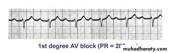
Prolonged pr (<0,2 sec.)
*rhaumatic fever
*first degree heart block .
*drugs
*electrolyte disturbance
QRS complex
Normal 0,06-0,10 s
Abnormalities of qrs1- qrs widening
Boundle branh block(bbb):
*RBBB
-normal varient
-rt ventricle pathology
-congenital heart disease (atrial septal defect)
-coronary artery disease
*LBBB
-coronary artery disease
-hypertension.
-aortic valve disease
-cardiomyopathy
RBBB
-wide qrs complex
-rsr pattern or m shape in v1
-slurred s in v5 v6
-inverted t in v1
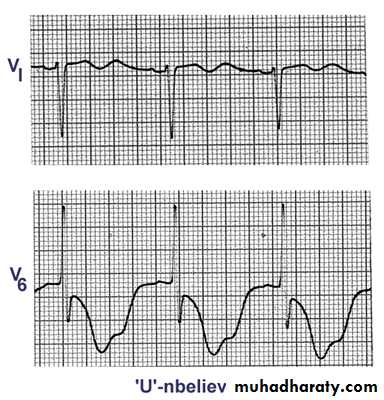
LBBB
-wide qrs complex
-small q in v1
-m shape qrs in v6
2- increase voltage
Ventricular hypertrophy:
*LVH
-hypertension
-hypertrophic cardiomyopathy
-aortic stenosis
*RVH
-pulmonary stenosis
-pulmonary hypertension
*LVH
-increase r in v5-6
- increase s in v1-2
sum. <35 s sq
-LV strain pattern (st,t inversion in: i,avl,v5-v6)
*RVH
-increase r voltage in v1-v2
-deep s in v5-6
-RV strain pattern (st,t inversion in v1-2 )
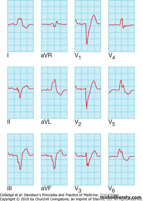
3- Abnormal Q wave
Myocardial infarction
*inferior mi: pathological q wave in leads ii, iii ,avf
*anterior mi: pathological q wave in leads i,avl,v1-6
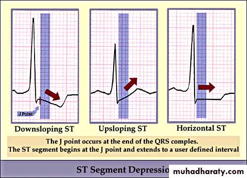
St segment
Start from s wave till the beginning of t wave
Abnormalities of st segment1.st elevation
St elevation convex upward
*acute mi
*prinzmetal angina
*ventricular aneurysm
*normal variant
St elevation concave upward: pericarditis
Other causes (lbbb,hyperkalemia)
2. St depression
IHD
*subendocardial ischemia
*non q wave mi
*reciprocal changes in acute mi
Non-ischemic causes
*vh, bbb, digoxine,
hypokalemia, mvp, cns diseases
T wave
Most labile wave in ecg
Amplitude >5mm
Abnormalities of t wave
t inversion
*mi
*ischemia
*pericarditis
*myocarditis
*cns dis
*vh (strain pat.)
*digoxine
*mvp
Peaked t wave
*anxiety
*hyperkalemia
U wave
Represent after depolarization of ventricles.Normal u has same polarity of t, amplitude 1/3 of t
abnormalities of u wave
Prominent upright u
*bradycardia
*hypokalemia
*quinidine
*mvp
*cns diseases
inverted u wave: IHD, LVH
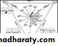
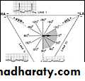
axis of the heart
Normal :(-30 )to (+110)
*>-30 :left axis deviation
*<+110:right axis deviation
LAD : LI up
L II,L III down
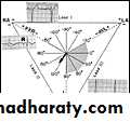
RAD : L II down
LII,III up