
Neurological examination

Objectives
☤Understand neurological examination
☤Perform a neurological examination
☤Higher function
☤Cranial nerves
☤Motor system
☤Sensory system
☤Interpret neurological examination
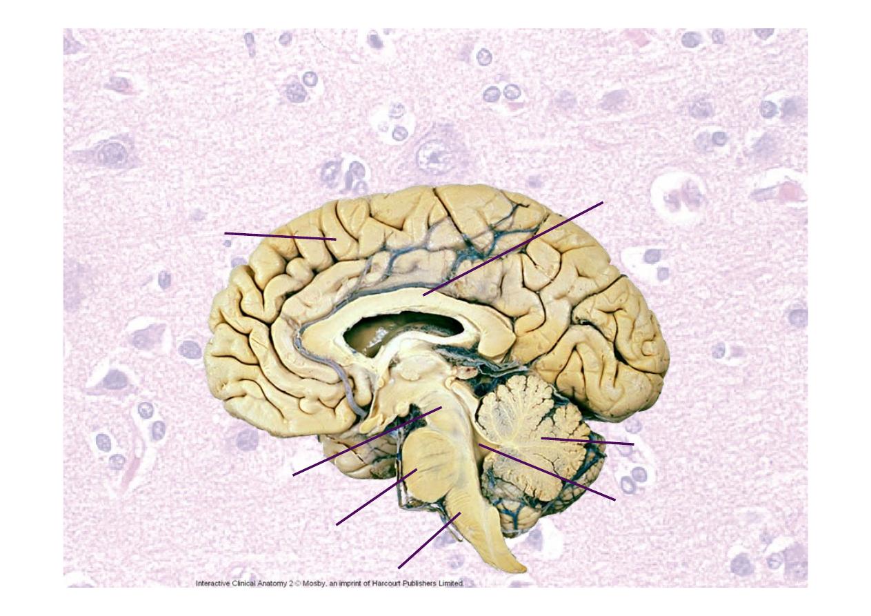
Neuroanatomy – parts of
nervous system
Cerebrum
Cerebellum
Medulla oblongata
Pons
Midbrain
IVth Ventricle
Corpus callosum
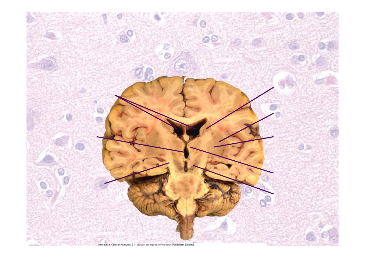
Neuroanatomy – parts of
nervous system
Lateral ventricles
Pyramidal tract
Caudate
Putamen
Globus
pallidus
IIIrd ventricle
Thalamus
Substantia
nigra
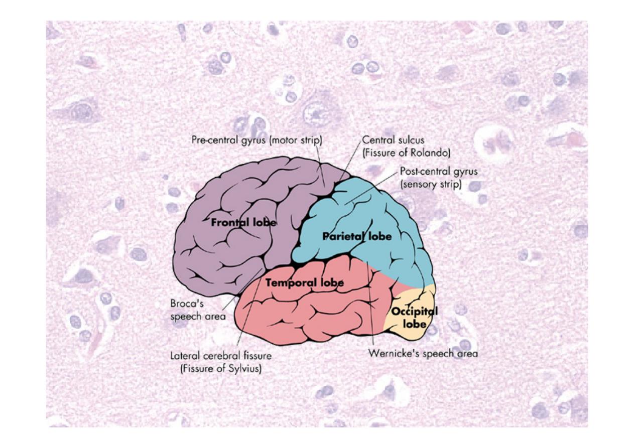
Neuroanatomy - lobes
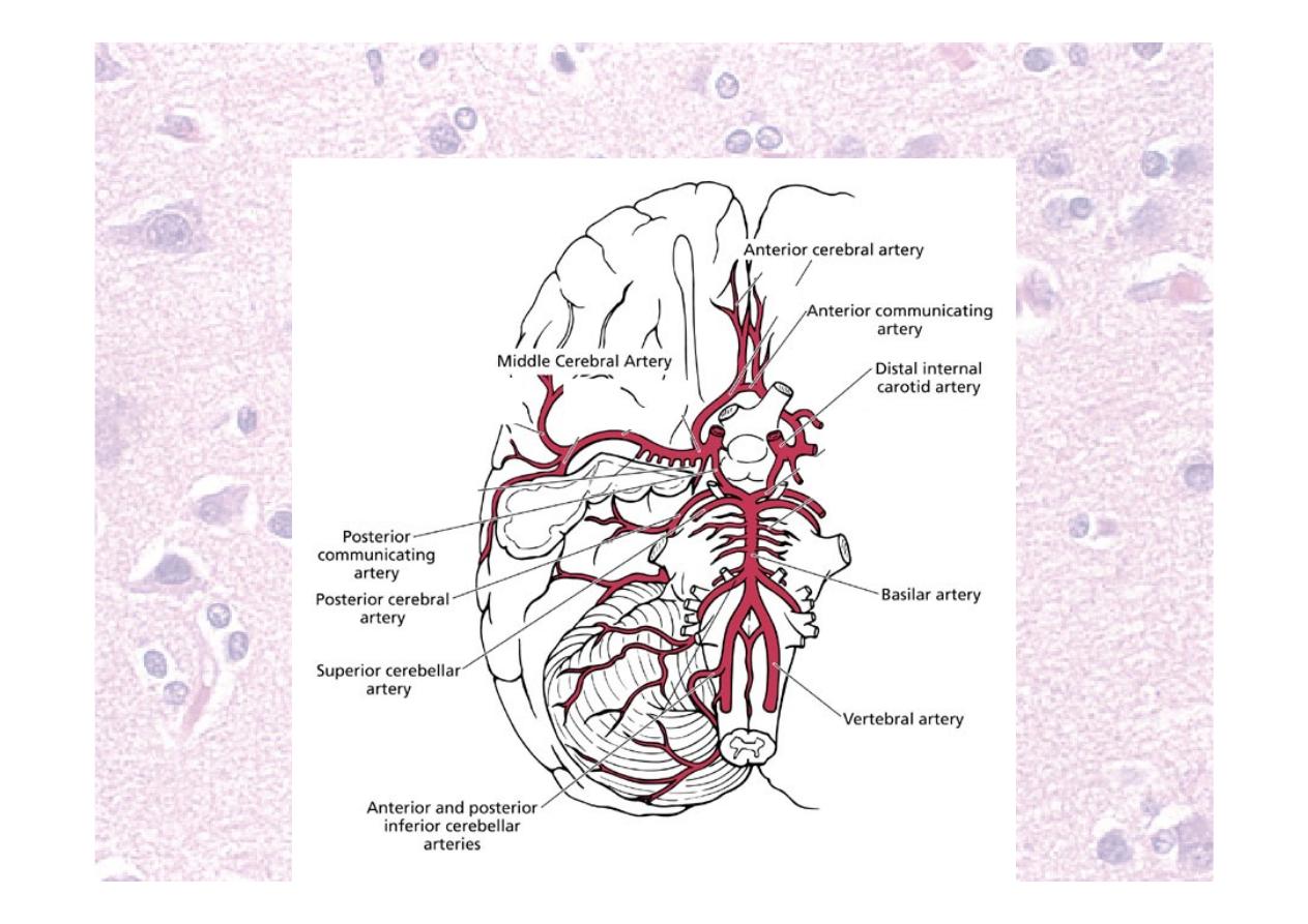
Neuroanatomy – blood supply
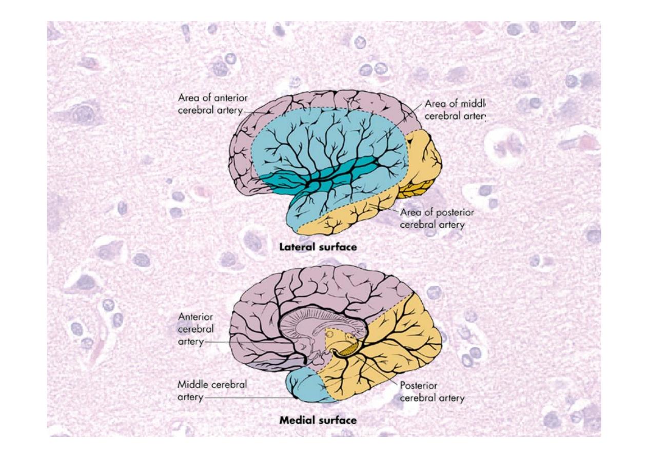
Neuroanatomy – blood supply

Neuroanatomy – parts of
nervous system
☤Don’t forget
☤Spinal cord
☤Peripheral nerves
☤Autonomic nerves
☤Aim of neurological examination is to locate
the lesion
☤Aim of neurological history is to determine
lesion type
☤Don’t rely on CT or MRI – you need to
know what part to scan!
General inspection
Neurological examination is part
of a full patient assessment

Things to look for
☤Observations
☤Neck stiffness, Kernig sign
☤Skin e.g. café-au-lait spots
☤Anal sensation and tone
☤With spinal problems especially e.g. cauda equina
☤General examination e.g.
☤Cyanosis (impaired consciousness)
☤Lymphadenopathy (malignancy, infection)
☤Murmurs, carotid bruit or AF (source of emboli)
☤Teeth, ears (source of infection)
☤Breasts, abdomen (malignancy)
Higher mental function -
cognition

Higher mental function -
cognition
☤Language: dysphasia,
dyslexia, dysgraphia
☤Verbal, reading and
writing
☤Gnosis “knowing
things”: agnosia
☤Geography, objects,
people
☤Praxis “doing things”:
dyspraxia
☤Dress, draw, write
☤Number skills:
dyscalculia
☤Memory: amnesia
☤Immediate, recent,
remote
☤visual, verbal
☤retrograde,
anterograde
☤Reasoning
☤Emotion
☤Personality
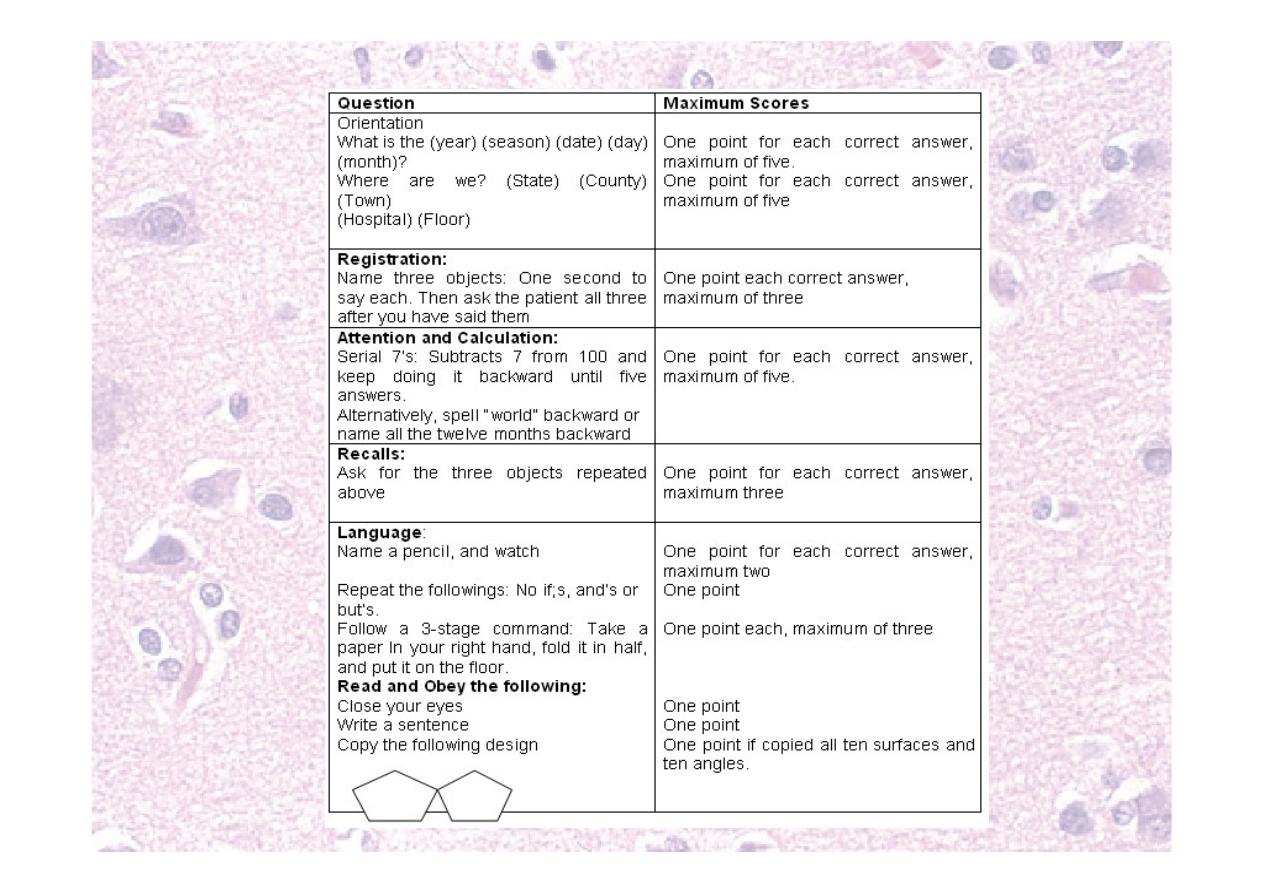
Testing higher mental function
☤Mini-mental state examination
(MMSE)
☤30 point score
☤Assesses multiple aspects of
cognition
☤Abbreviated mental test score
☤10 point easy bedside test

Abbreviated mental test score
1. Age
2. Time to nearest hour
3. An address to be repeated at the end of the test
4. Where are you now? (name of hospital etc)
5. Year
6. Recognition of 2 people e.g. doctor, nurse
7. Date of birth
8. Year second world war began
9. Name of present monarch
10. Count backwards from 20 to 1
☤
Remember to test recall of address
Unable to
follow
instruction
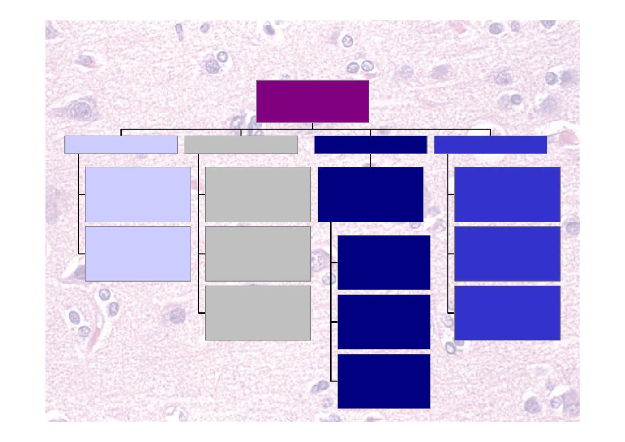
Speech
Fluent but
disorganised
Receptive
Wernicke
Unable to
repeat
Conductive
Arcuate fasciculus
Non-fluent
speech
Expressive
Broca
Unable to
name items
Nominal
Angular gyrus
Dysphasia
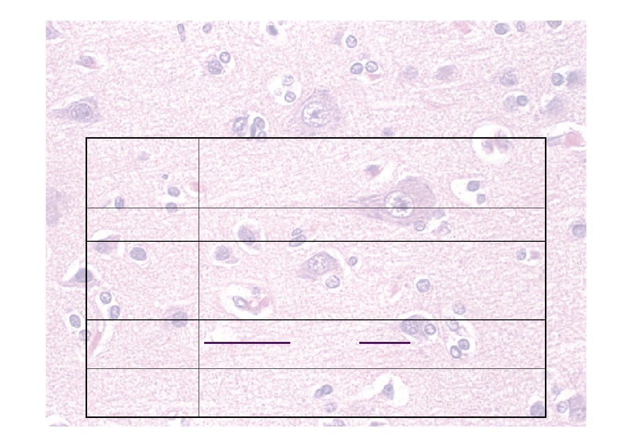
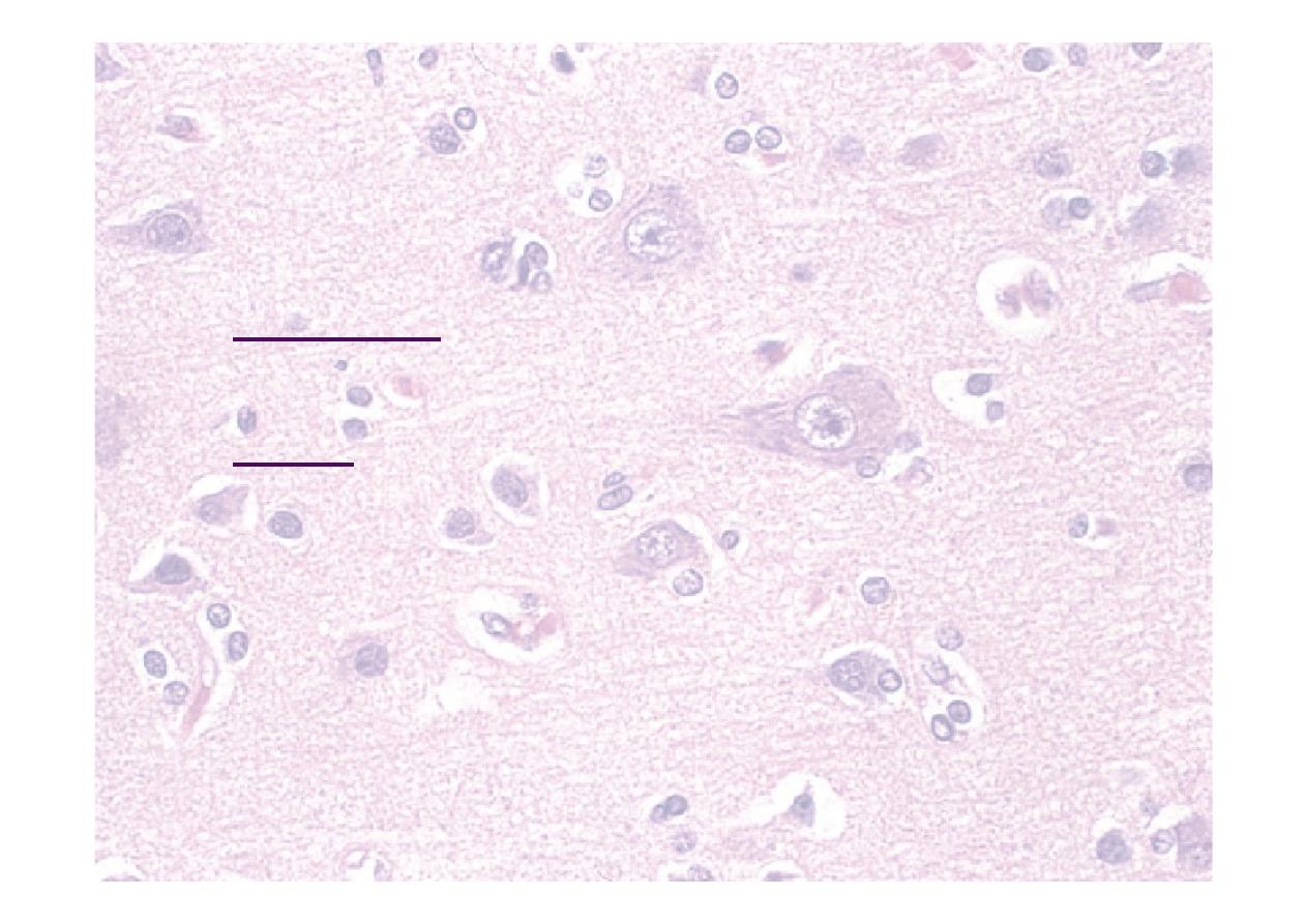
Slow
slurred
explosive
Cerebellar
Flaccid
Nasal
Bulbar
Spastic
"Donald Duck"
Pseudobulbar
Soft
monotone
Extrapyramidal
Articulation
Dysarthria
Hoarse voice
Bovine cough
Phonation
Dysphonia
Components
of speech disorder
Speech and
language

Test speech and language
☤ Listen to history
☤ Expression, reception and phonation
☤ Ask “take this paper with your right hand, fold in half and
place on the floor
☤ Reception
☤ Repeat “No ifs, ands or buts”
☤ Nonsense phrase to assess repetition
☤ Repeat “British constitution,” “Baby hippopotamus”
☤ Articulation
☤ Name the strap of a watch, or nib of a pen
☤ Nominal dysphasia
☤ Language disorders usually co-exist “Global”

Cognition summary
☤ 10 point MTS
☤ Age
☤ Time
☤ Address
☤ Place
☤ Year
☤ 2 people
☤ DoB
☤ WWII
☤ Monarch
☤ 20 to 1
☤ Recall
☤Speech and
language
☤Expression
☤Reception
☤Repetition
☤Articulation

Cranial nerves
Numbered for convenience…
…and named for what they do
(mostly)
1
st
(olfactory) nerve
Nuclei
None – terminates in cortex
Comes from Inferior frontal and temporal lobes
Goes
through
Cribriform plate
Supplies
Sense of smell
Tested by
Test smell in each nostril
separately e.g. coffee
1
st
(olfactory) nerve
☤Test smell in each
nostril with eyes closed.
☤Coffee
☤Vanilla
☤Perfumed soap
☤Avoid ammonia –
activates sensory nerves
Anosmia — Head injury, tumour, nasal
congestion.

Examination of Vision
☤
Optic (II) nerve – sensory
inputs from retina
☤
Oculomotor –III }
☤
Trochlear – IV } Motor
☤
Abducens – VI }
2
nd
(optic) nerve
Nuclei
Lateral geniculate nucleus in
thalamus (vision), superior
colliculus (reflexes)
Comes from
Thalamus
Goes through Optic chiasm to optic foramen
Supplies
Vision from retina
Tested by
AFRO – acuity, fields, reflexes,
ophthalmoscopy
2
nd
(optic) nerve
☤Visual Acuity
☤Snellen chart
Wear specs or correct with pinhole
☤Finger counting
☤Light-dark
Snellen visual acuity
Need good lighting
Patient is kept 6 metres (20ft) from the chart
Read as 6/5, 6/6, 6/10, 6/60
Test one eye at a time

☤Fields to confrontation
☤Test one eye at a time
☤Use red pin from each direction
☤Gross defect only
☤Central vision/blind spot – with red pin
faded colour with lesions with macula
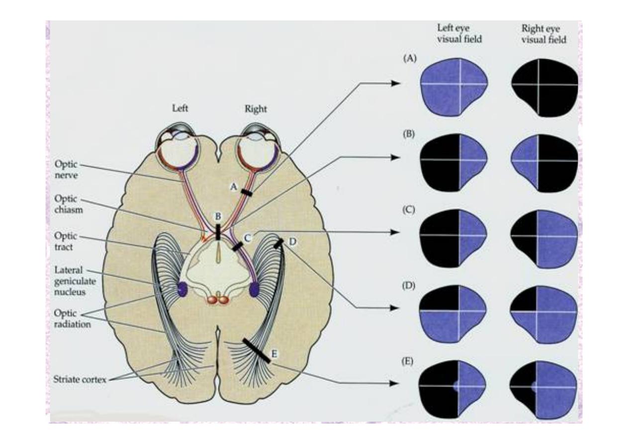
Optic pathways and
defects
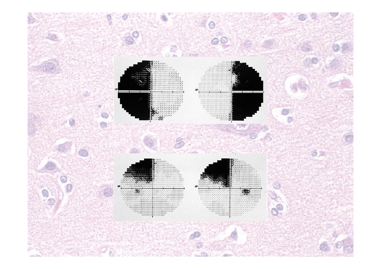
Optic nerve problems
Bitemporal hemianopia
Homonymous quadrantanopia

Cranial nerve nuclei
☤Different nuclei for different functions
☤One nerve can have more than one
nucleus
☤One nucleus can have more than one
nerve
☤Efferent means going out of the CNS
(like sewage effluent goes out to sea)
☤Afferent means going into the CNS

3
rd
(oculomotor) nerve
Nuclei
Oculomotor nucleus (motor),
Edinger-Westphal nucleus (PS)
Comes from
Midbrain
Goes through Superior orbital fissure
Supplies
Motor to extraocular muscles
except superior oblique and
lateral rectus, levator palpebrae
superioris. PS to eye
Tested by
Extraocular movements and
pupillary reflexes (motor)
4
th
(trochlear) nerve
Nuclei
Trochlear nucleus
Comes from
Midbrain - dorsum
Goes through Superior orbital fissure
Supplies
Motor to superior oblique
muscle
Tested by
Extraocular movements
6
th
(abducens) nerve
Nuclei
Abducens nucleus
Comes from
Junction of pons and medulla
Goes through Superior orbital fissure
Supplies
Motor to lateral rectus muscle
Tested by
Extraocular movements

III (oculomotor), IV (trochlear),
VI (abducens) nerves
☤Cn III supplies
☤All extraocular muscles except lateral
rectus, superior oblique
☤Levator palpebrae superioris
☤Parasympathetic supply to eye
☤Cn IV supplies superior oblique
☤Cn VI supplies lateral rectus
☤These nerves are tested together
Extraocular muscles and actions
Lateral rectus
Medial
rectus
Superior
rectus
Inferior
rectus
Superior oblique
Inferior
oblique
Right eye

Testing extra ocular muscles
☤Look for ptosis
☤Eye movements (ask about diplopia, watch
light reflection on cornea)
☤6 directions of muscle pull
☤Watch for nystagmus
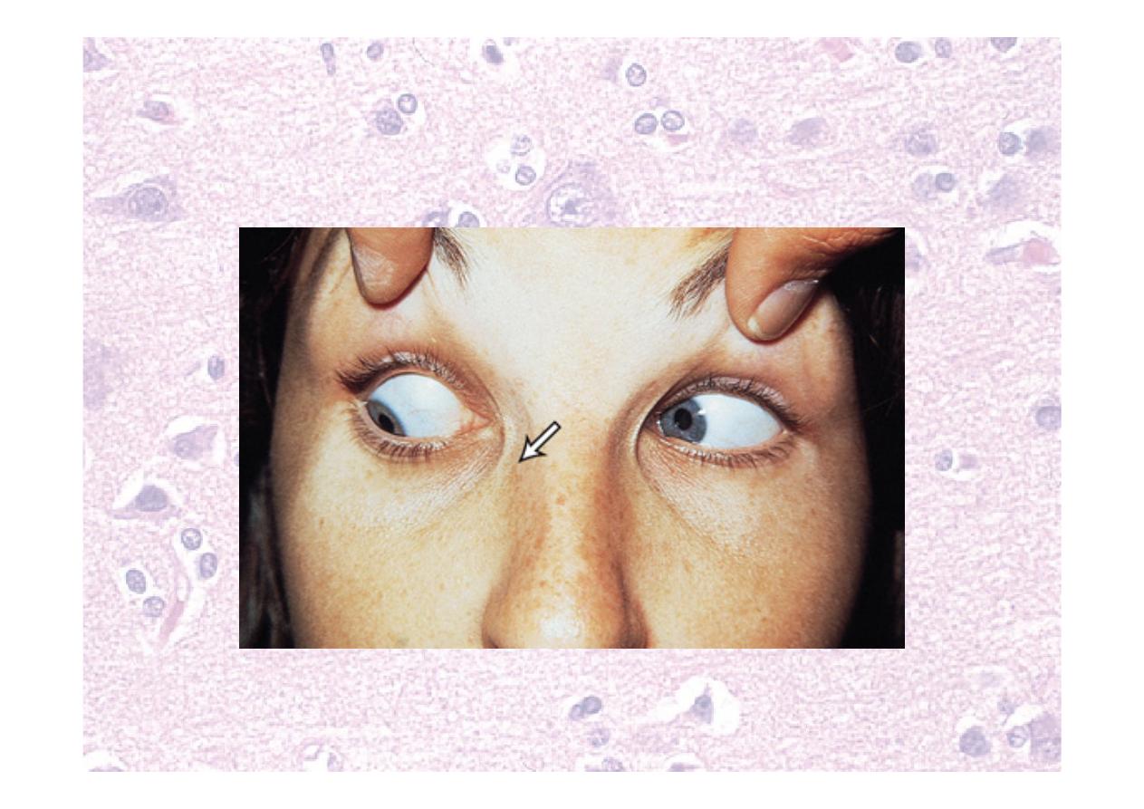
Cn III, IV, VI defects
Left abducens palsy
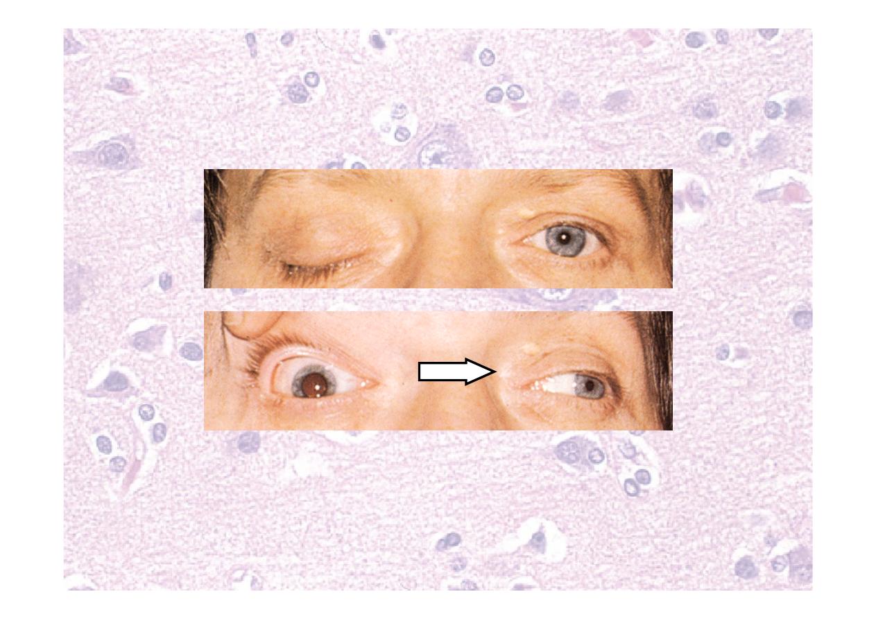
Cn III, IV, VI defects
Right oculomotor palsy
A painful, acute Cn III palsy is an emergency!
- could be an enlarging aneurysm
Cn III, IV, VI defects
Left trochlear palsy

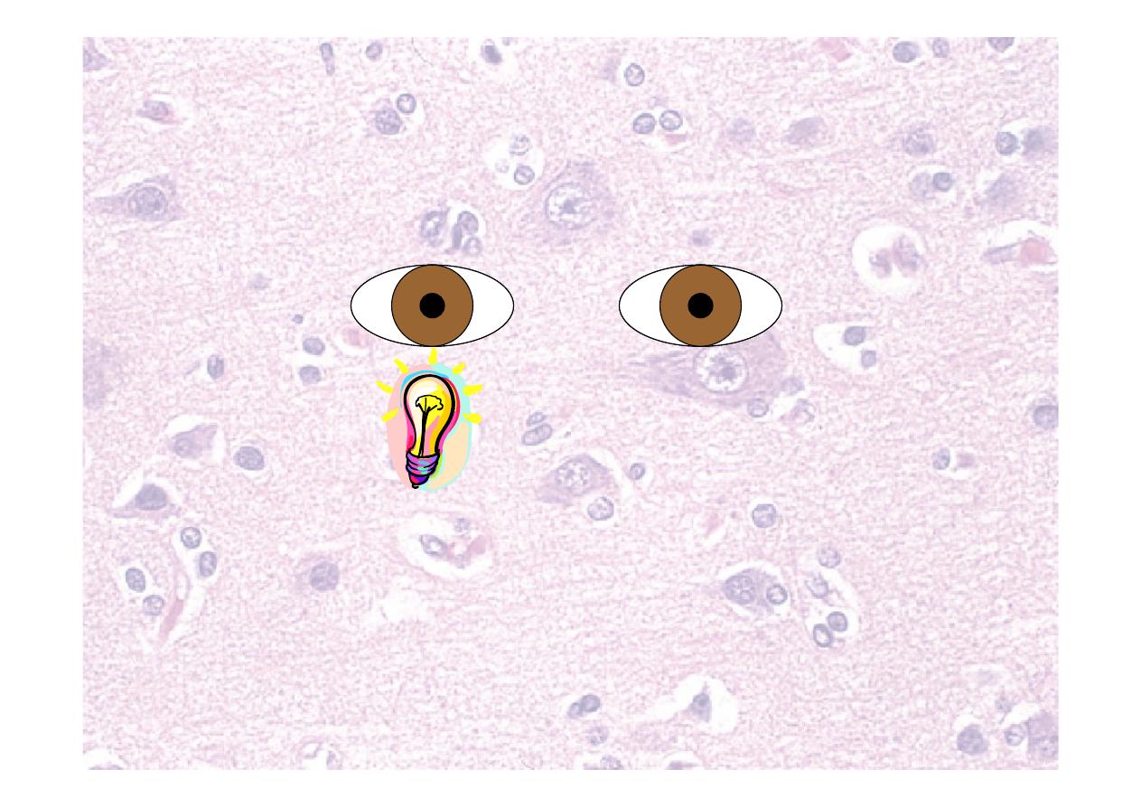
Pupillary reflexes
☤Light reflex
☤Fix the eyes on a distant point
☤bring torch / light onto pupil from side
☤Look for direct light reflex and consensual light
reflex
☤Is the defect afferent or efferent
☤Accommodation reflex
Check convergence and pupillary constriction
Relative afferent pupillary
defect (RAPD)
Swinging light test – shine
light first in one eye, then
swing to the other
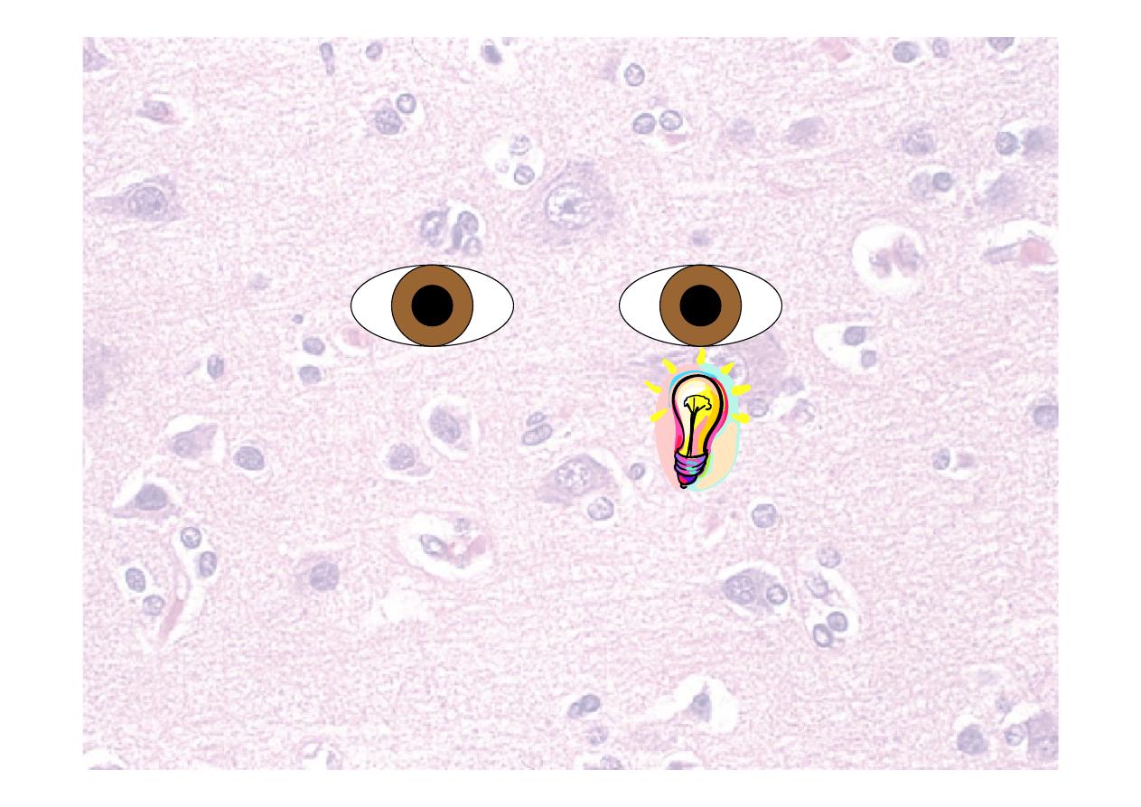
Relative afferent pupillary
defect (RAPD)
Left RAPD = left optic
nerve pathology

Summary of eyes
1.
Visual equity with Snellen chart
2.
Visual fields with confrontation
3.
Eye movements
4.
Pupillary reflexes
5.
Fundoscopy
Nystagmus
Jerk
Pendular
Type
Vertical
Horizontal
Rotatory
Direction of nystagmus
1st degree
Only on looking to:
one side
2nd degree
On looking to:
one side
straight ahead
3rd degree
On looking
all directions
Degree
Direction of fast phase
Ocular
Pendular
Vestibular
Never vertical
Central
Any direction
Can be just one eye
Can change direction
Causes
Examine:
Max 30 degrees
from midline
5
th
(trigeminal) nerve
Nuclei
Trigeminal motor nucleus, trigeminal
sensory nucleus (length of brainstem into
cervical cord)
Comes from Pons
Goes
through
V1 ophthalmic: superior orbital fissure
V2 maxillary: foramen rotundum
V3 mandibular: foramen ovale
Supplies
Sensation to face, motor to muscles of
mastication
Tested by
Corneal reflex (sensory), touch, pin,
clench teeth, open mouth, jaw jerk
5
th
(trigeminal) nerve
☤Sensation to face
☤Motor to muscles of mastication
☤Corneal reflex (sensory)
☤Jaw jerk (both components)

CN V - motor
1. Inspect for wasting
2. Ask the patient to clench the teeth and
feel for the bulk
3. Ask the patient to open the mouth
against resistance
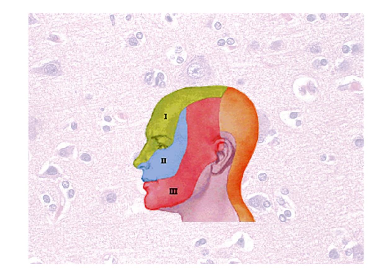
Trigeminal nerve divisions
Ophthalmic
Maxillary
Mandibular
☤Corneal reflex
Touch the cornea with a wisp of cotton
Look for direct and consensual reflexes
Touch sensation via ophthalmic branch of
Trigeminal Nerve
Then Motor nucleus of VII —Obicularis oculi
☤Jaw jerk
Loosely open the mouth
Place the fore finger above the chin
Tap with tendon hammer
7
th
(facial) nerve
Nuclei
Facial nucleus (nervus intermedius –
salivary nucleus, nucleus solitarius)
Comes from Junction of pons and medulla
Goes
through
Stylomastoid foramen, branches in
parotid (T,Z,B,M,C)
Supplies
Motor to muscles of facial expression,
taste to anterior 2/3 of tongue, sensation
to ear canal and palate, PS to salivary
and lacrimal glands (nervus intermedius)
Tested by
Corneal reflex (motor), inspect face at
rest, wrinkle forehead, close eyes, blow
out cheeks, show teeth (taste: sweet,
sour, salt, bitter)
7
th
(facial) nerve
Supplies:
☤ Motor to muscles of facial
expression
☤ Also carries:
☤ taste to anterior 2/3 of
tongue
☤ sensation to ear canal and
palate
☤ PS to salivary and lacrimal
glands (nervus intermedius)
Tested by:
☤Inspect face at rest
☤Corneal reflex (motor)
☤Wrinkle forehead
☤Close eyes
☤Blow out cheeks
☤Show teeth
☤(Taste: sweet, sour,
salt, bitter)
Upper face has bi-cortical
representation
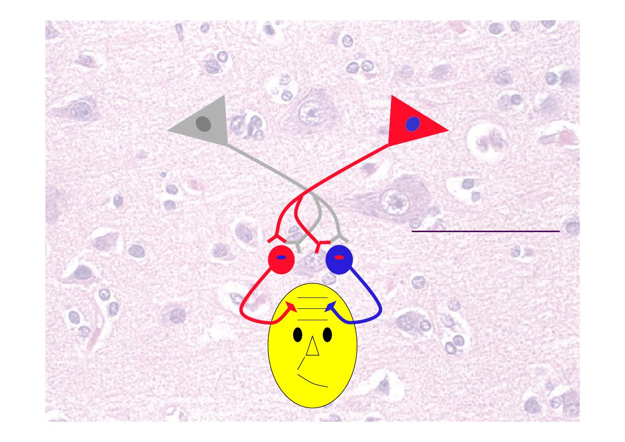
UMN damage does not affect
upper face
Orbicularis oculi
is sometimes
represented
bilaterally, so
may be spared
or involved in
UMN lesions
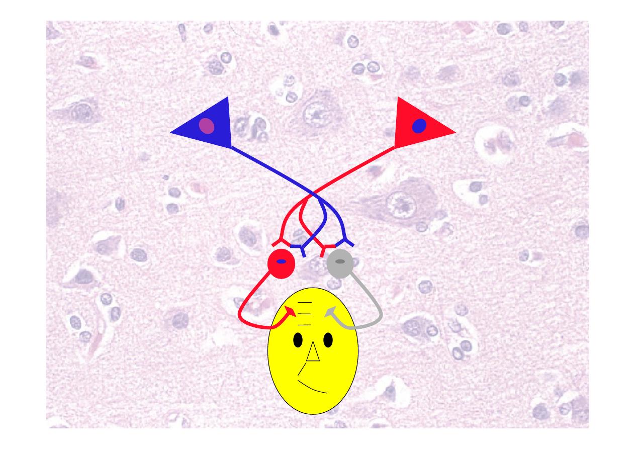
LMN lesion affects whole side
of face
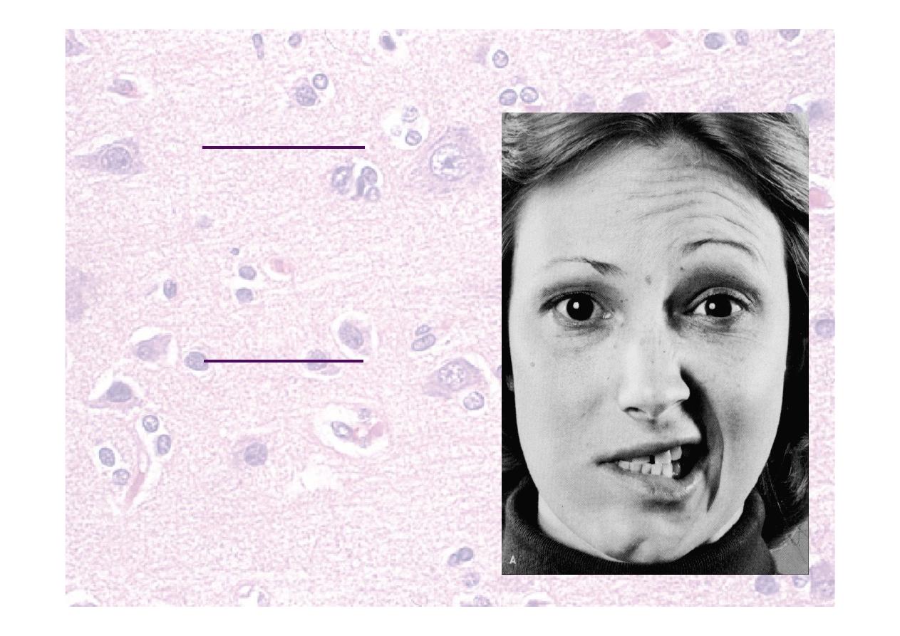
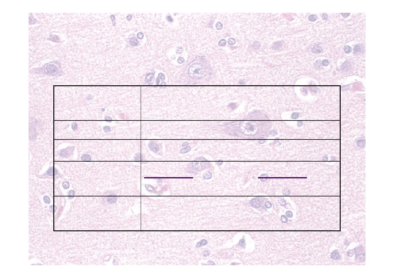
7
th
(facial) nerve - testing
UMN lesion
☤Spares forehead as:
☤Bilateral cortical
representation
☤LMN intact
LMN lesion
☤Can involve forehead as:
☤LMN only innervates one
side
☤Beware: can be partial
CnVII palsy!
8
th
(vestibulocochlear) nerve
Nuclei
Vestibular and cochlear nuclei
on floor of IVth ventricle
Comes from
Junction of pons and medulla
Goes through Internal acoustic meatus
Supplies
Hearing (cochlea), balance
organs
Tested by
Otoscopy, whispered speech,
Rinné, Weber (vestibular tests)
8
th
(vestibulocochlear) nerve
☤Supplies:
☤Hearing (cochlea)
☤Balance organs
☤Tested by:
☤Otoscopy
☤Whispered speech
☤Rinné, Weber tests
☤Vestibular tests e.g. Hallpike manoeuvre

Rinné test
☤Use 512Hz tuning fork
☤Place base firmly on mastoid process
☤Ask patient to tell you when sound
disappears
☤Hold fork tips 2cm from EAM
☤Can the patient hear it now?

Interpreting Rinné test
☤Tests whether bone (BC) or air
conduction (AC) is better
☤If can be heard in front of ear, AC>BC
☤Normal
☤Rinné positive
☤If cannot be heard in front of ear,
AC<BC
☤Indicates conductive deafness
☤Rinné negative

Weber test
☤Use the same tuning fork again
☤Hold somewhere on the head in the
midline
☤Usually vertex or forehead
☤Which side is louder?

Interpreting Weber test
☤Conductive deafness
☤localises to abnormal ear
☤Sensorineural deafness
☤localises to normal ear
☤Try it on yourself with a finger in an ear
☤Where does the sound localise?
☤Have you caused conductive or
sensorineural deafness?
9
th
(glossopharyngeal) nerve
Nuclei
Nucleus solitarius and nucleus
ambiguus of medulla
Comes from
Medulla
Goes through Jugular foramen
Supplies
Sensation to pharynx, middle &
inner ear, post 1/3 tongue, taste
post 1/3 tongue, PS to parotid,
visceral sense from carotid
body and sinus
Tested by
Gag reflex (sensory), (taste) –
with CnX
10
th
(vagus) nerve
Nuclei
Nucleus solitarius and nucleus
ambiguus of medulla, dorsal
vagal (motor) nucleus
Comes from
Medulla
Goes through Jugular foramen
Supplies
Sensation to pharynx and
larynx. Motor to pharynx,
larynx, palate (from CnXI). PS to
thoracic & abdominal organs
Tested by
Say “Ah” – uvula moves towards
normal side, gag reflex (motor),
phonation/cough, swallow
IX (glossopharyngeal) and X
(vagus) nerves
☤Tested together
☤Glossopharyngeal supplies:
☤Sensation to pharynx, middle & inner ear, post
1/3 tongue
☤Taste post 1/3 tongue
☤PS to parotid
☤Visceral sense from carotid body and sinus
☤Vagus supplies:
☤Sensation to pharynx and larynx
☤Motor to pharynx, larynx, palate (from Cn XI)
☤PS to thoracic & abdominal organs
IX (glossopharyngeal) and X
(vagus) nerves
☤Tested together
☤Say “Ah” – uvula moves towards
normal side
☤Phonation/cough
☤Gag reflex (IX sensory, X motor)
☤Swallow – only if rest of IX and X
normal
☤(taste – not routine)
11
th
(accessory) nerve
Nuclei
Spinal – C1-5 anterior horns
(Cranial – nucleus ambiguus, with CnX)
Comes from
Medulla
Goes through Jugular foramen
Supplies
Motor to sternocleidomastoid
and trapezius muscles (and
motor to vagus)
Tested by
Turn head against resistance,
shrug shoulders
11
th
(accessory) nerve
☤Motor to sternocleidomastoid and
trapezius muscles
☤Examine bulk of SCM and
trapezius
☤Turn head against resistance
☤Shrug shoulders
12
th
(hypoglossal) nerve
Nuclei
Hypoglossal nucleus
Comes
from
Medulla
Goes
through
Hypoglossal foramen
Supplies
Motor to muscles of tongue except
palatoglossus
Tested by Look at tongue at rest, poke out
tongue and move side-to-side
12
th
(hypoglossal) nerve
☤Motor to muscles of tongue except
palatoglossus
☤Look at tongue at rest
☤Fasciculation
☤Poke out tongue
☤Move side-to-side
☤Press into cheek against resistance

Practical – cranial nerves
☤2nd (optic) nerve
☤AFRO – acuity,
fields, reflexes,
ophthalmoscopy
☤3rd (oculomotor),
4th (trochlear), 6th
(abducens) nerve
☤Test light reflex and
extraocular muscles
☤5th (trigeminal)
nerve
☤Motor and sensory
☤7th (facial) nerve
☤8th (vestibulocochlear)
nerve
☤Rinné, Weber test
☤9th (glossopharyngeal)
nerve
☤10th (vagus) nerve
☤11th (accessory) nerve
☤12th (hypoglossal)
nerve

Trunk and limbs
Not just peripheral nervous
system

Inspection
☤ Posture – clawing hands, pes cavas
☤ Wasting/Hypertrophy – feel for muscle bulk
☤ Proximal or distal?
☤ Symmetrical?
☤ Specific muscle group?
☤ Fasciculation
☤ Abnormal movements
☤ Tremor
☤ Chorea
☤ Other uncontrolled movement
☤ Scars – tracheotomy scars,
☤ Surrounding – wheel chair, walking stick or urinary
catheter ,NG, PEG

Tone - testing
☤Patient relaxed, movements by examiner
☤Rapid alternating movements
☤Upper limb: rotate wrist, flex and extend elbow
☤Lower limb: roll leg and watch foot
☤Lift leg briskly at knee
☤Does heel lift or drag along bed?
☤Test for clonus at:
☤Ankle by rapid dorsiflexion
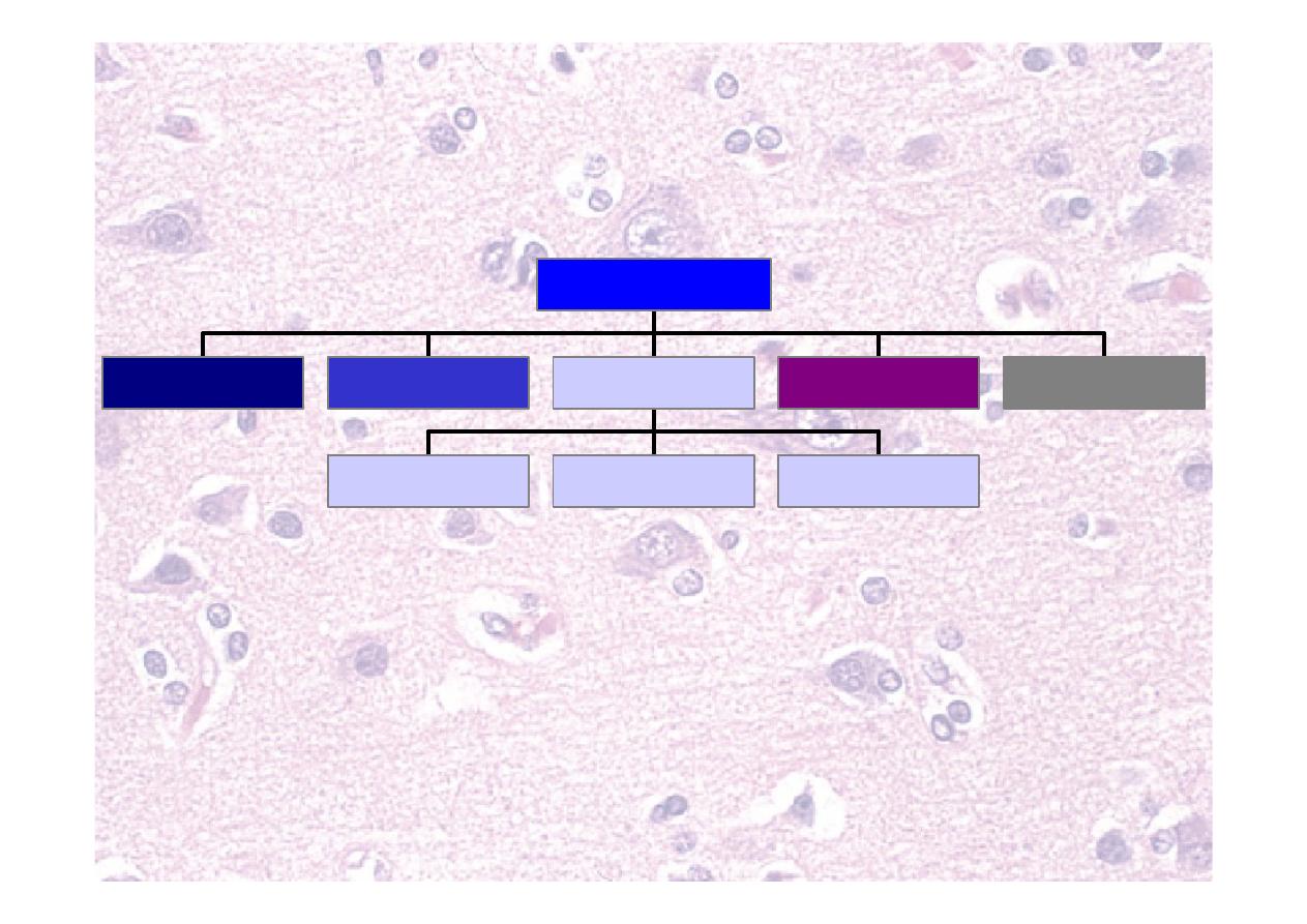
Tone - abnormalities
Normal
Barely perceptible
resistance to movement
Decreased Reduced resistance to
movement
LMN or
cerebellar
lesion
“Lead
pipe”
↑
Increased resistance
throughout movement
UMN lesion
“Clasp
knife”
↑
High resistance that
suddenly releases
Extrapyramidal
lesion
“Cogwheel
rigidity”
Jerky resistance
Extrapyramidal
lesion (esp. PD)

Power
☤
Arm drift
☤
↓Pyramidal lesion, ↑Parietal/ cerebellar lesion
☤
Compare with
☤
other side
☤
yourself
☤
Test each muscle group
☤
Is the deficit:
☤
Proximal or distal?
☤
Flexor or extensor?
☤
Cortical, nerve root, peripheral nerve
distribution?
MRC power rating
0
No movement
1
Flicker only
2
Moves with gravity eliminated (test
horizontal movement)
3
Moves against gravity, but not resistance
4
Moves against resistance (- for only just, +
for nearly normal)
5
Normal power
Myotomes
C5
Shoulder abduction
C6
Elbow flexion
C7
Elbow extension
C8
Finger flexion
T1
Finger abduction
L2
Hip flexion
L3
Knee extension
L4
Dorsiflexion at ankle
L5
Extensor hallucis longus
S1
Plantar flexion
Reflexes – what do they test?
☤Spinal arc modulated by higher centres
Biceps
C5,6
Radial (supinator, brachioradialis) C5,6
Triceps
C6,7,8
Finger jerk/ Hoffman
C8
Abdominal (4 quadrants)
T7-12
Knee
L3,4
Ankle
S1,2
Plantar
L5,S1,2

Tendon reflexes – how do I
test them?
☤Everything needs to be floppy
☤Patient’s limb
☤Your tendon hammer wrist
☤Find tendon with finger (upper limb)
☤Hit tendon or finger on tendon
☤Try reinforcement if no response
☤Clench teeth
☤Pull hands apart
☤Abdominal –stroke each quadrant towards
umbilicus
☤Plantar – stroke outer sole and ball of foot
Interpreting reflexes
☤Reduced in
☤LMN lesion (including same level spine)
☤Muscle weakness
☤Early UMN lesion
☤Slow in hypothyroidism
☤Increased in
☤UMN lesion
☤Anxiety, thyrotoxicosis, youth

Recording reflexes
☤0 = none, even with reinforcement
☤+ = only present with reinforcement
☤++ = normal
☤+++ = hyper-reflexia
☤++++ = with clonus
☤Clonus is rhythmical beating in
response to muscle stretch

UMN lesion
¾ Normal muscle bulk
¾ Increased tone
¾ Reduced power
¾ Brisk reflexes
LMN lesion
¾ Wasting and atrophy
¾ Reduced tone
¾ Reduced power
¾ Reduced or absent
reflexes
Coordination
Upper Limb
☤Finger-nose
☤Dysmetria or past-
pointing
☤Clap with alternating
palm and dorsum
☤Dysdiadochokinesia
☤“Play piano”
☤Rebound
☤Test at same time as
drift
Lower limb
☤Heel-shin test
☤Run heel down other
shin, lift up and repeat
☤Tapping toes

Cerebellar signs
☤ D
ysdiadochokinesia
☤ A
taxia – present with eyes open/close (broad base
gait)
☤ N
ystagmus- fast component towards the lesion
☤ I
ntention tremor (=dysmetria)
☤ S
peech – slow, slurred, explosive, scanning
☤ H
ypotonia
☤ Pendular jerks-muscle contraction and relaxation is
slow
☤ Past pointing
☤ Rebound phenomenon
Ipsilateral signs. If central (vermis, alcohol) only truncal
ataxia may be present
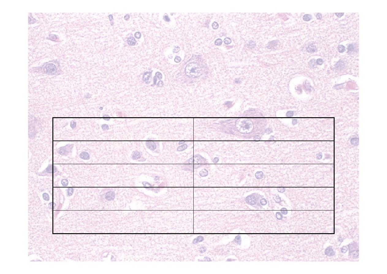
Sensory modalities and testing
Pain
Pin (neurotip)
Spinothalamic
Cross–spinal cord
Temperature
Ice, cold tuning fork
(not routine)
Spinothalamic
Cross–spinal cord
Proprioception
Eyes closed, move
joint
Dorsal column
Cross - brainstem
Light Touch
Wisp of cotton wool
Dorsal column
Cross - brainstem
2 point
discrimination
Two-point
discriminator
Dorsal column
Cross - brainstem
Vibration
128 Hz tuning fork
on bony prominence
Dorsal column
Cross - brainstem
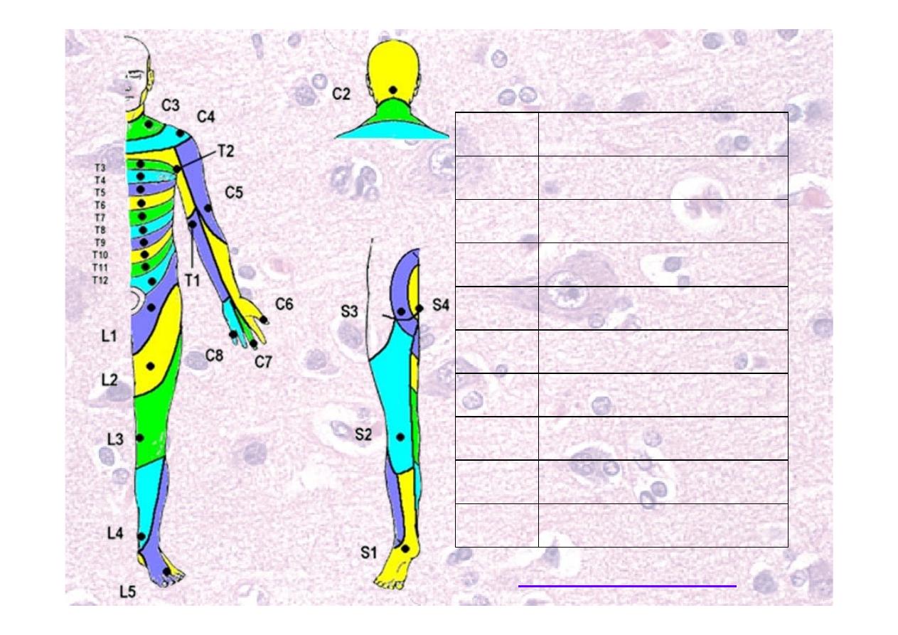
Dermatomes
C4
Cape
C7
Middle finger
T2
Arm pit
T5
Nipple
T10
Umbilicus
T12
Inguinal ligament
L2
Hands in pockets
L3
Knee
L5
Big toe
S1
Lateral foot

Finally….
☤Romberg Test
Positive if sways when eyes closed
☤Gait
Gait assessment
☤Walk a few metres, turn and return
☤Watch width, rhythm and step length
☤If easy, try tandem walking
Tone
Strength
Visual
Vestibular
Proprioception
Balance
Sensation
Coordination
Gait depends on:
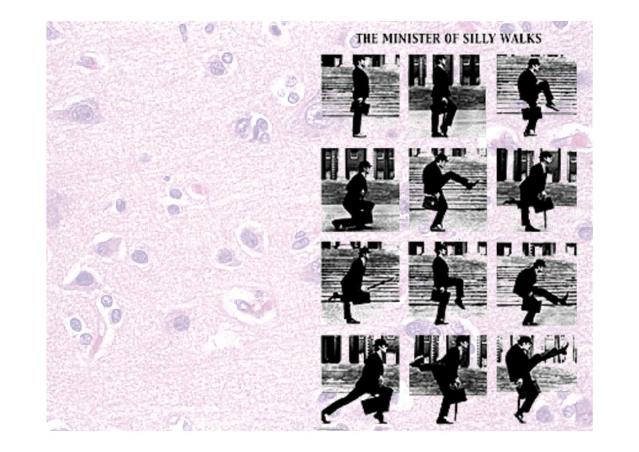
Gait
☤Hemiparetic/spastic
☤Festinant
☤PD, short shuffling steps,
leaning forward
☤Wide-based
☤Cerebellar (staggering)
☤Frontal lobe
☤Stamping
☤
↓proprioception
☤Steppage/high stepping
☤Foot drop
☤Proximal myopathy
☤waddling

Extra tests with spinal
problems
☤Straight leg raise (root entrapment, Lasegue)
☤Pain at <60º
☤Eased by bending knee
☤Can be “crossed” – pain on other side
☤Femoral stretch
☤Perianal sensation
☤Touch and pin
☤Anal tone
☤Anal “wink”

Practical - limbs
☤ Inspection
☤ Tone
☤ Power
☤ UL: Drift, SAb/Ad,EF,EE,WE,FF,FAb/Ad, opposition
☤ LL: HF, HE, KF, KE, AInv/Ev, DF, EHL, PF
☤ Sensory modalities and testing
☤ Pin, touch, proprioception, vibration
☤ Reflexes
☤ UL: Biceps, brachioradialis, triceps
☤ LL: Knee, ankle, plantar
☤ Coordination
☤ Finger-nose, heel-shin, alternating movements, Romberg
☤ Spinal tests, SLR, femoral stretch if needed

Neurological Examination of
the unconscious patient
¾ ABC, any sedative drugs, C-spine protection if
trauma
¾ Pattern of breathing –
¾ ↑RR -pontine lesions, respiratory causes, Metabolic
causes
¾ Cheyne-stokes – central medullary lesions,
Resp/CVS
¾ Irregular pattern – medullary lesions
¾ Glasgow coma scale
¾ Pulse and blood pressure ? ↑ICP
¾ Blood glucose

¾ Temperature
¾ Hypothermia
¾ Pyrexia- CNS infection, medullary lesion
¾ Neck stiffness –meningitis, SAH,↑ICP
¾ Pupils- size
¾ Small- pontine lesion, opioids
¾ Unresponsive midbrain lesions
¾ Pupils – asymmetry
¾ Large/unreactive lll nerve palsy
¾ Small slow to dilate- Horner's
¾ Light reflex
¾ Fundoscopy – papilloedema

¾ Eye movements
¾ primary position - squint
¾ Nystagmus
¾ Dolls eye movements –
horizontal –lll / Vl,pons
vertical – lll , lV, midbrain
¾ Corneal reflex- V,Vll, pontine lesion
¾ Gag reflex
¾ Posture
¾ Decorticate – pyramidal tracts from cortex to
internal capsule
¾ Decerebrate – midbrain, thalamus and
subthalamic nuclei
¾ Check the tone of the limbs

¾Limb Reflexes
¾Planters
¾Monitoring the progress at regular time
interval – improving/deteriorating

AVPU score
☤A
lert
☤Responds to
V
erbal stimulus
☤Responds to
P
ainful stimulus
☤U
nresponsive
Glasgow coma scale–eyes (4)
Open spontaneously
4
Open to command
3
Open to pain
2
No opening
1
Closed due to swelling C
☤Record best score for each section
☤Record in components, not just sum
GCS – verbal (5)
Orientated
5
Disorientated
4
Inappropriate words
3
Sounds
2
None
1
Intubated
T
GCS – motor (6)
To command
6
Localises pain
5
Withdraws from pain
4
Abnormal flexion to pain 3
Extension to pain
2
None
1
☤Start with command
☤If no response, squeeze side of nails
☤Sternal rub and nail bed pressure bruise

System for neurological
examination
☤General inspection
☤Glasgow coma
scale
☤Higher mental
function
☤Speech and
language
☤Cranial nerves
☤Trunk and limbs
☤Inspection
☤Tone
☤Power
☤Sensation
☤Reflexes
☤Coordination
☤Gait