1
General Musculoskeletal
Screening: Upper Extremities
Gregory Crovetti, M.D.
Sports Medicine Program
West Suburban Health Care
Trinity Orthopaedics
8/27/02
Gregory Crovetti, M.D.
2
General Approach
History
Inspection
Range of Motion (ROM)
Palpation
Muscular and neurological exams
8/27/02
Gregory Crovetti, M.D.
3
History
An accurate history is essential
Will give you diagnosis 80-90% of time
How symptoms started (mechanism of
injury)?
Duration of complaint?
Location, nature of pain, or symptoms?
Exacerbating or relieving maneuvers?
8/27/02
Gregory Crovetti, M.D.
4
General Inspection
Observe how the patient moves as they
go into the room or move from chair to
table
General appearance
Body proportions
8/27/02
Gregory Crovetti, M.D.
5
Inspection of Specific Area
Look for asymmetry between sides
Swelling
Deformities
Atrophy
Erythema
8/27/02
Gregory Crovetti, M.D.
6
Range of Motion (Active)
Have patient range the joints
Watch for decreased or increased
movement of the joint compared to the
other side as well as the norm
Watch for pain with movement
Listen for crepitus or “popping”
Watch for abnormal movements
8/27/02
Gregory Crovetti, M.D.
7
Range of Motion (Passive)
Next range the joints passively,
comparing the end points to the active
Again note any decreased or increased
movement
Pain with the movement
Crepitus or “popping”
8/27/02
Gregory Crovetti, M.D.
8
Palpation
When palpating a structure, you need
to know the anatomy of that structure
Palpate for swelling
Palpate for warmth
Palpate each area of the structure in
turn evaluating for pain, and
abnormalities as compared to the other
side
8/27/02
Gregory Crovetti, M.D.
9
Muscular and Neurological
Check the following comparing one side
to the other:
–
Grade strength (0-5)
–
Grade reflexes (0-4)
–
Sensory exam
8/27/02
Gregory Crovetti, M.D.
10
Generalized Screening Exam
If any abnormalities,
a more thorough
exam of the joint
needs to be done.
Each joint is:
–
Inspected (look for
abnormalities)
–
Palpated
–
Examined
8/27/02
Gregory Crovetti, M.D.
11
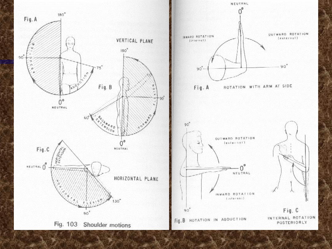
Neck: Active Range of Motion
Chin to chest (flexion)
“look at ceiling” (extension)
Chin to each shoulder (lateral rotation)
Ear to each shoulder (lateral flexion,
i.e., head tilt)
8/27/02
Gregory Crovetti, M.D.
12
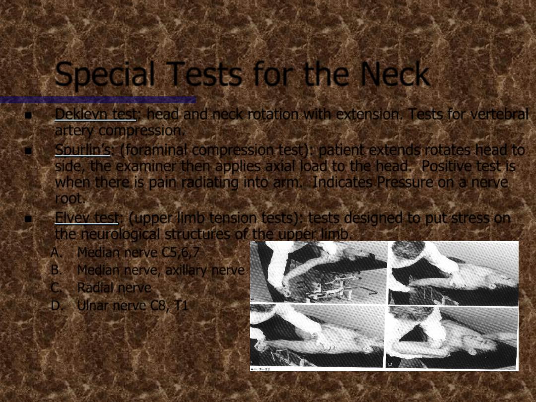
Special Tests for the Neck
Dekleyn test: head and neck rotation with extension. Tests for vertebral
artery compression.
Spurlin’s: (foraminal compression test): patient extends rotates head to
side, the examiner then applies axial load to the head. Positive test is
when there is pain radiating into arm. Indicates Pressure on a nerve
root.
Elvey test: (upper limb tension tests): tests designed to put stress on
the neurological structures of the upper limb.
A. Median nerve C5,6,7
B. Median nerve, axillary nerve
C. Radial nerve
D. Ulnar nerve C8, T1
8/27/02
Gregory Crovetti, M.D.
13
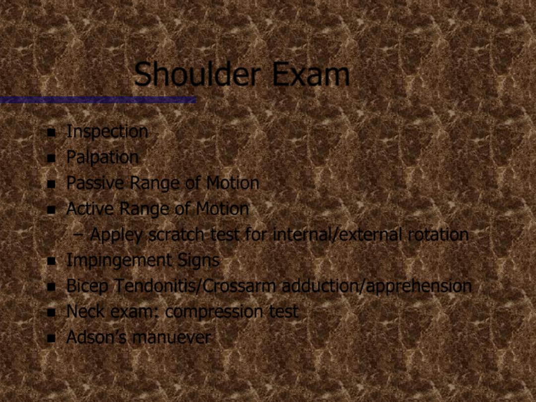
Shoulder Exam
Inspection
Palpation
Passive Range of Motion
Active Range of Motion
–
Appley scratch test for internal/external rotation
Impingement Signs
Bicep Tendonitis/Crossarm adduction/apprehension
Neck exam: compression test
Adson’s manuever
8/27/02
Gregory Crovetti, M.D.
14
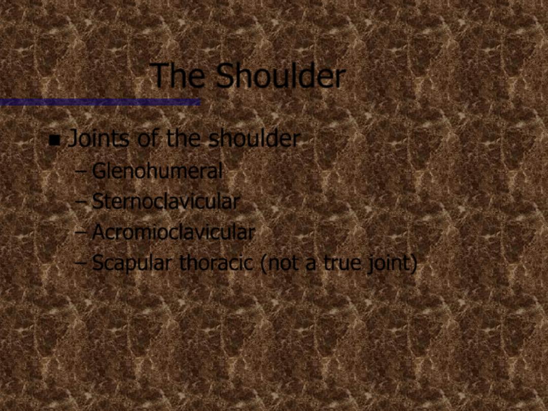
The Shoulder
Joints of the shoulder
–
Glenohumeral
–
Sternoclavicular
–
Acromioclavicular
–
Scapular thoracic (not a true joint)
8/27/02
Gregory Crovetti, M.D.
15
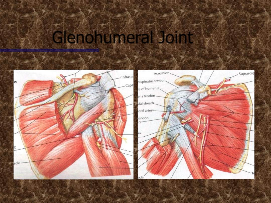
Glenohumeral Joint
8/27/02
Gregory Crovetti, M.D.
16
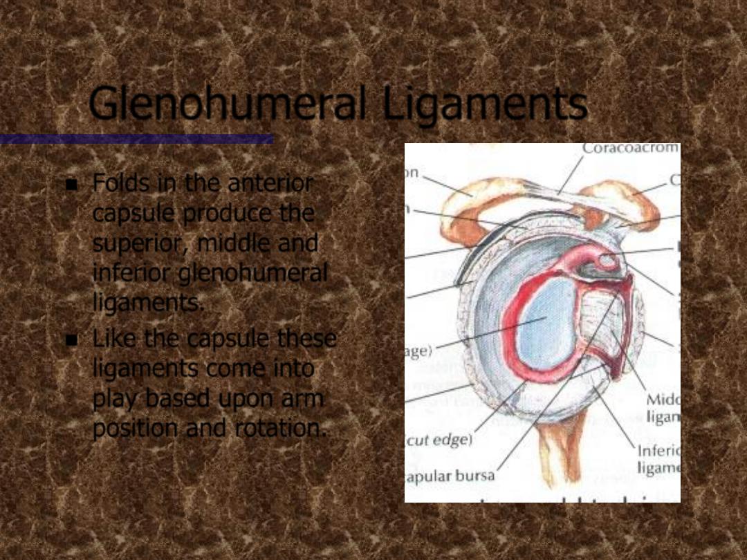
Glenohumeral Ligaments
Folds in the anterior
capsule produce the
superior, middle and
inferior glenohumeral
ligaments.
Like the capsule these
ligaments come into
play based upon arm
position and rotation.
8/27/02
Gregory Crovetti, M.D.
17
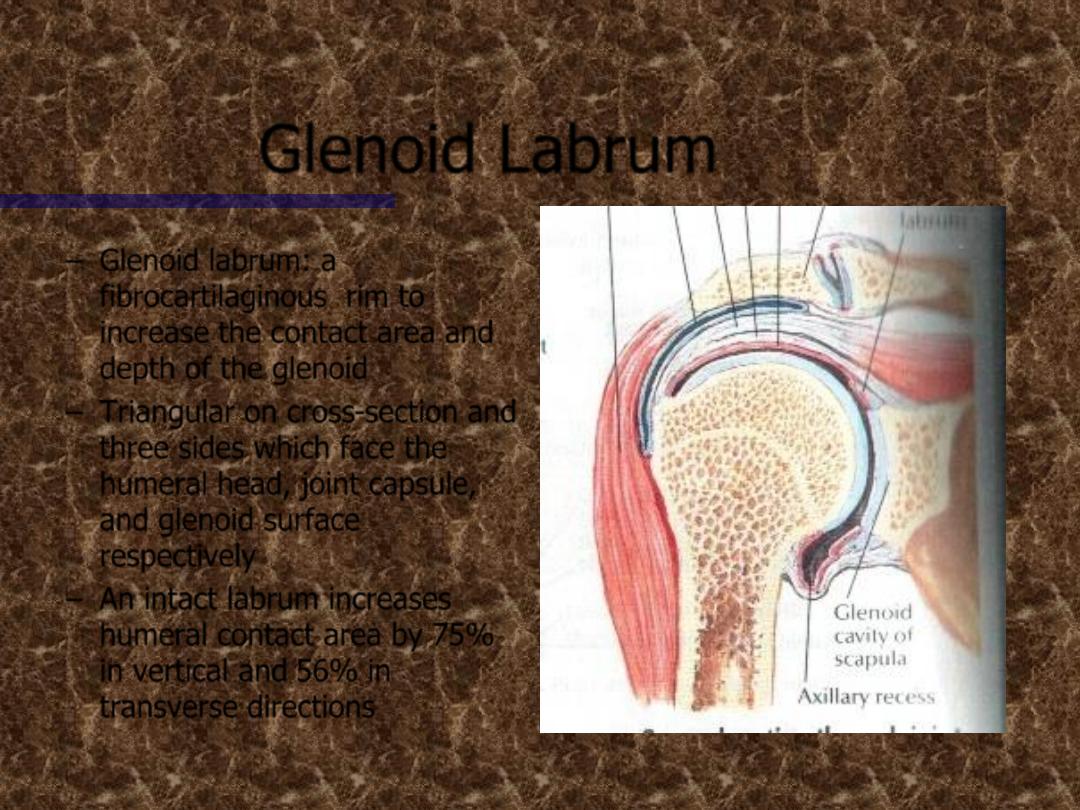
Glenoid Labrum
–
Glenoid labrum: a
fibrocartilaginous rim to
increase the contact area and
depth of the glenoid
–
Triangular on cross-section and
three sides which face the
humeral head, joint capsule,
and glenoid surface
respectively
–
An intact labrum increases
humeral contact area by 75%
in vertical and 56% in
transverse directions
8/27/02
Gregory Crovetti, M.D.
18
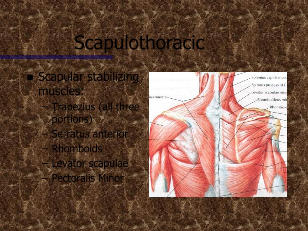
Scapulothoracic
Scapular stabilizing
muscles:
–
Trapezius (all three
portions)
–
Serratus anterior
–
Rhomboids
–
Levator scapulae
–
Pectoralis Minor
8/27/02
Gregory Crovetti, M.D.
19
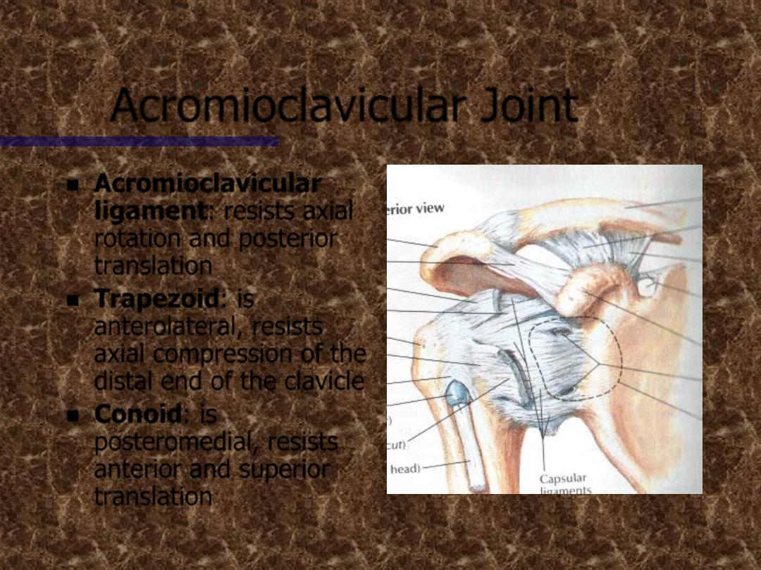
Acromioclavicular Joint
Acromioclavicular
ligament: resists axial
rotation and posterior
translation
Trapezoid: is
anterolateral, resists
axial compression of the
distal end of the clavicle
Conoid: is
posteromedial, resists
anterior and superior
translation

8/27/02
Gregory Crovetti, M.D.
20
Sternoclavicular Joint
These structures still
allow for 35 degrees
of elevation, 35
degrees of
translation, and 50
degrees of rotation
at the
sternoclavicular joint
8/27/02
Gregory Crovetti, M.D.
21
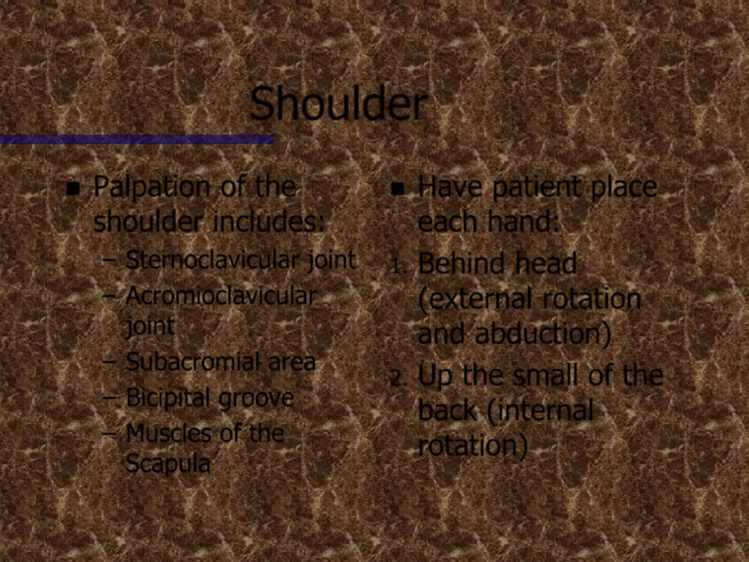
Shoulder
Palpation of the
shoulder includes:
–
Sternoclavicular joint
–
Acromioclavicular
joint
–
Subacromial area
–
Bicipital groove
–
Muscles of the
Scapula
Have patient place
each hand:
1.
Behind head
(external rotation
and abduction)
2.
Up the small of the
back (internal
rotation)
8/27/02
Gregory Crovetti, M.D.
22
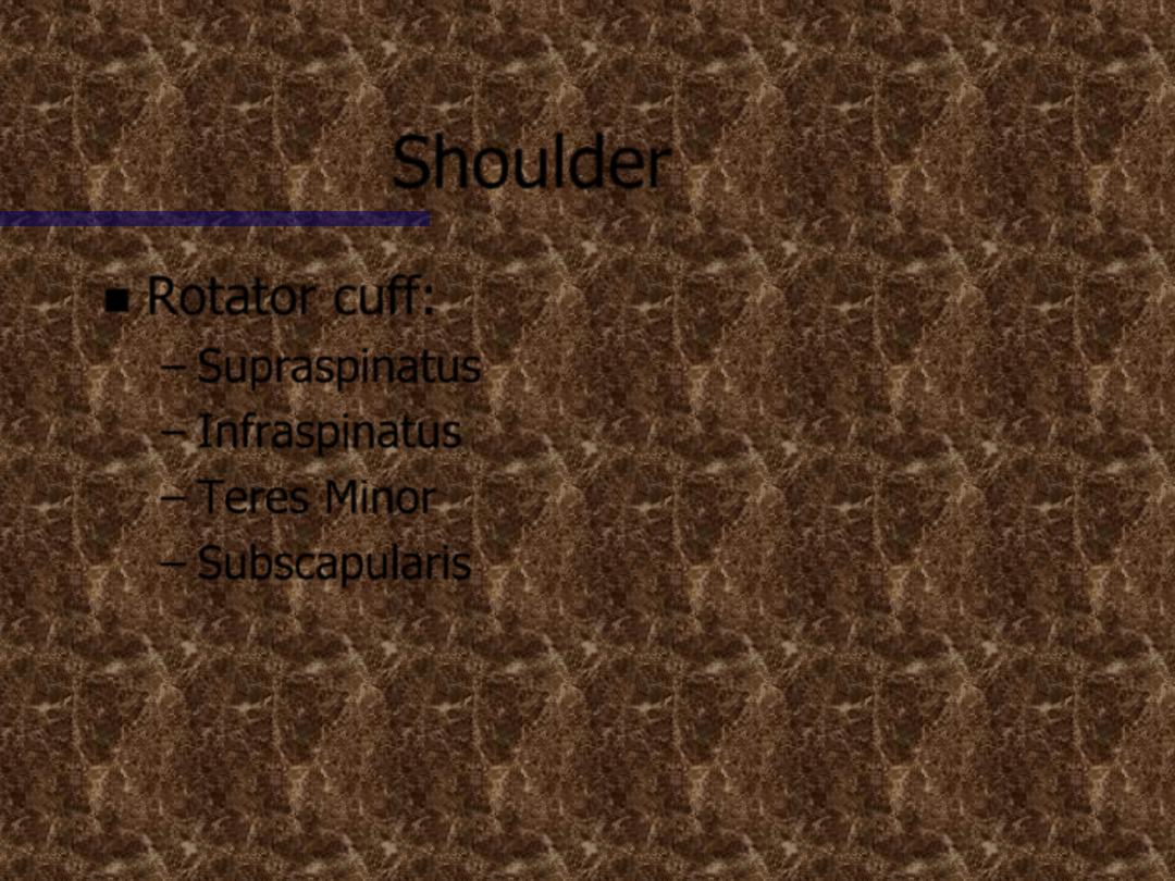
Shoulder
Rotator cuff:
–
Supraspinatus
–
Infraspinatus
–
Teres Minor
–
Subscapularis
8/27/02
Gregory Crovetti, M.D.
23
8/27/02
Gregory Crovetti, M.D.
24
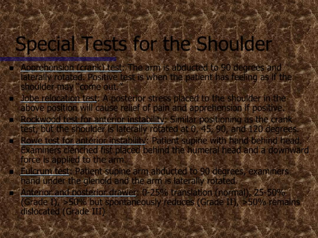
Special Tests for the Shoulder
Apprehension (crank) test: The arm is abducted to 90 degrees and
laterally rotated. Positive test is when the patient has feeling as if the
shoulder may “come out.”
Jobe relocation test: A posterior stress placed to the shoulder in the
above position will cause relief of pain and apprehension if positive.
Rockwood test for anterior instability: Similar positioning as the crank
test, but the shoulder is laterally rotated at 0, 45, 90, and 120 degrees.
Rowe test for anterior instability: Patient supine with hand behind head.
Examiners clenched fist placed behind the humeral head and a downward
force is applied to the arm.
Fulcrum test: Patient supine arm abducted to 90 degrees, examiners
hand under the glenoid and the arm is laterally rotated.
Anterior and posterior drawer: 0-25% translation (normal), 25-50%
(Grade I), >50% but spontaneously reduces (Grade II), >50% remains
dislocated (Grade III)
8/27/02
Gregory Crovetti, M.D.
25
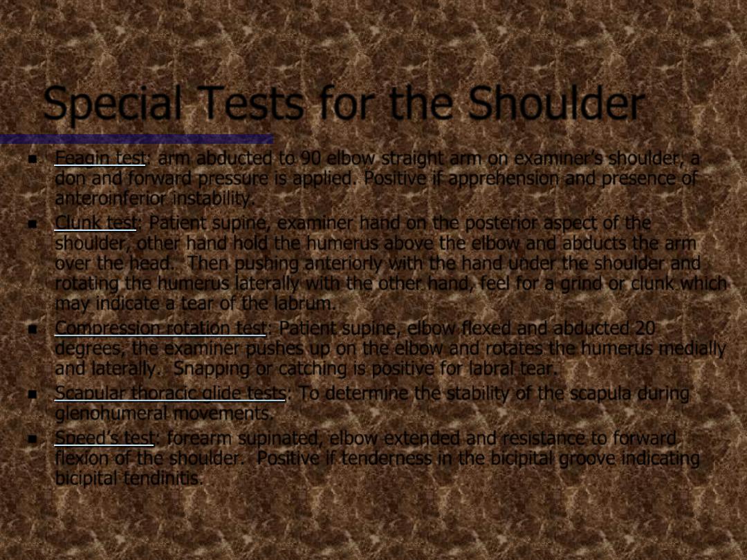
Special Tests for the Shoulder
Feagin test: arm abducted to 90 elbow straight arm on examiner’s shoulder, a
don and forward pressure is applied. Positive if apprehension and presence of
anteroinferior instability.
Clunk test: Patient supine, examiner hand on the posterior aspect of the
shoulder, other hand hold the humerus above the elbow and abducts the arm
over the head. Then pushing anteriorly with the hand under the shoulder and
rotating the humerus laterally with the other hand, feel for a grind or clunk which
may indicate a tear of the labrum.
Compression rotation test: Patient supine, elbow flexed and abducted 20
degrees, the examiner pushes up on the elbow and rotates the humerus medially
and laterally. Snapping or catching is positive for labral tear.
Scapular thoracic glide tests: To determine the stability of the scapula during
glenohumeral movements.
Speed’s test: forearm supinated, elbow extended and resistance to forward
flexion of the shoulder. Positive if tenderness in the bicipital groove indicating
bicipital tendinitis.
8/27/02
Gregory Crovetti, M.D.
26
Special Tests for the Shoulder
Yergason’s test: Elbow flexed to 90 degrees, forearm pronated,
resistance to supination is applied as the patient also laterally rotates
the arm. Positive if pain in the bicipital groove and indicates bicipital
tendinitis.
Supraspinatus (empty can/ Jobes) test: The shoulder is forward flexed
at 30 degrees, arms straight and thumbs pointing to ground, a
downward force is applied to the arms. Tests for tear or weakness of
the supraspinatus.
Codman’s (drop arm) test: shoulder is abducted to 90 degrees and
patient asked to lower the arm slowly. If drops or is painful, it is
positive and indicates tear in the rotator cuff.
Neer impingement test: Arm is elevated through forward flexion,
positive if painful.
Hawkins-Kennedy impingement test: Arm is forward flexed to 90 then
internally rotated, positive if painful.
8/27/02
Gregory Crovetti, M.D.
27
Special Tests for the Shoulder
Impingement test: Arm is abducted to 90 and full lateral rotation,
positive if painful.
Military brace (Costoclavicular Syndrome) test: Palpate the radial pulse
as the shoulder is drawn down and back. Positive if a decreased pulse
and indicates possible thoracic outlet syndrome.
Adson Maneuver: radial pulse palpated as arm is rotated laterally and
elbow is extended as the patient extends and rotates head to test
shoulder.
Allen test: Elbow is flexed to 90, shoulder abducted and laterally
rotated and patient rotates head away for the test side.
Halstead maneuver: Radial pulse felt as arm is pulled down as the
patients neck is hyperextended and rotated to the opposite side.
8/27/02
Gregory Crovetti, M.D.
28
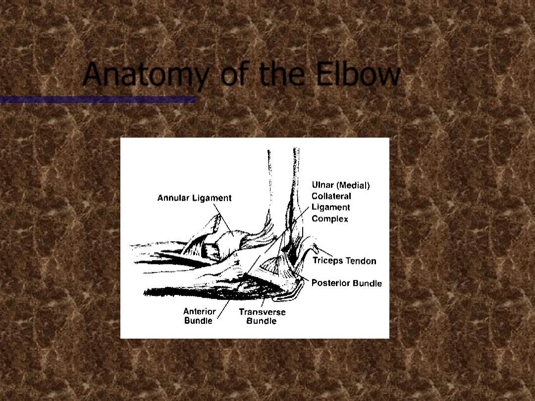
The Elbow
Palpation: lateral and medial
epicondyles, olecranon, radial head,
groove on either side of the olecranon
Inspect the carrying angle, and any
nodules or swelling
8/27/02
Gregory Crovetti, M.D.
29
8/27/02
Gregory Crovetti, M.D.
30
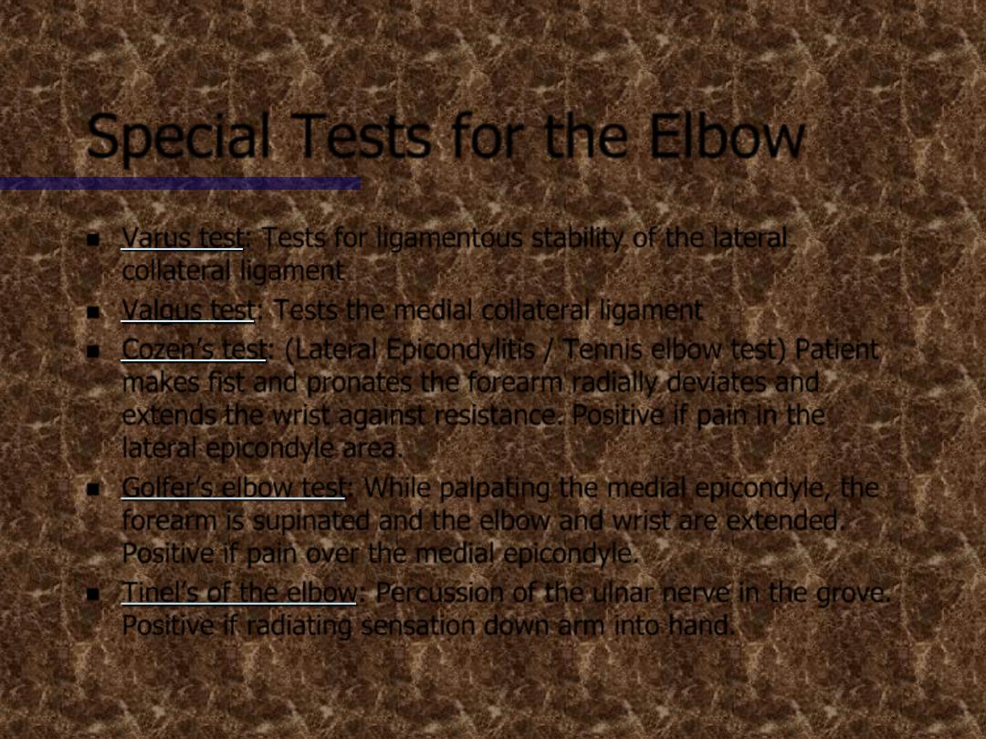
Special Tests for the Elbow
Varus test: Tests for ligamentous stability of the lateral
collateral ligament
Valgus test: Tests the medial collateral ligament
Cozen’s test: (Lateral Epicondylitis / Tennis elbow test) Patient
makes fist and pronates the forearm radially deviates and
extends the wrist against resistance. Positive if pain in the
lateral epicondyle area.
Golfer’s elbow test: While palpating the medial epicondyle, the
forearm is supinated and the elbow and wrist are extended.
Positive if pain over the medial epicondyle.
Tinel’s of the elbow: Percussion of the ulnar nerve in the grove.
Positive if radiating sensation down arm into hand.
8/27/02
Gregory Crovetti, M.D.
31
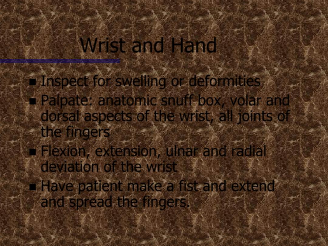
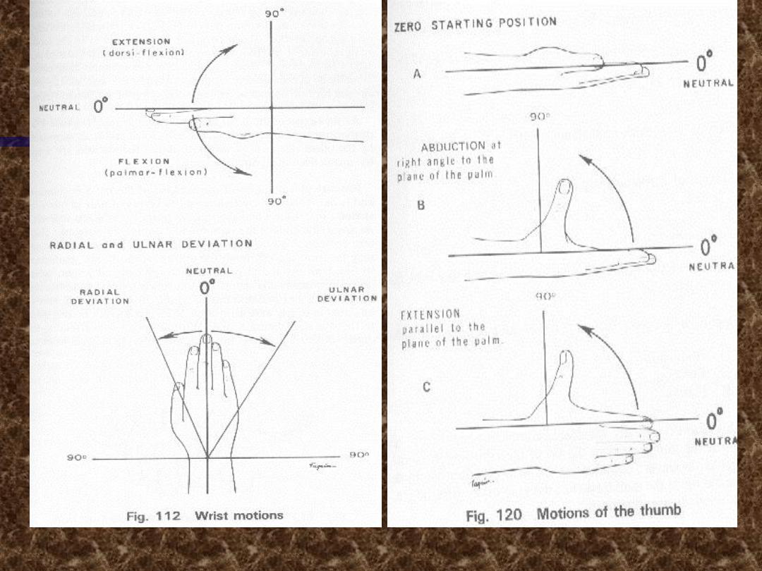
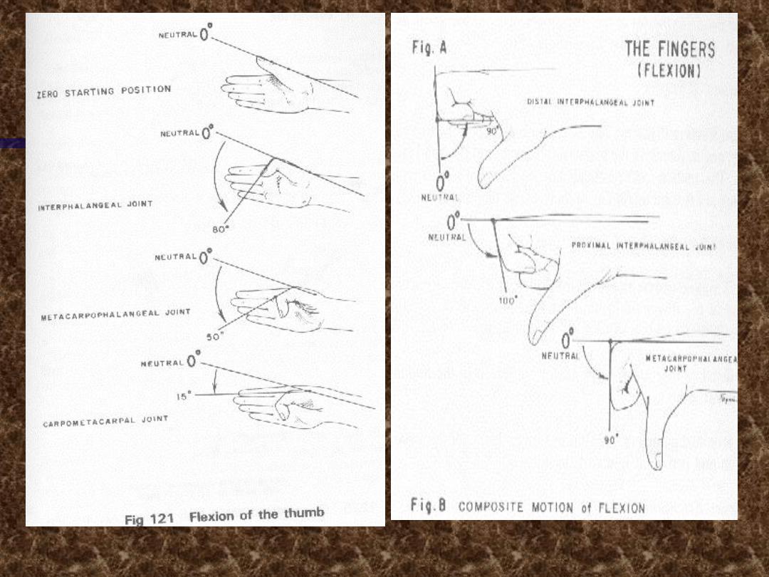
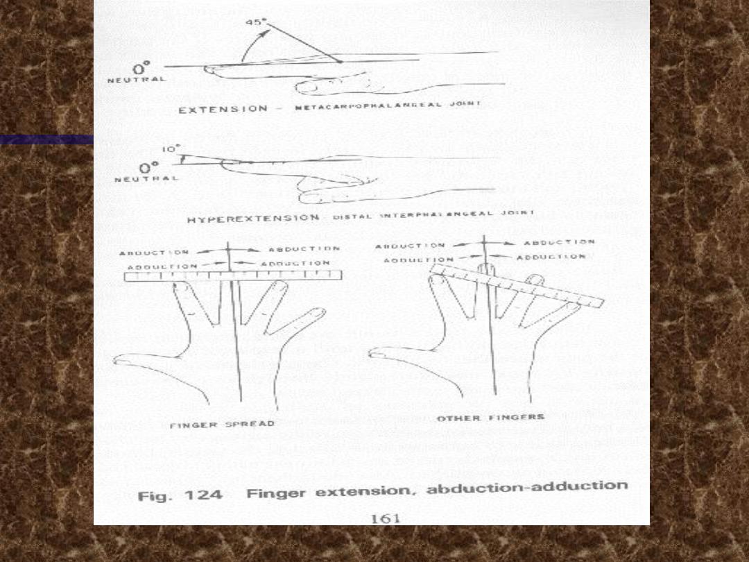
Wrist and Hand
Inspect for swelling or deformities
Palpate: anatomic snuff box, volar and
dorsal aspects of the wrist, all joints of
the fingers
Flexion, extension, ulnar and radial
deviation of the wrist
Have patient make a fist and extend
and spread the fingers.
8/27/02
Gregory Crovetti, M.D.
32
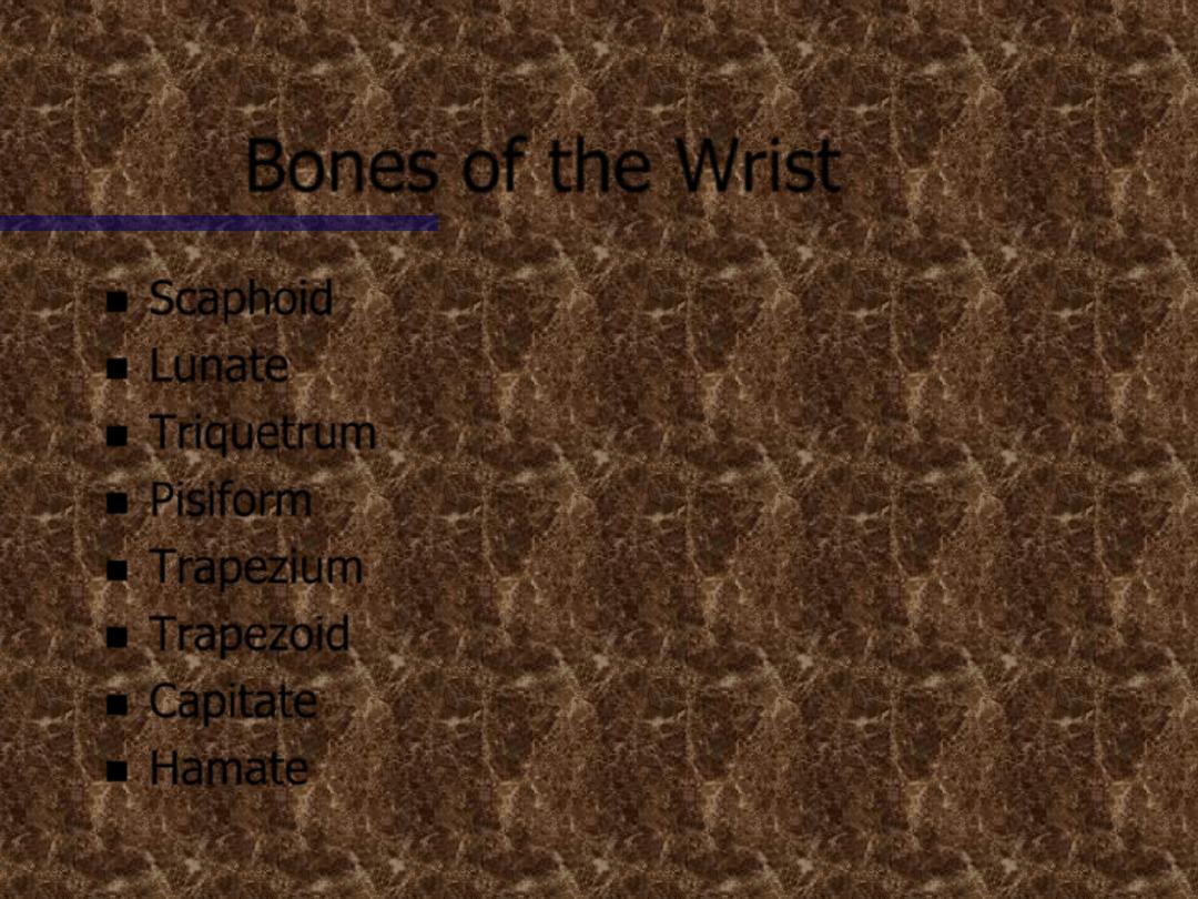
Bones of the Wrist
Scaphoid
Lunate
Triquetrum
Pisiform
Trapezium
Trapezoid
Capitate
Hamate
8/27/02
Gregory Crovetti, M.D.
33
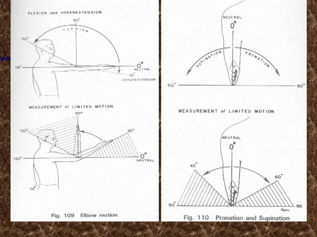
Anatomy of the Elbow
8/27/02
Gregory Crovetti, M.D.
34
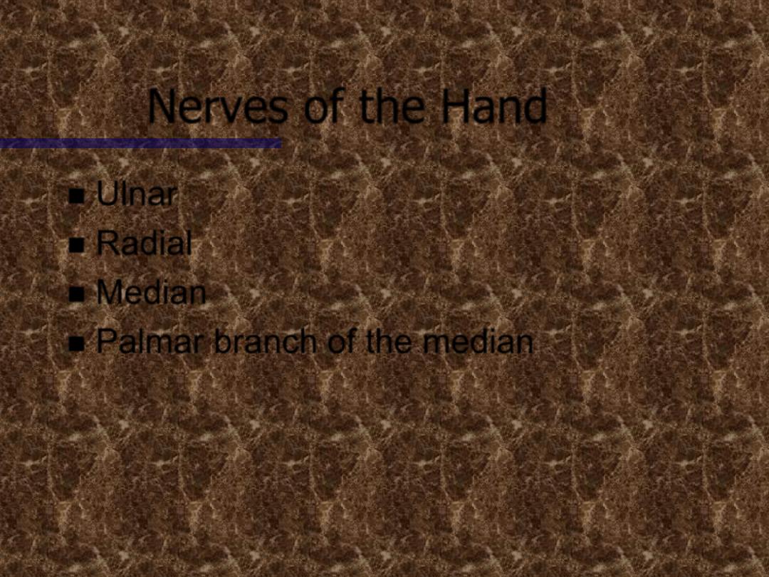
Nerves of the Hand
Ulnar
Radial
Median
Palmar branch of the median
8/27/02
Gregory Crovetti, M.D.
35
8/27/02
Gregory Crovetti, M.D.
36
8/27/02
Gregory Crovetti, M.D.
37
8/27/02
Gregory Crovetti, M.D.
38
Special Tests of Hand and Wrist
Cascade sign: Patient flexes the fingers, the tips should all converge toward the
scaphoid tubercle. If they do not, it may indicate a fracture in that finger.
Boutonniere deformity: Extension of the MCP and DIP joints and flexion of the
PIP joint. This is due to a rupture of the central tendinous slip of the extensor
hood.
Swan-neck deformity: Flexion of the MCP and DIP joints, with extension of the
PIP joint. This is due to contracture of the intrinsic muscles. Seen after trauma
or in RA.
Ulnar drift: Ulnar deviation of the digits most commonly due to RA.
Dupuytren’s contracture: This is due to contracture of the palmar fascia. Most
common in the ring finger or little finger, men more then women, ages 50-70.
Claw fingers: This deformity is a form a combination of a ulnar and median
nerve palsy. This causes loss of intrinsic muscle function and over action of the
extrinsic extensors. This causes hyperextension of the MCP joints and flexion of
the PIP and DIP joints. If the intrinsic function of the hand is lost, it is then
called an intrinsic minus hand.
8/27/02
Gregory Crovetti, M.D.
39
Special Tests of Hand and Wrist
Trigger finger: Results from a thickening of the flexor tendon sheath, causing
sticking of the tendon. At later stages the finger can become stuck in flexion,
needing to be passively extended. Associated with RA.
Bishop’s Hand: (Benediction Hand) Secondary to ulnar nerve palsy. There is
wasting of the hypothenar, interossei, and the two medial lumbrical muscles.
Flexion of the 4
th
and 5
th
fingers is the most noticeable deformity.
“Z” deformity of the thumb: May be secondary to RA or heredity. The thumb
is flexed at the MCP and hyperextended at the IP joint.
Drop- wrist: Secondary to radial nerve palsy.
Mallet finger: The distal phalanx remains in flexion when the finger is
extended. This is the result of rupture or avulsion of the extensor tendon from
the distal phalanx.
Clubbing: Can be caused by many medical problems such as pulmonary or
cardiac diseases, as well as genetic.
Heberden’s nodes: Swelling of the DIP joints secondary to OA.
Bouchard’s nodes: Swelling of the PIP joints secondary to RA.
8/27/02
Gregory Crovetti, M.D.
40
Special Tests of Hand and Wrist
Ganglion cyst: Localized swelling usually on the dorsum of the hand.
Thumb ulnar collateral ligament test: (test for gamekeeper’s or skier’s
thumb) Valgus stress applied to the MCP joint, if 10-20 degrees there is
most likely a partial tear
Carpal Compression test: Pressure applied directly to the carpal tunnel
for 30 seconds. If positive, indicates carpal tunnel syndrome.
Froment’s sign: Patient holds piece of paper between the thumb and
index paper. If the distal phalanx flexes, it is a positive test and
indicates ulnar nerve palsy. If the MCP joint hyperextends, it is a
positive Jeanne’s sign and also indicates ulnar nerve palsy.
Allen test: Tests for competency of the ulnar and radial arteries.
Anatomic snuffbox: Lies between the extensor pollicis longus and
extensor pollicis brevis tendons. The scaphoid bone is palpated inside
the box as well as the radial styloid. Pain in the box should indicate
scaphoid fracture until proven otherwise.
8/27/02
Gregory Crovetti, M.D.
41
Special Tests of Hand and Wrist
Guyon’s canal: (pisohamate) Through this canal runs the ulnar nerve. If
compression of the canal occurs, there is sensation lose to the fingers and
muscle weakness in the hand of ulnar distribution.
>35 degrees indicates a torn ulnar and accessory collateral ligaments.
Murphy’s sign: Patient makes a fist, if the head of the third metacarpal is level
with the second and fourth metacarpals, it is a sign of a lunate dislocation.
Retinacular ligament test: Test for the structures around the PIP joint. The
patient is passive, the PIP joint is held in extension and the DIP is flexed. If the
DIP does not flex, the retinacular ligaments (collateral) or capsule is tight. The
PIP joint is the flexed, if the DIP now flexes easily, the retinacular ligaments are
tight and the capsule is normal.
Lunatotiquetral Ballottement (Reagan’s test): The triquetrum is grasped
between the thumb and second finger of one hand and the lunate between the
thumb and second finger of the other hand. The lunate is then moved up and
down, if any laxity, crepitus or pain it indicates a positive test for
Lunatotriquetral instability.
8/27/02
Gregory Crovetti, M.D.
42
Special Tests of Hand and Wrist
Watson (scaphoid shift) test: The patient’s hand is taken into full ulnar deviation
and slight extension. With the other hand the thumb is pressed against the
distal pole of the scaphoid to prevent it from moving. The patient’s hand is then
moved radially and slightly flexed. If the dorsal pole of the scaphoid subluxes
over the dorsal rim of the radius and there is pain, it is a positive test for
scaphoid and lunate instability.
Scaphoid stress test: Modification of Watson test in which the patient actively
radial deviates the wrist while scaphoid pressure is applied. If there is pain and
a clunk, it is a positive test.
“Piano Key” test: Patient’s arms are in pronation. Using the index finger while
stabilizing the hand with the other hand the distal ulna is pushed down. The test
is positive if there is pain and difference in mobility compared to the other side.
This indicates distal radioulnar joint instability.
Axial load test: Axial load to the thumb or fingers, if pain or crepitation it is a
positive test for metacarpal or adjacent carpal bone fracture or joint arthrosis.
Grind test: Grabbing the thumb below the metacarpophalangeal joint, an axial
load is applied with rotation. If there is pain the test is positive and indicates
DJG of the metacarpophalangeal or metacarpotrapezial joints.
8/27/02
Gregory Crovetti, M.D.
43
Special Tests of Hand and Wrist
Finkelstein test: Tests for De Quervain’s or Hoffmann’s disease. A positive test
indicates a tenosynovitis of the abductor pollicis longus and extensor pollicis
brevis tendons.
Sweater finger sign: When patient makes a fist, if one of the distal phalanx
(most often the ring finger) does not flex, the test is positive. It indicates a
ruptured flexor digitorum profundus tendon.
Bunnel-Littler test: (Finochietto-Bunnel test) The patient is passive during the
test. The test is for structures around the MCP joint. The MCP joint is held in
extension, while the PIP is flexed. If unable to flex the PIP, the test is positive
and indicates tight intrinsic muscle or contracture of the joint capsule. The
MCP is then slightly flexed, if the PIP now flexes easily it indicates tight
intrinsic muscles and that the capsule is normal. If the PIP still does not flex it
indicates a tight joint capsule.
Tinel’s sign: Positive if tingling into the fingers of the median nerve
distribution, indicating carpal tunnel syndrome.
Phalen’s test: Position must be held for one minute. If positive indicates carpal
tunnel syndrome. The dorsal aspect of the hands is pushed together to
maximal flexion of the wrists.
8/27/02
Gregory Crovetti, M.D.
44
Case
75-year old man comes in for yearly physical.
History of hypertension, elevated lipids, and
mild obesity
He has taken your advise and started an
exercise program, and now has a complaint
of right shoulder pain.
What do you want to know?
What do you do next?