
11/18/2015
Diseases of the heart valves | Davidson
’s Principles and Practice of…
data:text/html;charset=utf-8,%3Ch2%20id%3D%2277c45d9bcafc42838aebaa126c81e6e4%22%20style%3D%22margin%3A%201.3em%200px%200.5em%3B%20padding%3A%200px%3B%20border%3A%200px%3B%20font-fa
… 11/12
Infective endocarditis
This is caused by microbial infection of a heart valve (native or prosthetic), the lining of a cardiac chamber or blood vessel, or a
congenital anomaly (e.g. septal defect). The causative organism is usually a bacterium, but may be a rickettsia, chlamydia or
fungus.
Pathophysiology
Infective endocarditis typically occurs at sites of pre-existing endocardial damage, but infection with particularly virulent or
aggressive organisms (e.g. Staphylococcus aureus) can cause endocarditis in a previously normal heart; staphylococcal
endocarditis of the tricuspid valve is a common complication of intravenous drug misuse. Many acquired and congenital cardiac
lesions are vulnerable to endocarditis, particularly areas of endocardial damage caused by a high-pressure jet of blood, such as
ventricular septal defect, mitral regurgitation and aortic regurgitation, many of which are haemodynamically insignificant. In
contrast, the risk of endocarditis at the site of haemodynamically important low-pressure lesions, such as a large atrial septal
defect, is minimal.
Infection tends to occur at sites of endothelial damage because they attract deposits of platelets and fibrin that are vulnerable to
colonisation by blood-borne organisms. The avascular valve tissue and presence of fibrin and platelet aggregates help to protect
proliferating organisms from host defence mechanisms. When the infection is established, vegetations composed of organisms,
fibrin and platelets grow and may become large enough to cause obstruction or embolism. Adjacent tissues are destroyed and
abscesses may form. Valve regurgitation may develop or increase if the affected valve is damaged by tissue distortion, cusp
perforation or disruption of chordae. Extracardiac manifestations, such as vasculitis and skin lesions, are due to emboli or
immune complex deposition. Mycotic aneurysms may develop in arteries at the site of infected emboli. At autopsy, infarction of
the spleen and kidneys and, sometimes, an immune glomerulonephritis are found.
Microbiology
Over three-quarters of cases are caused by streptococci or staphylococci. The viridans group of streptococci (Streptococcus
mitis, Strep. sanguis) are commensals in the upper respiratory tract that may enter the blood stream on chewing or teeth-

11/18/2015
Diseases of the heart valves | Davidson
’s Principles and Practice of…
data:text/html;charset=utf-8,%3Ch2%20id%3D%2277c45d9bcafc42838aebaa126c81e6e4%22%20style%3D%22margin%3A%201.3em%200px%200.5em%3B%20padding%3A%200px%3B%20border%3A%200px%3B%20font-fa
… 22/12
brushing, or at the time of dental treatment, and are common causes of subacute endocarditis (
Box 18.113
). Other organisms,
including Enterococcus faecalis, E. faecium and Strep. bovis, may enter the blood from the bowel or urinary tract. Strep.
milleri and Strep. bovis endocarditis is associated with large-bowel neoplasms.
18.113 Microbiology of infective endocarditis
Pathogen
Of native
valve
(n = 280)
In IV drug
users
(n = 87)
Of prosthetic
valve
Early
(n =
15)
Late
(n =
72)
Staphylococci
124 (44%)
60 (69%)
10
(67%)
33
(46%)
Staph. aureus
106 (38%)
60 (69%)
3
(20%)
15
(21%)
Coagulase-negative
18 (6%)
0
7
(47%)
18
(25%)
Streptococci
86 (31%)
7 (8%)
0
25
(35%)
Oral
59 (21%)
3 (3%)
0
19
(26%)
Others (non-
enterococcal)
27 (10%)
4 (5%)
0
6 (8%)
Enterococcus spp.
21 (8%)
2 (2%)
1 (7%)
5 (7%)
HACEK group (see
text)
12 (4%)
0
0
1 (1%)

11/18/2015
Diseases of the heart valves | Davidson
’s Principles and Practice of…
data:text/html;charset=utf-8,%3Ch2%20id%3D%2277c45d9bcafc42838aebaa126c81e6e4%22%20style%3D%22margin%3A%201.3em%200px%200.5em%3B%20padding%3A%200px%3B%20border%3A%200px%3B%20font-fa
… 33/12
Polymicrobial
6 (2%)
8 (9%)
0
1 (1%)
Other bacteria
12 (4%)
4 (5%)
0
2 (3%)
Fungi
3 (1%)
2 (2%)
0
0
Negative blood
culture
16 (6%)
4 (5%)
4
(27%)
5 (7%)
Adapted from Moreillon P, Que YA. Lancet 2004; 363:139–149.
Staph. aureus has now overtaken streptococci as the most common cause of acute endocarditis. It originates from skin
infections, abscesses or vascular access sites (e.g. intravenous and central lines), or from intravenous drug use. It is highly
virulent and invasive, usually producing florid vegetations, rapid valve destruction and abscess formation. Other causes of acute
endocarditis include Strep. pneumoniae and Strep. pyogenes.
Post-operative endocarditis after cardiac surgery may affect native or prosthetic heart valves or other prosthetic materials. The
most common organism is a coagulase-negative staphylococcus (Staph. epidermidis), a normal skin commensal. There is
frequently a history of wound infection with the same organism. Staph. epidermidisoccasionally causes endocarditis in patients
who have not had cardiac surgery, and its presence in blood cultures may be erroneously dismissed as contamination. Another
coagulase-negative staphylococcus, Staph. lugdenensis,causes a rapidly destructive acute endocarditis that is associated with
previously normal valves and multiple emboli. Unless accurately identified, it may also be overlooked as a contaminant.
In Q fever endocarditis due to Coxiella burnetii, the patient often has a history of contact with farm animals. The aortic valve is
usually affected and there may also be hepatitis, pneumonia and purpura. Life-long antibiotic therapy may be required.
Gram-negative bacteria of the so-called HACEK group (Haemophilus spp., Actinobacillus
actinomycetemcomitans,Cardiobacterium hominis, Eikenella spp. and Kingella kingae) are slow-growing, fastidious
organisms that are only revealed after prolonged culture and may be resistant to penicillin.
Brucella is associated with a history of contact with goats or cattle and often affects the aortic valve.

11/18/2015
Diseases of the heart valves | Davidson
’s Principles and Practice of…
data:text/html;charset=utf-8,%3Ch2%20id%3D%2277c45d9bcafc42838aebaa126c81e6e4%22%20style%3D%22margin%3A%201.3em%200px%200.5em%3B%20padding%3A%200px%3B%20border%3A%200px%3B%20font-fa
… 44/12
Yeasts and fungi (Candida, Aspergillus) may attack previously normal or prosthetic valves, particularly in immunocompromised
patients or those with indwelling intravenous lines. Abscesses and emboli are common, therapy is difficult (surgery is often
required) and mortality is high. Concomitant bacterial infection may be present.
Incidence
The incidence of infective endocarditis in community-based studies ranges from 5 to 15 cases per 100 000 per annum. More than
50% of patients are over 60 years of age (
Box 18.114
). In a large British study, the underlying condition was rheumatic heart
disease in 24% of patients, congenital heart disease in 19%, and other cardiac abnormalities (e.g. calcified aortic valve, floppy
mitral valve) in 25%. The remaining 32% were not thought to have a pre-existing cardiac abnormality.
18.114 Endocarditis in old age
•
Symptoms and signs: may be non-specific, e.g. confusion, weight loss, malaise and weakness, and the
diagnosis may not be suspected.
•
Common causative organisms: often enterococci (from the urinary tract) and Strep. bovis (from a
colonic source).
•
Morbidity and mortality: much higher.
Clinical features
Endocarditis can take either an acute or a more insidious ‘subacute’ form. However, there is considerable overlap because the
clinical pattern is influenced not only by the organism, but also by the site of infection, prior antibiotic therapy and the presence
of a valve or shunt prosthesis. The subacute form may abruptly develop acute life-threatening complications, such as valve
disruption or emboli.
Subacute endocarditis
This should be suspected when a patient with congenital or valvular heart disease develops a persistent fever, complains of
11/18/2015
Diseases of the heart valves | Davidson
’s Principles and Practice of…
data:text/html;charset=utf-8,%3Ch2%20id%3D%2277c45d9bcafc42838aebaa126c81e6e4%22%20style%3D%22margin%3A%201.3em%200px%200.5em%3B%20padding%3A%200px%3B%20border%3A%200px%3B%20font-fa
… 55/12
....,+
-----
'
unusual tiredness, night sweats or weight loss, or develops new signs of valve dysfunction or heart failure. Less often, it presents
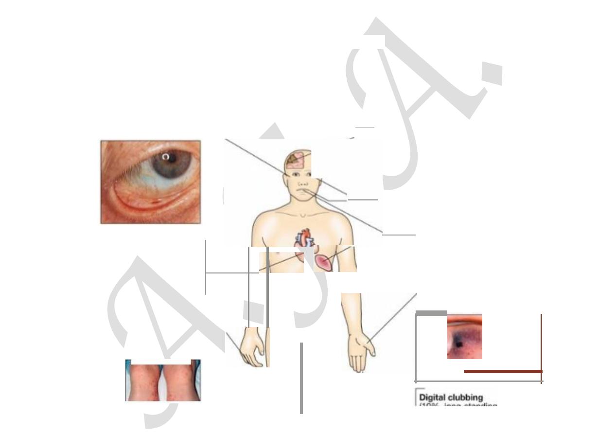
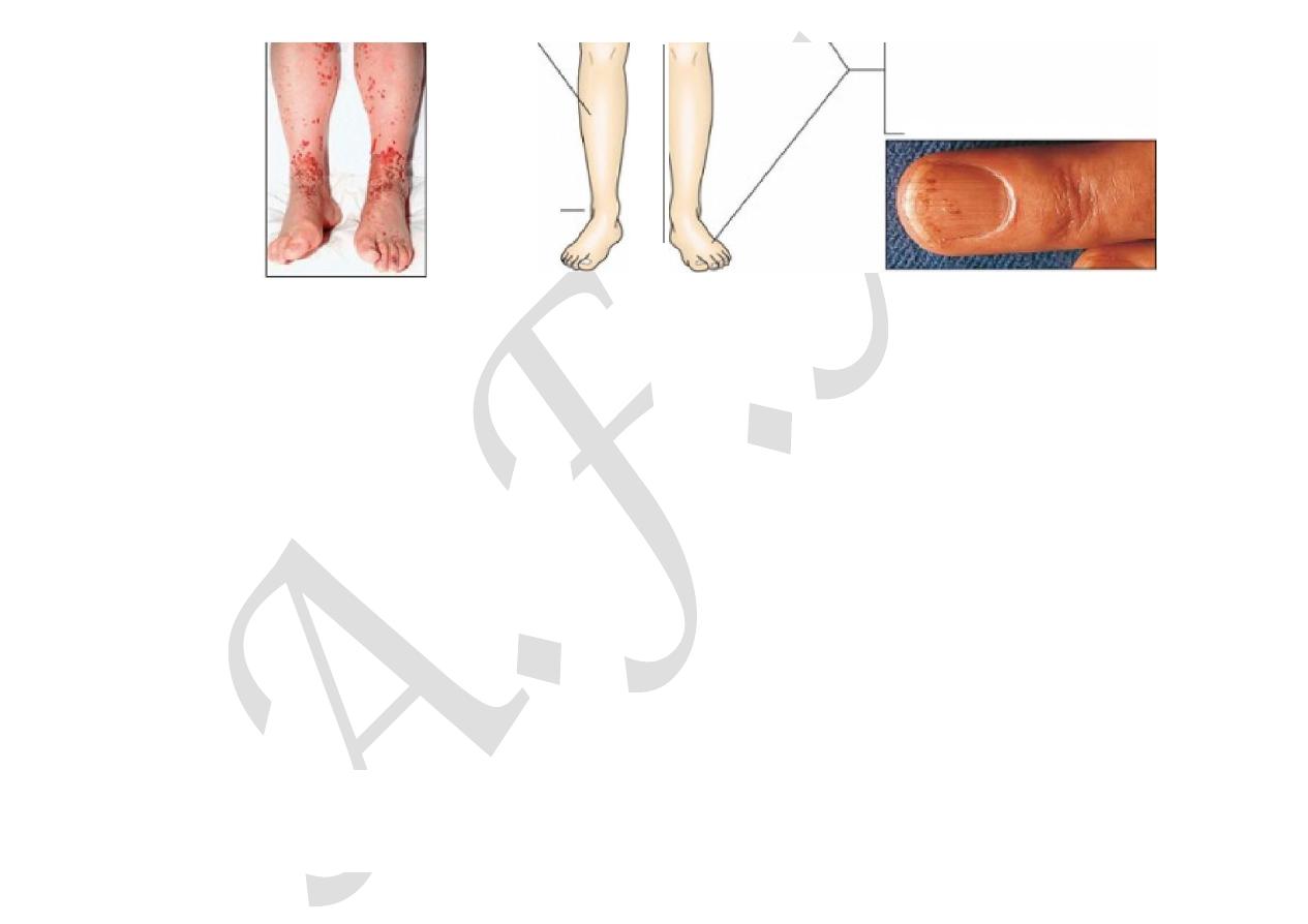
as an embolic stroke or peripheral arterial embolism. Other features (
Fig. 18.93
) include purpura and petechial
haemorrhages in the skin and mucous membranes, and splinter haemorrhages under the fingernails or toe nails. Osler’s nodes
are painful tender swellings at the fingertips that are probably the product of vasculitis; they are rare. Digital clubbing is a late
sign. The spleen is frequently palpable; in Coxiella infections, the spleen and the liver may be considerably enlarged.
Microscopic haematuria is common. The finding of any of these features in a patient with persistent fever or malaise is an
indication for re-examination to detect hitherto unrecognised heart disease.
Subcon
jun
cU
v
al haemorrhages
---.
(2
-
5%)
(90% ne
w
o
r
ch
ang
ed
mum,
I.W)
Condu
c
tion d
i
so
r
de
r
(
10-20
%)
(
40-50
%)
H
aematuria
=-
----
+--1
1"
"-)
I
(
60-7
0%)
O
s
i
e
r
'
s nodes
--,
(5%)
P
etec
h
l
al rash
(
4
().5()%,
m
a
y
be
t
ronsie
nt)
,.-----
~rebral e
mbo
li
(
1
5%)
---
R
olh
'
s
spo
t
s I
n fun
d
l
(
r
ar
e
.<
5
%)
P
e
t
ech
l
al hae
m
orr
h
ages on
mucous me
mb
ranes and fund
i
(20-30%)
P
oor den
l
i
t
i
on
!,,-
-----
Splenomegal
y
(3
0-40%
,
long
-
s
t
an
d
in
g
endocarditis
on
ly
)
S
ystemic
o
mb
o
li
(7%)
Nail-
f
old
i
n
fa
rc
t
..
•
•
11/18/2015
Diseases of the heart valves | Davidson
’s Principles and Practice of…
data:text/html;charset=utf-8,%3Ch2%20id%3D%2277c45d9bcafc42838aebaa126c81e6e4%22%20style%3D%22margin%3A%201.3em%200px%200.5em%3B%20padding%3A%200px%3B%20border%3A%200px%3B%20font-fa
… 66/12
-
•
l
U
J?l)
.
1
o
n
9~~,:ang1r19
e
n
docarditis ont¥)
Sp
li
nter h.aemor-rhages
(1
0
%
)
loss olf
pu
l$
e
s
F I G.
1 8 . 9 3
Clinical features which may be present in endocarditis. Insets (Petechial rash, nail-fold i…
Acute endocarditis
This presents as a severe febrile illness with prominent and changing heart murmurs and petechiae. Clinical stigmata of chronic
endocarditis are usually absent. Embolic events are common, and cardiac or renal failure may develop rapidly. Abscesses may
be detected on echocardiography. Partially treated acute endocarditis behaves like subacute endocarditis.
Post-operative endocarditis
This may present as an unexplained fever in a patient who has had heart valve surgery. The infection usually involves the valve
ring and may resemble subacute or acute endocarditis, depending on the virulence of the organism. Morbidity and mortality are
high and redo surgery is often required. The range of organisms is similar to that seen in native valve disease, but when
endocarditis occurs during the first few weeks after surgery, it is usually due to infection with a coagulase-negative
staphylococcus that was introduced during the peri-operative period. A clinical diagnosis of endocarditis can be made on the
presence of two major, one major and three minor, or five minor criteria (
Box 18.115
).
18.115 Diagnosis of infective endocarditis (modified Duke criteria)
Major criteria
Positive blood culture

11/18/2015
Diseases of the heart valves | Davidson
’s Principles and Practice of…
data:text/html;charset=utf-8,%3Ch2%20id%3D%2277c45d9bcafc42838aebaa126c81e6e4%22%20style%3D%22margin%3A%201.3em%200px%200.5em%3B%20padding%3A%200px%3B%20border%3A%200px%3B%20font-fa
… 77/12
•
Typical organism from two cultures
•
Persistent positive blood cultures taken > 12 hrs apart
•
Three or more positive cultures taken over > 1 hr
Endocardial involvement
•
Positive echocardiographic findings of vegetations
•
New valvular regurgitation
Minor criteria
•
Predisposing valvular or cardiac abnormality
•
Intravenous drug misuse
•
Pyrexia ≥ 38°C
•
Embolic phenomenon
•
Vasculitic phenomenon
•
Blood cultures suggestive: organism grown but not achieving major criteria
•
Suggestive echocardiographic findings
Definite endocarditis = two major, or one major and three minor, or five minor
Possible endocarditis = one major and one minor, or three minor
Investigations
Blood culture is the crucial investigation because it may identify the infection and guide antibiotic therapy. Three to six sets of
blood cultures should be taken prior to commencing therapy and should not wait for episodes of pyrexia. The first two
specimens will detect bacteraemia in 90% of culture-positive cases. Aseptic technique is essential and the risk of contaminants
should be minimised by sampling from different venepuncture sites. An in-dwelling line should not be used to take cultures.
Aerobic and anaerobic cultures are required.

11/18/2015
Diseases of the heart valves | Davidson
’s Principles and Practice of…
data:text/html;charset=utf-8,%3Ch2%20id%3D%2277c45d9bcafc42838aebaa126c81e6e4%22%20style%3D%22margin%3A%201.3em%200px%200.5em%3B%20padding%3A%200px%3B%20border%3A%200px%3B%20font-fa
… 88/12
Echocardiography is key for detecting and following the progress of vegetations, for assessing valve damage and for detecting
abscess formation. Vegetations as small as 2–4 mm can be detected by transthoracic echocardiography, and even smaller ones
(1–1.5 mm) can be visualised by transoesophageal echocardiography (TOE), which is particularly valuable for identifying
abscess formation and investigating patients with prosthetic heart valves. Vegetations may be difficult to distinguish in the
presence of an abnormal valve; the sensitivity of transthoracic echo is approximately 65% but that of TOE is more than 90%.
Failure to detect vegetations does not exclude the diagnosis.
Elevation of the ESR, a normocytic normochromic anaemia, and leucocytosis are common but not invariable. Measurement of
serum CRP is more reliable than the ESR in monitoring progress. Proteinuria may occur and microscopic haematuria is usually
present.
The ECG may show the development of AV block (due to aortic root abscess formation) and occasionally infarction due to
emboli. The chest X-ray may show evidence of cardiac failure and cardiomegaly.
Management
The case fatality of bacterial endocarditis is approximately 20% and even higher in those with prosthetic valve endocarditis and
those infected with antibiotic-resistant organisms. A multidisciplinary approach, with cooperation between the physician,
surgeon and microbiologist, increases the chance of a successful outcome. Any source of infection should be removed as soon as
possible; for example, a tooth with an apical abscess should be extracted.
Empirical treatment depends on the mode of presentation, the suspected organism, and whether the patient has a prosthetic
valve or penicillin allergy (
Box 18.116
). If the presentation is acute, flucloxacillin and gentamicin are recommended, while for
a subacute or indolent presentation, benzyl penicillin and gentamicin are preferred. In those with penicillin allergy, a prosthetic
valve or suspected meticillin-resistant Staph. aureus (MRSA) infection, triple therapy with vancomycin, gentamicin and oral
rifampicin should be considered. Following identification of the causal organism, determination of the minimum inhibitory
concentration (MIC) for the organism is essential to guide antibiotic therapy.
18.116 Antimicrobial treatment of common causative organisms in infective

11/18/2015
Diseases of the heart valves | Davidson
’s Principles and Practice of…
data:text/html;charset=utf-8,%3Ch2%20id%3D%2277c45d9bcafc42838aebaa126c81e6e4%22%20style%3D%22margin%3A%201.3em%200px%200.5em%3B%20padding%3A%200px%3B%20border%3A%200px%3B%20font-fa
… 99/12
endocarditis
Duration
Organism
Antimicrobial
Dose
Native
valve
Prosthetic
valve
Viridans streptococci and Strep. bovis
MIC ≤ 0.1
mg/L
Benzyl
penicillin IV
and
gentamicin IV
1.2 g 6 times daily
1 mg/kg 2–3 times
daily
4 wks
1
6 wks
2 wks
2 wks
MIC > 0.1 to <
0.5 mg/L
Benzyl
penicillin IV
and
gentamicin IV
1.2 g 6 times daily
1 mg/kg 2–3 times
daily
4 wks
6 wks
2 wks
4–6 wks
MIC ≥ 0.5
mg/L
Benzyl
penicillin IV
and
gentamicin IV
1.2 g 6 times daily
1 mg/kg 2–3 times
daily
4 wks
6 wks
4 wks
4–6 wks
Enterococci
Ampicillin-
sensitive
Ampicillin IV
and
gentamicin
IV
2
2 g 6 times daily
1 mg/kg 2–3 times
daily
4 wks
6 wks
4 wks
6 wks
Ampicillin-
resistant
Vancomycin
IV
and
1 g twice daily
1 mg/kg 2–3 times
daily
4 wk
6 wks

11/18/2015
Diseases of the heart valves | Davidson
’s Principles and Practice of…
data:text/html;charset=utf-8,%3Ch2%20id%3D%2277c45d9bcafc42838aebaa126c81e6e4%22%20style%3D%22margin%3A%201.3em%200px%200.5em%3B%20padding%3A%200px%3B%20border%3A%200px%3B%20font-fa
…
1010/12
gentamicin
IV
2
4 wks
6 wks
Staphylococci
Penicillin-
sensitive
Benzyl
penicillin IV
1.2 g 6 times daily
4 wks
6 wks
Penicillin-
resistant
Flucloxacillin
IV
2 g 6 times daily (<
85 kg 6-hourly)
4 wks
6 wks
3
Meticillin-
sensitive
Penicillin-
resistant
Vancomycin
IV and
1 g twice daily
4 wks
6 wks
3
Meticillin-
resistant
gentamicin IV
1 mg/kg 3 times
daily
4 wks
6 wks
3
(MIC = minimum inhibitory concentration)
A 2-week treatment regimen may be sufficient for fully sensitive strains of Strep. viridans and Strep. bovis, provided specific
conditions are met (
Box 18.117
). For the empirical treatment of bacterial endocarditis, penicillin plus gentamicin is the
regimen of choice for most patients; when staphylococcal infection is suspected, however, vancomycin plus gentamicin is
recommended.
18.117 Conditions for the short-course treatment of Strep.
viridans/bovis endocarditis
•
Native valve infection

11/18/2015
Diseases of the heart valves | Davidson
’s Principles and Practice of…
data:text/html;charset=utf-8,%3Ch2%20id%3D%2277c45d9bcafc42838aebaa126c81e6e4%22%20style%3D%22margin%3A%201.3em%200px%200.5em%3B%20padding%3A%200px%3B%20border%3A%200px%3B%20font-fa
…
1111/12
•
MIC ≤ 0.1 mg/L
•
No adverse prognostic factors (e.g. heart failure, aortic regurgitation, conduction defect)
•
No evidence of thromboembolic disease
•
No vegetations > 5 mm diameter
•
Clinical response within 7 days
Cardiac surgery (débridement of infected material and valve replacement) is advisable in a substantial proportion of patients,
particularly those with Staph. aureus and fungal infections (
Box 18.118
). Antimicrobial therapy must be started before
surgery.
18.118 Indications for cardiac surgery in infective endocarditis
•
Heart failure due to valve damage
•
Failure of antibiotic therapy (persistent/uncontrolled infection)
•
Large vegetations on left-sided heart valves with evidence or ‘high risk’ of systemic emboli
•
Abscess formation
N.B. Patients with prosthetic valve endocarditis or fungal endocarditis often require cardiac surgery.
Prevention
Until recently, antibiotic prophylaxis was routinely given to people at risk of infective endocarditis undergoing interventional
procedures. However, as this has not been proven to be effective and the link between episodes of infective endocarditis and
interventional procedures has not been demonstrated, antibiotic prophylaxis is no longer offered routinely for defined
interventional procedures.
Valve replacement surgery

11/18/2015
Diseases of the heart valves | Davidson
’s Principles and Practice of…
data:text/html;charset=utf-8,%3Ch2%20id%3D%2277c45d9bcafc42838aebaa126c81e6e4%22%20style%3D%22margin%3A%201.3em%200px%200.5em%3B%20padding%3A%200px%3B%20border%3A%200px%3B%20font-fa
…
1212/12
Diseased heart valves can be replaced with mechanical or biological prostheses. The three most commonly used types of
mechanical prosthesis are the ball and cage, tilting single disc and tilting bi-leaflet valves. All generate prosthetic sounds or clicks
on auscultation. Pig or allograft valves mounted on a supporting stent are the most commonly used biological valves. They
generate normal heart sounds. All prosthetic valves used in the aortic position produce a systolic flow murmur.
All mechanical valves require long-term anticoagulation because they can cause systemic thromboembolism or may develop valve
thrombosis or obstruction (
Box 18.119
); the prosthetic clicks may become inaudible if the valve malfunctions. Biological valves
have the advantage of not requiring anticoagulants to maintain proper function; however, many patients undergoing
valve replacement surgery, especially mitral valve replacement, will have atrial fibrillation that requires anticoagulation
anyway. Biological valves are less durable than mechanical valves and may degenerate 7 or more years after implantation,
particularly when used in the mitral position. They are more durable in the aortic position and in older patients, so are
particularly appropriate for patients over 65 undergoing aortic valve replacement.
18.119 Prosthetic heart valves: optimal anticoagulant control
Mechanical valves
Target INR
Ball and cage (e.g. Starr–Edwards)
Tilting disc (e.g. Bjork–Shiley)
3.5
Bi-leaflet (e.g. St Jude)
3.0
Biological valves with atrial fibrillation
2.5
Symptoms or signs of unexplained heart failure in a patient with a prosthetic heart valve may be due to valve dysfunction, and
urgent assessment is required. Biological valve dysfunction is usually associated with the development of a regurgitant murmur.