1
Medicine Dr.Sabah
HEART FAILURE (2)
Types and Clinical presentation:
1. Acute and chronic heart failure
2. Left, right and biventricular heart failure
3. Diastolic and systolic dysfunction(backward-congestive x foreward-output)
4. High-output failure
1. Acute and chronic heart failure
+ suddenly, =MI
+ Gradually=progressive valvular heart disease.
+ gradual impairment of cardiac function
variety of compensatory changes.
='compensated HF =impaired cardiac function =adaptive changes have prevented
development of overt heart failure.
+ minor event (intercurrent infection or development of atrial fibrillation)
precipitate
overt or acute heart failure .
+ Acute left heart failure : de novo or acute decompensated episode on background of
chronic heart failure=acute-on-chronic heart failure
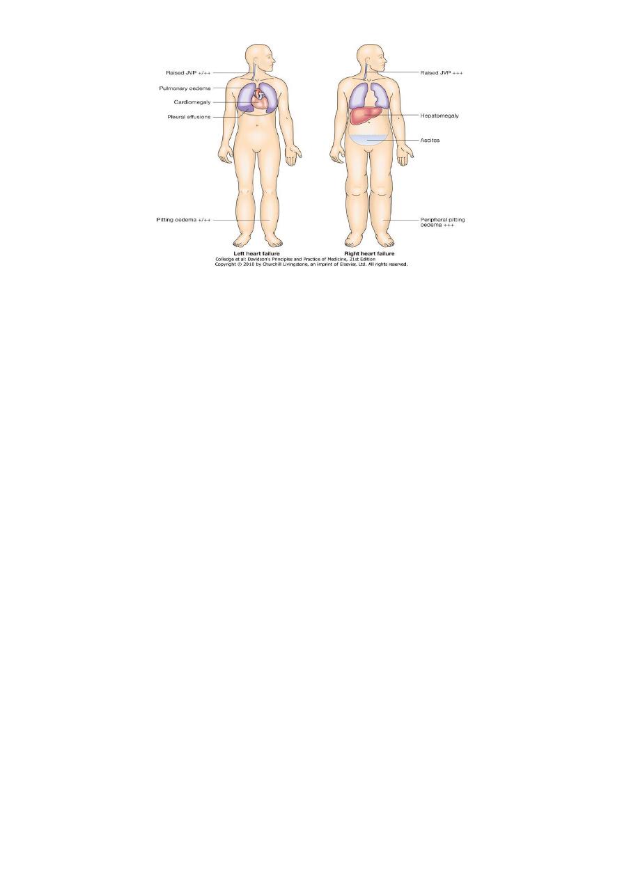
2. Left, right and biventricular heart failure
* left side of the heart = functional unit of LA and LV, together with mitral and aortic valves;
* right heart = RA, RV, and tricuspid and pulmonary valve
2
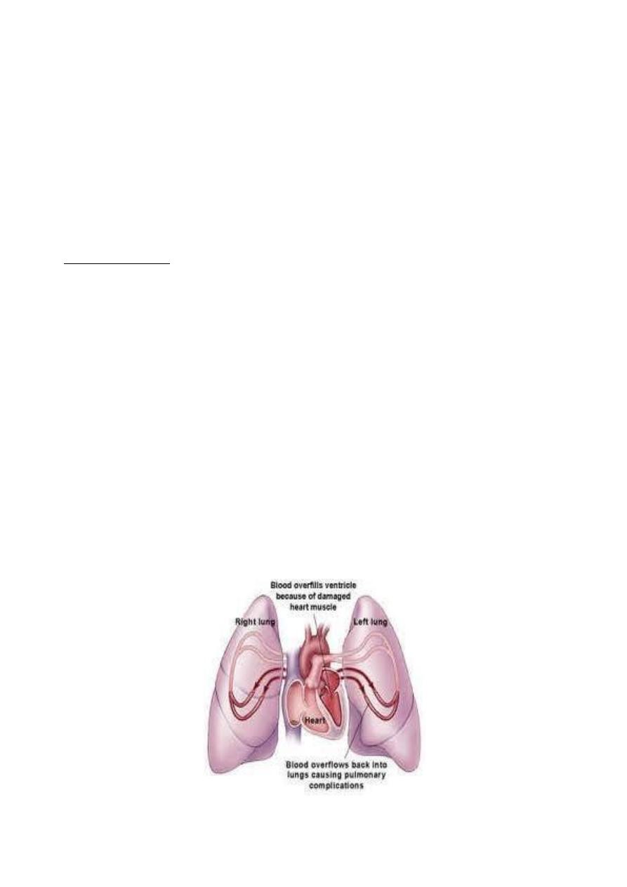
* Left-sided heart failure= reduction LV output and increase LA or pulmonary venous pressure.
Acute increase LA pressure
pulmonary congestion or P. oedema; more gradual increase in
LA pressure, (mitral stenosis)
reflex pulmonary vasoconstriction,=protects patient from
pulmonary oedema at the cost of increasing pulmonary hypertension.
* Right-sided heart failure. =reduction in RV output for any given RA pressure. Causes of
isolated right heart failure include chronic lung disease (cor pulmonale), multiple pulmonary
emboli and pulmonary valvular stenosis.
* Biventricular heart failure. disease process, (dilated cardiomyopathy or ischaemic heart
disease), affects both ventricles . Disease of left heart leads to chronic elevation of left atrial
pressure, pulmonary hypertension and right heart failure.
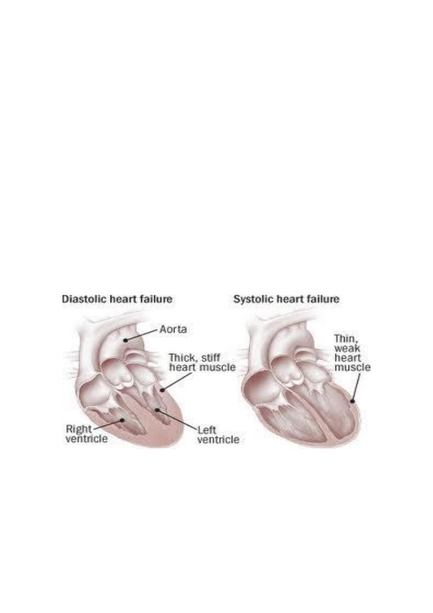
3. Diastolic and systolic dysfunction:
1- impaired myocardial contraction =systolic dysfunction
2- poor ventricular filling and high filling pressures (abnormal ventricular
relaxation)=diastolic dysfunction,caused ( stiff non-compliant ventricle) commonly found
in patients with LV hypertrophy.
d Systolic and diastolic dysfunction often coexist, particularly in patients with coronary
artery disease.
4. High-output failure
Conditions such as large arteriovenous shunt, beri-beri ,severe anaemia or thyrotoxicosis can
occasionally cause heart failure due to an excessively high cardiac output.

3
Factors that precipitate or aggravate HF in patients with pre-existing heart
disease:
1) Myocardial ischaemia or infarction
2) Intercurrent illness, e.g. infection
3) Arrhythmia, e.g. atrial fibrillation
4) Inappropriate reduction of therapy
5) Administration of a drug with negative inotropic properties (e.g. β-blocker) or fluid-retaining
properties (e.g. non-steroidal anti-inflammatory drugs (NSAIDs), corticosteroids)
6) Pulmonary embolism
7) Conditions associated with increased metabolic demand, e.g. pregnancy, thyrotoxicosis,
anaemia
8) I.v. fluid overload, e.g. post-operative i.v. infusion
Clinical Manifestations:
??ACUTE OR CHRONIC
??RIGHT SIDE , LEFT SIDE OR MIXED
??BACKWARD(congestive-diastolic) OR FOREWARD (systolic-output) OR MIXED
Symptoms:
a. cardinal symptoms of HF = fatigue and shortness of breath
b. Orthopnea
c. Paroxysmal Nocturnal Dyspnea (PND)
d. Cheyne-Stokes Respiration- advanced HF and usually associated with low cardiac output
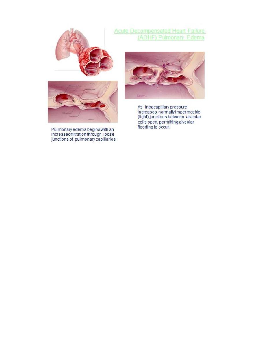
e. Acute Pulmonary Edema
f. Other symptoms= gastrointestinal symptoms.Cerebral symptoms
1. General Appearance
2. pulse
3. BP
4. Jugular Veins
5. Pulmonary Examination
6. Cardiac Examination
7. Abdomen and Extremities,Cardiac Cachexia
4
Clinical assessment:
Acute and Chronic heart failure:
j
Acute left (ventricular) heart failure
PRESENTATION
sudden onset of dyspnoea at rest that rapidly progresses to acute respiratory
distress-
orthopnoea and prostration. precipitant, e.g.acute MI,
EXAM:
- General
agitated, pale and clammy.
- peripheries are cool to touch and pulse is rapid.
- Pulse=Inappropriate bradycardia or excessive tachycardia ( precipitant for acute episode
of heart failure.)
- BP= usually high ( sympathetic nervous system activation, normal or low if patient is in
cardiogenic shock.
- Jugular venous pressure (JVP)= usually elevated, particularly with associated fluid
overload or right heart failure.
Precordium:
- Apex= acute de novo HF,=no time for V dilatation =apex not displaced.
- Auscultation occasionally murmur of catastrophic valvular or septal rupture, or triple
'gallop' rhythm.
- Crepitations at lung bases, consistent with pulmonary oedema.
5
U
Chronic heart failure (LT&RT)
O relapsing and remitting course=periods of stability and episodes of decompensation
worsening symptoms = necessitate hospitalisation.
O clinical picture depends =nature of underlying heart disease, type of heart failure, and
neurohumoral changes developed .
low cardiac output fatigue, listlessness and poor effort tolerance; peripheries - cold and BP
is low.
blood flow diverted away from skeletal muscle(to maintain perfusion of vital
organs)=contribute to fatigue and weakness.
Poor renal perfusion oliguria and uraemia.
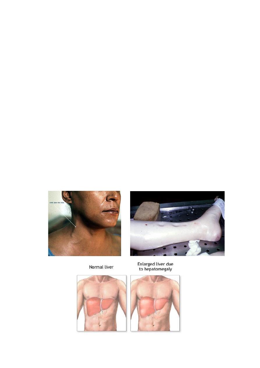
Congestion
Left = Pulmonary oedema (left HF )+ inspiratory crepitations -lung bases.
Right = high JVP + hepatic congestion + dependent peripheral oedema. ambulant patients,
oedema affects the ankles, whereas in bed-bound patients it collects around the thighs and
sacrum.
Ascites or pleural effusion -some cases
Chronic heart failure =marked weight loss (cardiac cachexia) { combination of anorexia and
impaired absorption } (gastrointestinal congestion, poor tissue perfusion due to a low cardiac
output, and skeletal muscle atrophy due to immobility
6
Complications:
In advanced heart failure
= Renal failure : caused by poor renal perfusion (low cardiac output ). Exacerbated by diuretic
therapy, ACEI and ARB
= Hypokalaemia : potassium-losing diuretics or hyperaldosteronism (activation of RA system +
impaired aldosterone met. -hepatic congestion). Most of body's potassium is intracellular
substantial depletion of potassium stores, even when the plasma potassium concentration is
in normal range.
= Hyperkalaemia: effects of drug treatment, esp. combination of ACE inhibitors and
spironolactone (both promote potassium retention), renal dysfunction.
= Hyponatraemia: feature of severe heart failure poor prognostic sign. diuretic therapy,
inappropriate water retention (high ADH secretion), or failure of the cell membrane ion
pump.
= Impaired liver function: hepatic venous congestion and poor arterial perfusion mild
jaundice and abnormal liver function tests; reduced synthesis of clotting factors can make
anticoagulant control difficult.
= Thromboembolism: Deep vein thrombosis and pulmonary embolism ( effects of a low
cardiac output and enforced immobility), systemic emboli =related to arrhythmias, atrial
flutter or fibrillation, or intracardiac thrombus complicating mitral stenosis, MI or LV
aneurysm).
= Atrial and ventricular arrhythmias =very common electrolyte changes (e.g. hypokalaemia,
hypomagnesaemia), underlying structural heart disease, pro-arrhythmic effects of increased
circulating catecholamines or drugs.
= Sudden death occurs in up to 50% of patients with heart failure often due to a ventricular
arrhythmia. Frequent ventricular ectopic beats and runs of non-sustained ventricular
tachycardia are common findings in patients with heart failure and are associated with an
adverse prognosis.
7
INVESTIGATIONS:
1- DIAGNOSIS
2- DETECT CAUSE
3- DETECT COMPLICATIONS
4- ASSESS SEVERITY
SIMPLE VS.INVASIVE:
Serum urea and electrolytes, haemoglobin, thyroid function, ECG and chest X-ray may
Brain natriuretic peptide (BNP) =elevated in HF and marker of risk;
Echocardiography : determine aetiology . detect hitherto unsuspected valvular heart disease,
such as occult mitral stenosis, and other conditions that may be amenable to specific remedies
identify patients who benefit from long-term therapy with drugs, e.g.ACEI
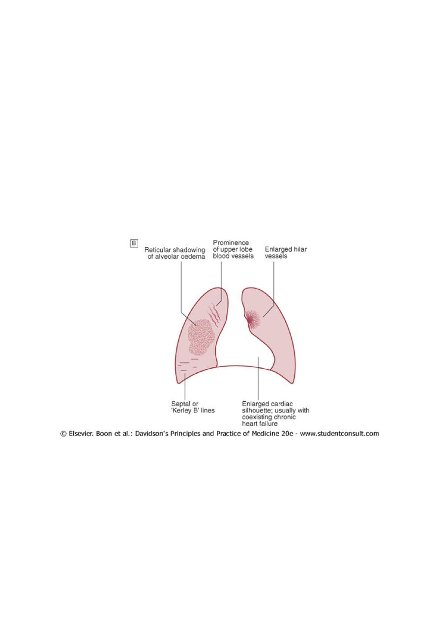
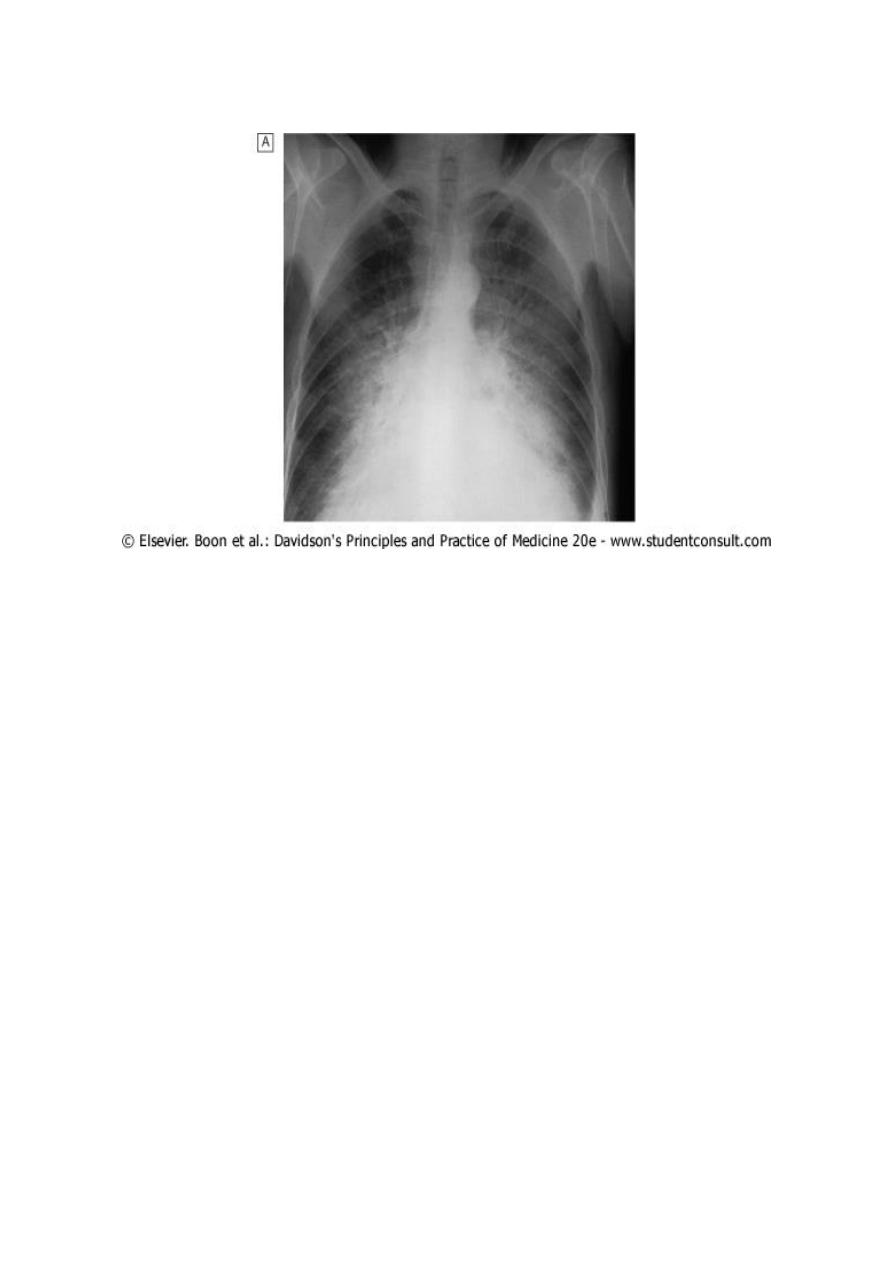
Chest X-ray:
chest X-ray in left heart failure:
rise in pulmonary venous pressure from left-sided cardiac failure abnormal distension of
upper lobe pulmonary veins (in erect position).
vascularity of lung fields
more prominent: right and left pulmonary arteries
dilate.

8
Subsequently, interstitial oedema thickened interlobular septa and dilated lymphatics.
=evident as horizontal lines in the costophrenic angles (septal or 'Kerley B' lines).
9
More advanced changes =alveolar oedema hazy opacification spreading from hilar regions,
and pleural effusions
Management of HF:
General measures
Management of acute pulmonary oedema
Management of chronic heart failure
Invasive measures= (ICD,CRT,CR,HT)
1. General measures for the management of heart failure
Education: Explanation of nature of disease, treatment and self-help strategies
Diet : Good general nutrition and weight reduction for obese . Avoidance of high-salt
foods and added salt, especially for patients with severe congestive heart failure
Alcohol: Moderation or elimination of alcohol consumption. Alcohol-induced
cardiomyopathy requires abstinence
Smoking: Cessation
Exercise: Regular moderate aerobic exercise within limits of symptoms
Vaccination: Influenza and pneumococcal vaccination should be considered
2. Management of acute pulmonary oedema:
C Sit patient up (reduce pulmonary congestion).initially -strict bed rest continuous
monitoring of cardiac rhythm, BP and pulse oximetry

11
C oxygen (high-flow, high-concentration): Non-invasive positive pressure ventilation
(continuous positive airways pressure (CPAP) of 5-10 mmHg) by a tight-fitting facemask
C Nitrates-i.v. glyceryl trinitrate 10-200 μg/min or buccal glyceryl trinitrate 2-5 mg, titrated
upwards every 10 minutes, until clinical improvement occurs or systolic BP falls to < 110
mmHg.
C loop diuretic e.g.furosemide 50-100 mg i.v.
C IV opiates -cautiously used when patients are in extremis. reduce sympathetically mediated
peripheral vasoconstriction but may cause respiratory depression and exacerbation of
hypoxaemia and hypercapnia
C inotropic agents may be required to augment CO, esp. in hypotensive pat. Insertion of intra-
aortic balloon pump -in patients with acute cardiogenic pulmonary oedema, especially when
secondary to myocardial ischaemia
3. Management of chronic heart failure:
Non-pharmocological- General measures
Pharmocological
- DIURETICS
- VASODILATERS
- INOTROPIC AGENTS
# General measures
Education of patients and relatives about causes and treatment of HF adherence to a
management plan . Some patients may need to weigh themselves daily adjust their
diuretic therapy accordingly. In patients with CAD, secondary preventative measures, (low-
dose aspirin and lipid-lowering therapy). ( statins not effective in patients with severe heart
failure).
# Drug therapy
o Cardiac function improved by
- increasing contractility,
- optimising preload or
- decreasing afterload .
o Drugs that reduce preload are appropriate in patients with high end-diastolic filling
pressures and evidence of pulmonary or systemic venous congestion.
o Those that reduce afterload or increase myocardial contractility are more useful in patients
with signs and symptoms of a low cardiac output.
11
1. Diuretic therapy
A- Loop diuretic
1- increase urinary Na and water excretion,
reduction in blood & plasma volume.
2- reduces preload
improves pulmonary and systemic venous congestion.
3- may reduce afterload and ventricular volume,
a fall in wall tension and increased
cardiac efficiency.
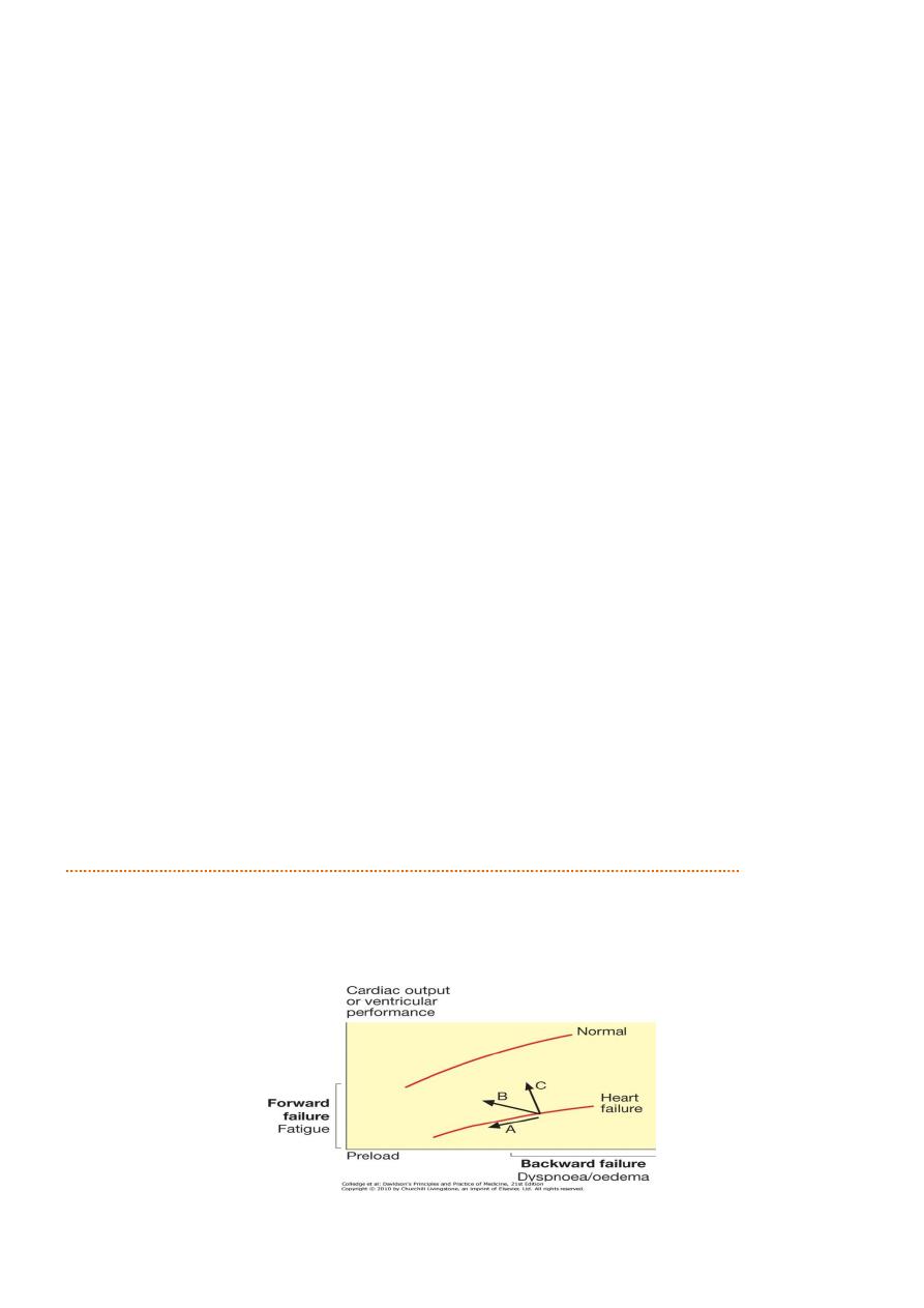
Diuretic and cardiac output
1- Although a fall in preload (V filling pressure) tends to reduce CO, 'Starling curve' in HF is flat,
==substantial and beneficial fall in filling pressure with little change in cardiac output
2- excessive diuretic therapy
undesirable fall in CO, with a rising serum urea, hypotension
and increasing lethargy, especially in patients with a marked diastolic component to their HF
f
High dose In some patients with severe chronic HF, (in presence of chronic renal impairment,)
oedema may persist despite oral loop diuretics. IV infusion of furosemide 10 mg/hr may
initiate a diuresis.
B- Combining a loop diuretic with a thiazide (e.g. bendroflumethiazide 5 mg daily) or a thiazide-
like diuretic (e.g. metolazone 5 mg daily) = prove effective,(cause excessive diuresis.
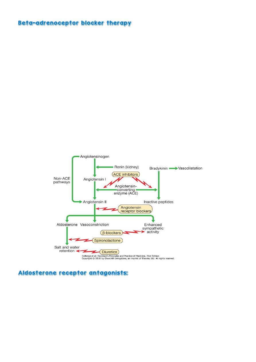
C- Aldosterone receptor antagonists:
1- Ex.spironolactone and eplerenone,
2- potassium-sparing diuretics
3- of particular benefit in patients with heart failure.
4- may cause hyperkalaemia, particularly when used with an ACE inhibitor.
5- improve long-term clinical outcome in patients with severe heart failure or heart failure
following acute MI
Effect of treatment on ventricular performance curves in Hf
a. Diuretics and venodilators
b. Angiotensin-converting enzyme (ACE) inhibitors and mixed vasodilators
c. Positive inotropic agents

12
2. Vasodilators:
valuable in chronic heart failure;
1- HYDRALLAZINE+NITRITES
2- ACE INHIBITORS
3- AR-BLOCKER
1. venodilators (e.g. nitrates) reduce preload, and arterial dilators (e.g. hydralazine) reduce
afterload . use is limited by pharmacological tolerance and hypotension.
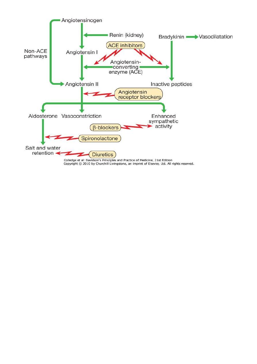
♧ Angiotensin-converting enzyme (ACE) inhibition therapy: interrupts vicious circle of
neurohumoral activation = preventing conversion of angiotensin I to angiotensin
II
preventing salt and water retention, peripheral arterial and venous vasoconstriction, and
activation of sympathetic nervous system .
- prevent undesirable activation of RAS caused by diuretic therapy.
- major benefit of ACE inhibition in HF =reduction in afterload, reduces preload causes
modest rise in plasma potassium concentrations.
- Treatment with combination of loop diuretic and ACE inhibitor = advantages.
- In moderate and severe heart failure
- ACE inhibitors =substantial improvement in effort tolerance and in mortality
- improve outcome and prevent onset of overt heart failure in patients with poor residual
left ventricular function following MI.
♧ SE:
- symptomatic hypotension
- impairment of renal function, especially in patients with bilateral renal artery stenosis or
those with pre-existing renal disease.
- Short-acting ACE inhibitors =marked falls in BP, particularly in elderly or when started in
presence of hypotension, hypovolaemia or hyponatraemia.
- In stable patients without hypotension (systolic BP > 100 mmHg), ACE inhibitors can
usually be safely started in community.
- in other patients, ,advisable to withhold diuretics for 24 hours before starting treatment
with a small dose of a long-acting agent, preferably given at night (Renal function must be
monitored and should be checked 1-2 weeks after starting therapy.
Starting dose Target dose
Enalapril 2.5 mg 12-hr. 10 mg 12-hr.
Lisinopril 5 mg daily 20 mg daily
Ramipril 1.25 mg 12-hr. 5 mg 12-hr.
13
♧ Angiotensin receptor blocker (ARB) therapy:
h losartan 50-100 mg once daily,
h Candesartan 4-16 mg daily or
h valsartan 40-160 mg daily
h act by blocking action of angiotensin II on heart, peripheral vasculature and kidney.
h produce beneficial haemodynamic changes - similar to effects of ACE inhibitors but-generally
better tolerated.
h have comparable effects on mortality and are a useful alternative for patients who cannot
tolerate ACE inhibitors .
h share all the more serious adverse effects of ACE inhibitors, including renal dysfunction and
hyperkalaemia.
h may be considered in combination with ACE inhibitors, especially in those with recurrent
hospitalisations for heart failure.
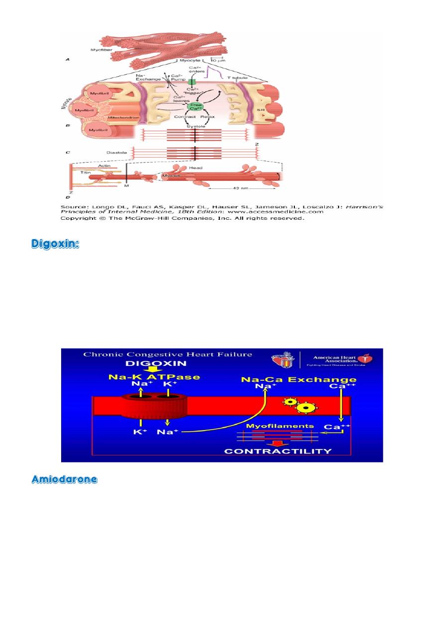
3. Inotropic agents:
1- Digoxin
2- floseqinon
3- milrinone
4- vesnarinone & Xamterol
5- Ca-sensitizing agent-levo-simendan-
14
first-line therapy heart failure and atrial fibrillation,( adequate control of ventricular rate +a
small positive inotropic effect.)
less certain patients with heart failure and sinus rhythm
no effect on overall survival but did reduce the need for hospitalisation.
In patients with severe heart failure (NYHA class III-IV) reduces likelihood of
hospitalisation --no effect on long-term survival
- potent anti-arrhythmic drug that has little negative inotropic effect and may be valuable
in patients with poor left ventricular function.
- only effective in the treatment of symptomatic arrhythmias, should not be used as a
preventative agent in asymptomatic patients.
15
USES:
1- helps to counteract deleterious effects of enhanced sympathetic stimulation
2- reduces risk of arrhythmias and sudden death.
DOSE:
1- When initiated in standard doses
may precipitate acute-on-chronic heart failure,
2- when given in small incremental doses (e.g. bisoprolol started at a dose of 1.25 mg daily, and
increased gradually over a 12-week period to a target maintenance dose of 10 mg daily),
BENIFIT:
1- increase ejection fraction,
2- improve symptoms,
3- reduce the frequency of hospitalisation and
4- reduce mortality in patients with chronic heart failure (Beta-blockers are more effective at
reducing mortality than ACE inhibitors: relative risk reduction of 33% versus 20% respectively
- spironolactone and eplerenone
potassium-sparing diuretics of particular benefit in patients with heart failure. may cause
hyperkalaemia, particularly when used with an ACE inhibitor. improve long-term clinical
outcome , patients with severe heart failure . heart failure following acute myocardial
infarction.

16
4. Implantable cardiac defibrillators and resynchronisation therapy:
j ICD: Patients with symptomatic ventricular arrhythmias and heart failure have a very poor
prognosis. Irrespective of their response to anti-arrhythmic drug therapy, all should be
considered for implantation of a cardiac defibrillator .
j CRT
- In patients with marked intraventricular conduction delay, prolonged depolarisation may
lead to uncoordinated left ventricular contraction.
- When associated with severe symptomatic heart failure, cardiac resynchronisation
therapy should be considered.
- both LV and RV are paced simultaneously in an attempt to generate a more coordinated
left ventricular contraction and improve cardiac output.
j Coronary revascularisation
Coronary artery bypass surgery or percutaneous coronary intervention may improve
function in areas of myocardium that are 'hibernating' because of inadequate blood supply,
can be used to treat carefully selected patients with heart failure and coronary artery
disease. ('hibernating' myocardium can be identified by stress echocardiography and
specialised nuclear or MR imaging).
Heart transplantation:
n Cardiac transplantation=established and successful form of treatment for patients with
intractable heart failure.
n Coronary artery disease and dilated cardiomyopathy are the most common indications. The
introduction of ciclosporin for immunosuppression) has improved survival, which is around
80% at 1 year.
n use of transplantation is limited by the efficacy of modern drug and device therapies, as well
as the availability of donor hearts,
n contraindicated in patients with pulmonary vascular disease due to long-standing left heart
failure, complex congenital heart disease (e.g. Eisenmenger's syndrome) or primary
pulmonary hypertension because the RV of the donor heart may fail in the face of high
pulmonary vascular resistance.
n heart-lung transplantation can be successful in patients with Eisenmenger's syndrome. Lung
transplantation has been used for primary pulmonary hypertension.
n complications
1. Rejection., episodes of rejection are common and may present with heart failure,
arrhythmias or subtle ECG changes; cardiac biopsy is often used to confirm the diagnosis
before starting treatment with high-dose corticosteroids.

17
2. Accelerated atherosclerosis. Recurrent heart failure is often due to progressive
atherosclerosis in the coronary arteries of the donor heart.
This is not confined to patients who were transplanted for coronary artery disease and is
probably a manifestation of chronic rejection. Angina is rare because the heart has been
denervated.
3. Infection. Opportunistic infection with organisms such as cytomegalovirus or Aspergillus
remains a major cause of death in transplant recipients.
Ventricular assist devices:
O Because of limited supply of donor organs, ventricular assist devices (VADs) employed as:
O abridge to cardiac transplantation
O potential long-term 'destination' therapy
O short-term restoration therapy following a potentially reversible insult such as viral
myocarditis.
O VADs assist cardiac output by using a roller, centrifugal or pulsatile pump that in some cases
is implantable and portable.
O They withdraw blood through cannulae inserted in the atria or ventricular apex and pump it
into the pulmonary artery or aorta.
O They are designed not only to unload the ventricles but also to provide support to the
pulmonary and systemic circulations.
O Their more widespread application is limited by high complication rates (haemorrhage,
systemic embolism, infection, neurological and renal sequelae), although some
improvements in survival and quality of life have been demonstrated in patients with severe
heart failure