
Pediatrics Lec 6 Dr. Ziyad
Fatima Ehsan Avci
1
Fetal Birth Injuries
Definition
The term birth injury is used to denote:
avoidable and unavoidable mechanical, hypoxic and ischemic injury affecting
the infant during labor and delivery.
Predisposing factors:
1. Macrosomia,
2. Prematurity,
3. Cephalopelvic disproportion,
4. Dystocia,
5. Prolonged labor, and
6. Breech presentation.
Incidence
Has been estimated at 2-7/1,000 live births.
Cranial Injuries
Erythema, abrasions, ecchymoses,
Of facial or scalp soft tissues may be seen after forceps or vacuum-assisted
deliveries.
Their location depends on the area of application of the forceps.
Subconjunctival ,retinal hemorrhages and petechiae of the skin of the head and
neck
All are common.
All are probably secondary to a sudden increase in intrathoracic pressure
during passage of the chest through the birth canal.
Parents should be assured that they are temporary and the result of
normal hazards of delivery.
Pediatrics Lec 6 Dr. Ziyad
Fatima Ehsan Avci
2
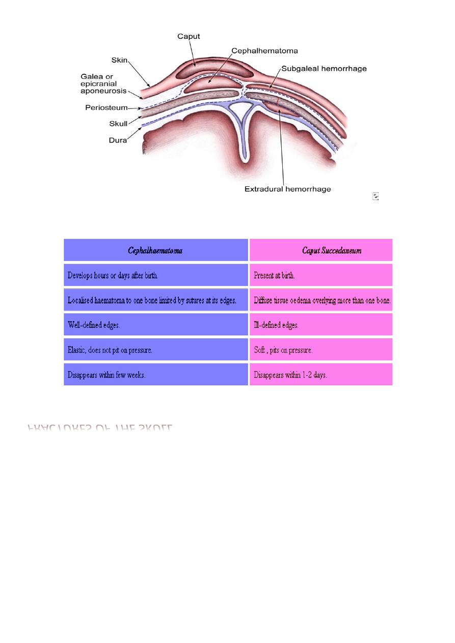
Molding
Molding of the head and overriding of the parietal bones are frequently
associated with caput succedaneum and become more evident after the
caput has receded but disappear during the first weeks of life.
Molding
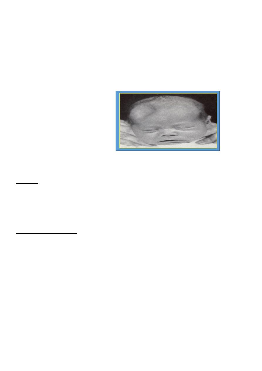
Caput succedaneum
Diffuse, sometimes ecchymotic, edematous swelling of the soft tissues of
the scalp involving the portion presenting during vertex delivery.
It may extend across the midline and across suture lines.
The edema disappears within the first few days of life.
Analogous swelling, discoloration, and distortion of the face are seen in
face presentations.
No specific treatment is needed, but if there are extensive ecchymoses,
phototherapy for hyperbilirubinemia may be indicated.

Pediatrics Lec 6 Dr. Ziyad
Fatima Ehsan Avci
3
Cephalhaematoma
It is a subperiosteal haematoma most commonly lies over one parietal bone.
It may result from difficult vacuum or forceps extraction .
Is a subperiosteal hemorrhage, so it is always limited to the surface of one
cranial bone.
There is no discoloration of the overlying scalp, and swelling is usually not
visible until several hours after birth, because subperiosteal bleeding is a
slow process.
An underlying skull fracture, usually linear and not depressed, is
occasionally associated with cephalohematoma.
A sensation of central depression suggesting( but not indicative )of an
underlying fracture or bony defect is
Cephalohematomas require no treatment, although phototherapy may be
necessary to ameliorate hyperbilirubinemia.
Cephalohematoma is differentiated from Cranial meningocele by:
1. Pulsation,
2. Increased pressure on crying, and the
3. Radiologic evidence of bony defect.
Most cephalohematomas are resorbed within 2 wk-3 mo, depending on
their size.
They may begin to calcify by the end of the 2nd wk.
Incision and drainage are contraindicated because of the risk of
introducing infection in a benign condition.
A massive cephalohematoma may rarely result in blood loss severe
enough to require transfusion.
It may also be associated with a skull fracture, coagulopathy, and
intracranial hemorrhage.
Pediatrics Lec 6 Dr. Ziyad
Fatima Ehsan Avci
4
Diagnosis and Differential Diagnosis
FRACTURES OF THE SKULL
May occur as a result of pressure from forceps, maternal symphysis pubis, sacral
promontory, or ischial spines. It may be:
(1) Vault fracture:
Usually affecting the frontal or parietal bone.
It may be linear or depressed fracture.
It needs no treatment unless there is intracranial haemorrhage.
Affected infants may be asymptomatic unless there is associated intracranial
injury.
It is advisable to elevate severe depressions to prevent cortical injury from
sustained pressure.
Pediatrics Lec 6 Dr. Ziyad
Fatima Ehsan Avci
5
2) Fracture base:
Usually associated with intracranial haemorrhage.
Fracture of the Occipital bone almost causes fatal hemorrhage due to disruption
of the underlying vascular sinuses.
It may result during breech deliveries from traction on the hyperextended spine
of the infant with the head fixed in the maternal pelvis.
Depressed fractures
Ping-Pong ball
Intracranial-Intraventricular Hemorrhage
Causes:
1. Sudden compression and decompression of the head as in breech and
precipitate labour.
2. Marked compression by forceps or in cephalopelvic disproportion.
3. Fracture skull.
Predisposing factors:
1. Prematurity due to physiological hypoprothrombinaemia, fragile
blood vessels and liability to trauma.
2. Asphyxia due to anoxia of the vascular wall .
3. Blood diseases.
Intracranial Haemorrhage Sites:
1. Subdural :
2. Subarachnoid:
3. Intraventricular :into the brain ventricles.
4. Intracerebral : into the brain tissues .
In (1) and (2) it is usually due to birth trauma,
in (3) and (4) the foetus is usually a premature exposed to hypoxia.

Pediatrics Lec 6 Dr. Ziyad
Fatima Ehsan Avci
6
Clinical picture:
1- Altered consciousness.
2- Flaccidity.
3- Breathing is absent, irregular and periodic or gasping.
4- Eyes: no movement, pupils may be fixed and dilated.
5- Opisthotonus, rigidity, twitches and convulsions.
6- Vomiting .
7- High pitched cry.
8- Anterior fontanelle is tense and bulging.
9- Lumbar puncture reveals bloody C.S.F.
DIAGNOSIS
1. History,
2. Clinical manifestations,
3. Transfontanel cranial ultrasonography or
4. Computed tomography (CT).
5. Lumbar puncture: indicated in the presence of signs of:
Increased intracranial pressure or
Deteriorating clinical condition
to identify gross subarachnoid hemorrhage or to rule out the possibility of bacterial
meningitis
Intracranial Haemorrhage Treatment
Seizures are treated with anticonvulsant drugs.
Anemia-shock, requires transfusion with packed red blood cells or fresh frozen
plasma.
Acidosis is treated with slow administration of sodium bicarbonate.
PREVENTION
The incidence of traumatic intracranial hemorrhage may be reduced by judicious
management of cephalopelvic disproportion and operative delivery.
Fetal or neonatal hemorrhage due to Maternal idiopathic thrombocytopenic purpura
(ITP) or Alloimmune thrombocytopenia may be prevented by maternal treatment
with Steroids,Intravenous immunoglobulin, or Fetal platelet transfusion.
Vitamin K should be given before delivery to all women receiving phenobarbital or
phenytoin during the pregnancy.
Pediatrics Lec 6 Dr. Ziyad
Fatima Ehsan Avci
7
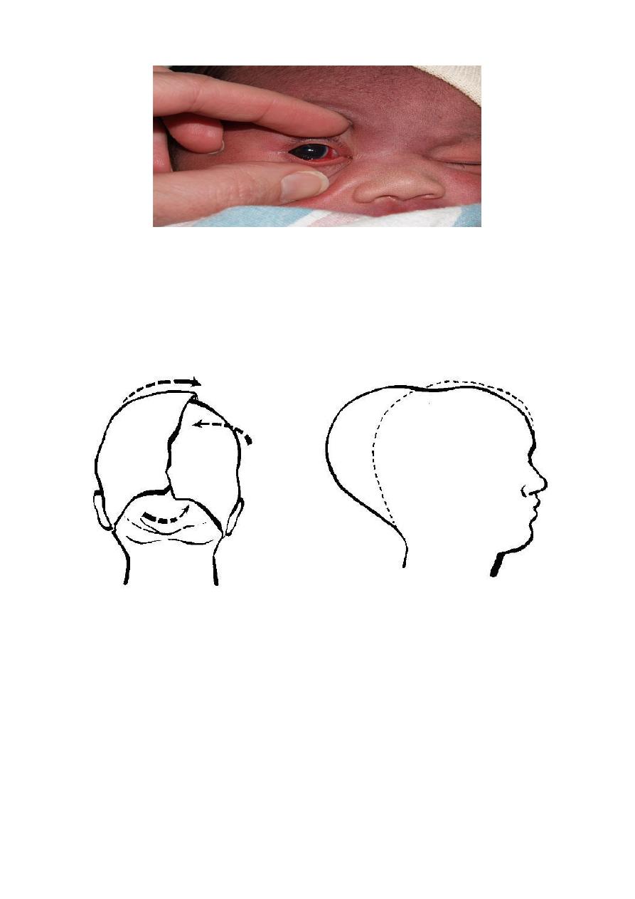
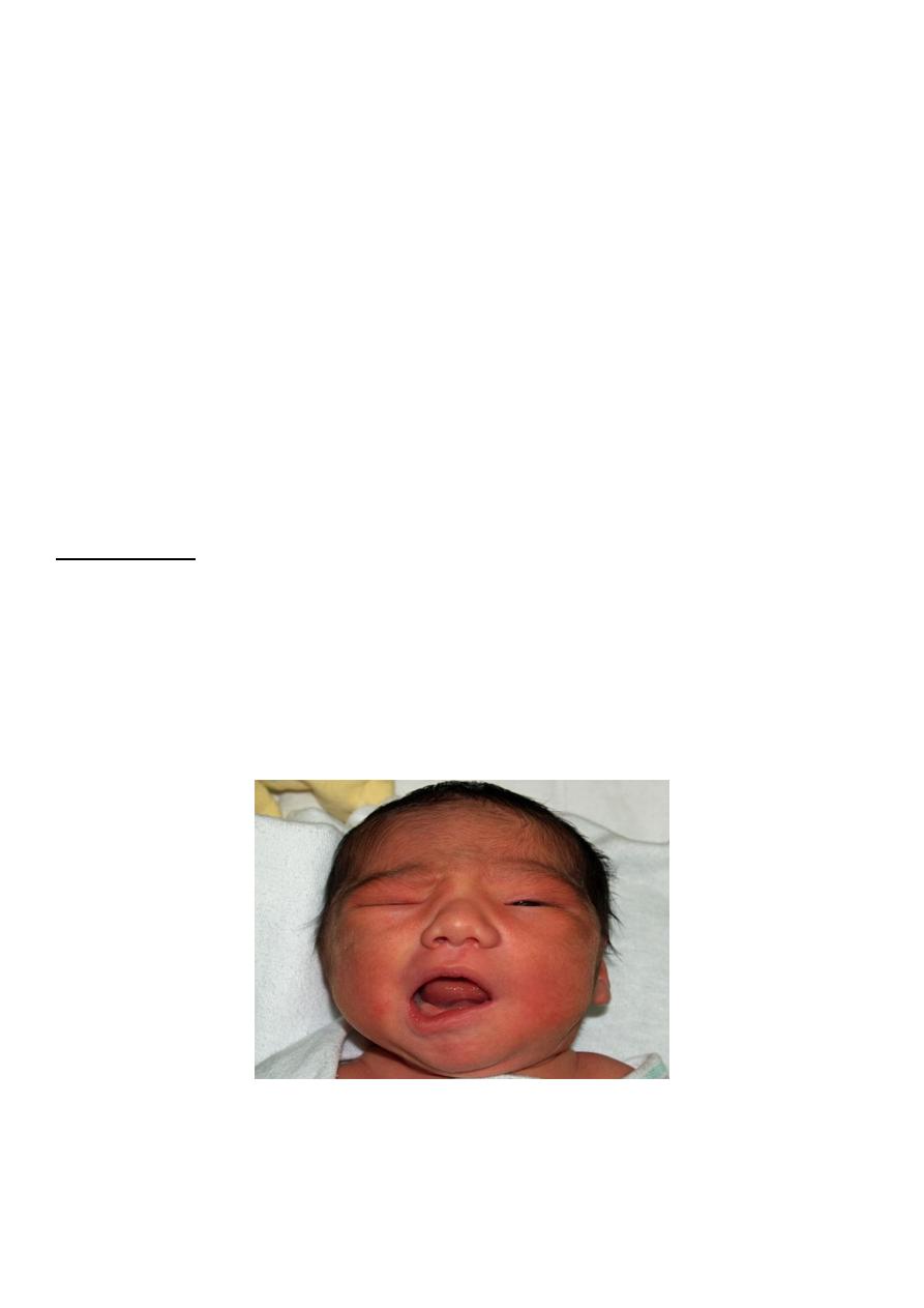
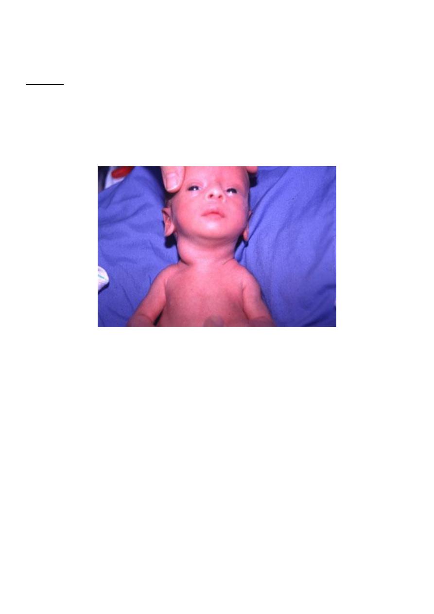
Facial Palsy (Bell’s palsy):
-
It is usually due to pressure by the forceps blade on the facial nerve along
its course
-
It appears within 1-2 days after delivery due to resultant oedema and
haemorrhage around the nerve.
Manifestations:
1. There is paresis of the facial muscles on the affected side with:
2. Partially opened eye and:
3. Flattening of the nasolabial fold.
4. The mouth angle is deviated towards the healthy side.
The prognosis depends on whether the nerve was injured by pressure or
whether the nerve fibers were torn.
Care of the exposed eye is essential.
Improvement occurs within few weeks.
Neuroplasty may be indicated when the paralysis is persistent.
Pediatrics Lec 6 Dr. Ziyad
Fatima Ehsan Avci
8
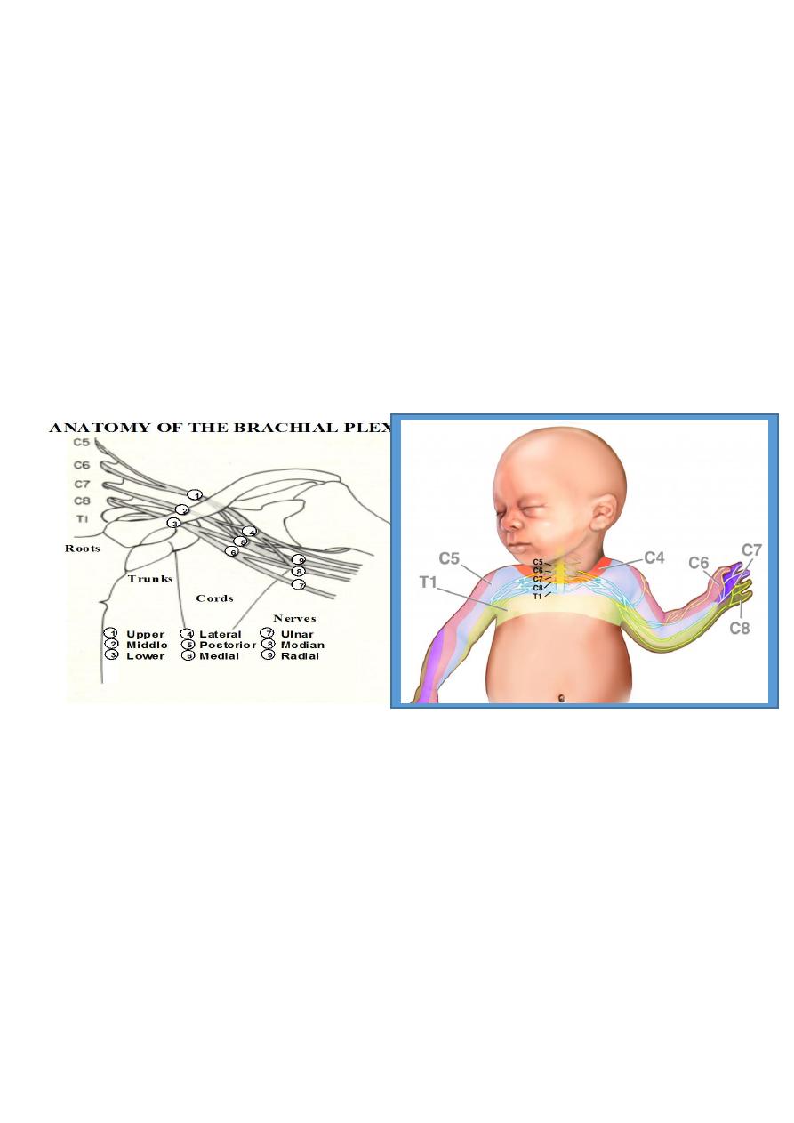
Peripheral Nerve Injuries
BRACHIAL PALSY:
Injury to the brachial plexus may cause paralysis of the upper arm with or
without paralysis of the forearm or hand or, more commonly, paralysis of the
entire arm. Approximately 45% are associated with shoulder dystocia.
These injuries occur in :
Macrosomic infants and when lateral traction is exerted on the head and neck
during delivery of the shoulder in a vertex presentation.
When the arms are extended over the head in a breech presentation.
When excessive traction is placed on the shoulders.
Klumpke's paralysis
Is a rarer form of brachial palsy;
Injury to the 7th and 8th cervical nerves and the 1st
thoracic nerve produces a paralyzed hand.
(Horner syndrome)
If the sympathetic fibers of the 1st thoracic root are also
injured : paralyzed hand and ipsilateral ptosis and
miosis.
The mild cases may not be detected immediately after birth.
Pediatrics Lec 6 Dr. Ziyad
Fatima Ehsan Avci
9
Brachial plexus palsy should Differentiated must be made
from :
1.
Cerebral injury;
2.
Fracture, dislocation, or epiphyseal separation of the
humerus;
3.
Fracture of the clavicle.
MRI demonstrates nerve root rupture or avulsion
Prognosis of BRACHIAL PALSY:
Depends on whether the nerve was merely injured or was lacerated.
If the paralysis was due to edema and hemorrhage about the nerve fibers,
function should return within a few months;
If due to laceration, permanent damage may result.
Involvement of the deltoid is usually the most serious problem and may result in
a shoulder drop secondary to muscle atrophy.
In general, paralysis of the upper arm has a better prognosis than paralysis of
the lower arm.
TREATMENT
Partial immobilization and appropriate positioning
to prevent development of contractures.
In upper arm paralysis:
the arm should be
abducted, with external rotation at the shoulder
and with full supination of the forearm and slight
extension at the wrist with the palm turned toward
the face.
Pediatrics Lec 6 Dr. Ziyad
Fatima Ehsan Avci
10
In lower arm or hand paralysis:
the wrist
should be splinted in a neutral position and
padding placed in the fist.
Gentle massage and range of motion
exercises may be started by 7-10 days of age.
If the paralysis persists without improvement for
3-6 months: neuroplasty, neurolysis, end-to-end
anastomosis, or nerve grafting offers hope for
partial recovery.
PHRENIC NERVE PARALYSIS
Phrenic nerve injury (3rd, 4th, 5th cervical nerves) with diaphragmatic
paralysis must be considered when cyanosis and irregular and labored
respirations develop.
Such injuries, usually unilateral, are associated with ipsilateral upper
brachial palsy.
DIAGNOSIS
is established by ultrasonography or fluoroscopic examination, which
reveals elevation of the diaphragm on the paralyzed side
There is no specific treatment, infants should be placed on the involved
side and given oxygen if necessary.
Recovery usually occurs spontaneously by 1-3 months; rarely, surgical
plication of the diaphragm may be indicated.

Pediatrics Lec 6 Dr. Ziyad
Fatima Ehsan Avci
11
Fractures
BONE INJURIES
These usually occur during difficult breech delivery.
(A) Vertebral Column Injuries:
These are fatal if associated with spinal cord
transection above C4 ,due to diaphragmatic
paralysis.
(B) Femur, Humerus and Clavicle:
Managed by splint to the long bone and a sling for
clavicular fracture.
CLAVICLE
This bone is fractured during labor and delivery more
frequently than any other bone; It is particularly
vulnerable when there is:
1.
Difficulty in delivery of the shoulder in vertex
presentations.
2.
Extended arms in breech deliveries.
The infant characteristically does not move the
arm freely on the affected side.
Crepitus and bony irregularity may be
palpated.
Discoloration is occasionally visible over the
fracture site.
Pediatrics Lec 6 Dr. Ziyad
Fatima Ehsan Avci
12
Treatment, consists of immobilization of the arm and
shoulder on the affected side.
A remarkable degree of callus develops at the site
within a week and may be the first evidence of
the fracture.
The prognosis is excellent.
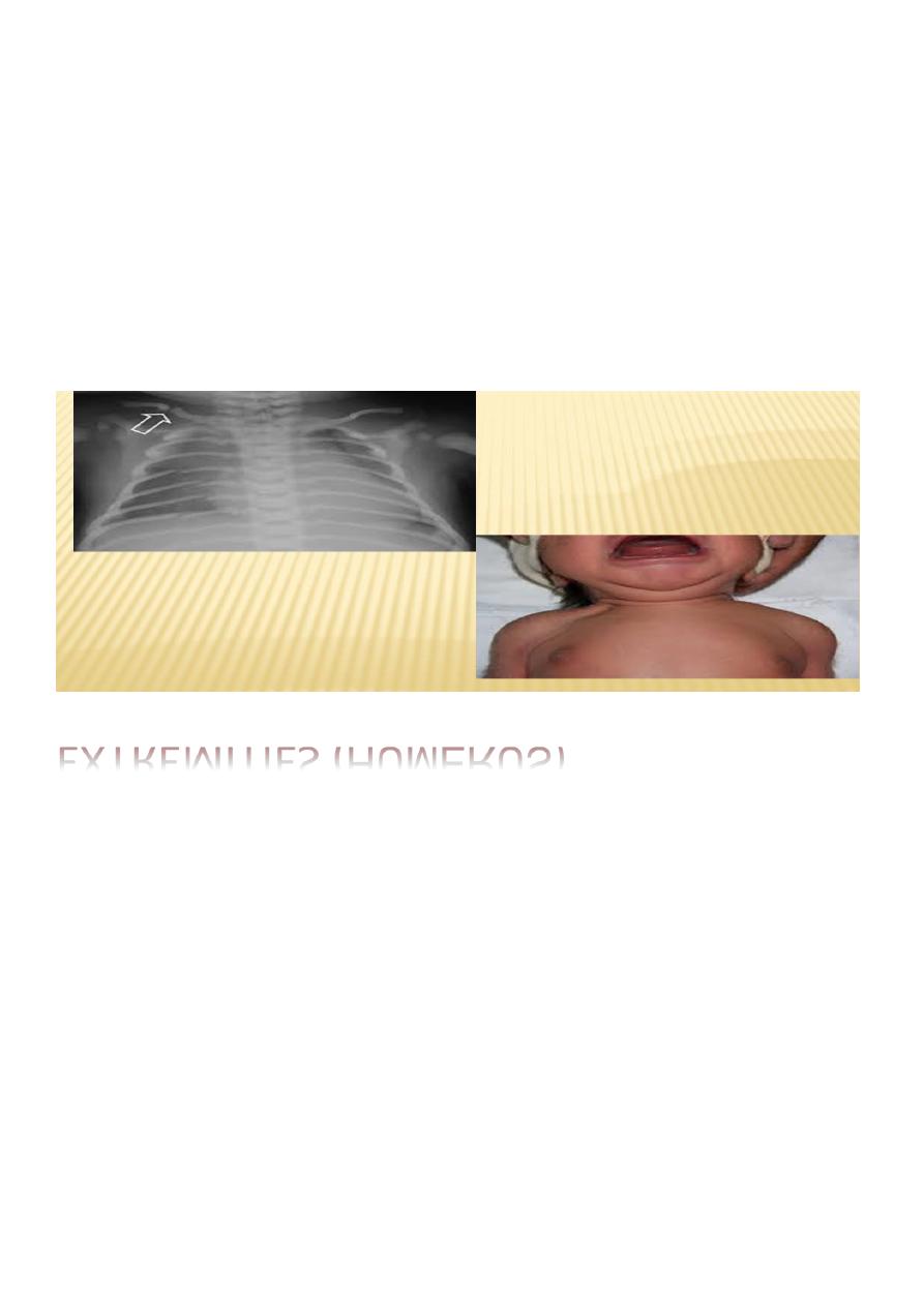
EXTREMITIES (
HUMERUS)
In fractures of the long bones, spontaneous movement of the
extremity is usually absent.
The Moro reflex is also absent from the involved extremity.
There may be associated nerve involvement.
Satisfactory results of treatment for a fractured humerus are
obtained with 2-4 wk of immobilization
(during which the arm is strapped to the chest).
A triangular splint and a bandage are applied, or a cast is
applied.
Healing is usually accompanied by excess callus formation.
The prognosis
is excellent for fractures of the extremities.
Fractures in preterm infants may be related to osteopenia
Pediatrics Lec 6 Dr. Ziyad
Fatima Ehsan Avci
13
MUSCLE INJURIES
Strenomastoid injury
Due to :
Exaggerated lateral flexion of the neck leading to torticollis and swelling in
the muscle.
It is usually improved within 2 weeks but permanent torticollis may
continue.
VISCERAL INJURIES
(Liver, spleen and kidney)
may be injured in breech delivery which should be avoided by holding
the fetus from its hips.
The liver is the only internal organ other than the brain that is injured
with any frequency during birth
.
The damage usually results from pressure on the liver during delivery
of the head in breech presentations.
Incorrect cardiac massage is a less frequent cause.

Pediatrics Lec 6 Dr. Ziyad
Fatima Ehsan Avci
14
Hepatic rupture may result in the formation of a subcapsular
hematoma.
The hematoma may be large enough to cause anemia.
Shock and death may occur if the hematoma breaks through the
capsule into the peritoneal cavity.
A mass may be palpable in the right upper quadrant; the abdomen
may appear blue.
Early suspicion by means of ultrasonographic diagnosis and prompt
supportive therapy can decrease the mortality of this disorder.
Surgical repair of a laceration may be required.
Rupture of the spleen:
May occur alone or in association with rupture of the liver.
The causes, complications, treatment, and prevention are similar.