
Obstetrics
Lec 4
Dr.
Aseil
1
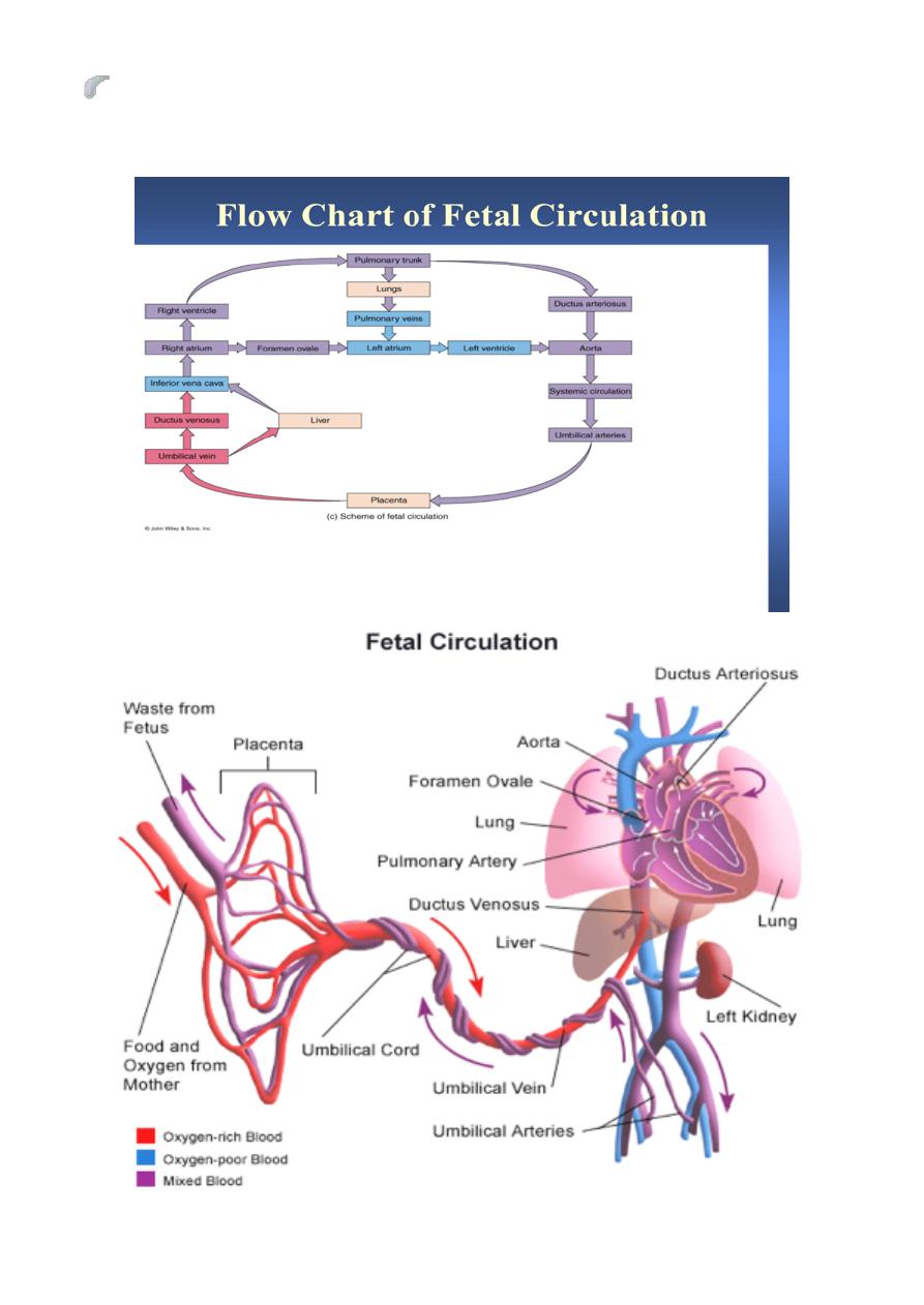
Fetal Circulation
Umbilical cord:
At term about 50 cm long 2 cm in diameter, contain :
2 umbilical arteries: return non-oxygenated blood, fecal waste, CO2 to
placenta
1umbilical vein: brings oxygenated blood and nutrients to the fetus
There is no nerve in cord or placenta .The arteries are spiral and give a cord-like
shape. The vessels are packed and protected by a viscous fluid which is Wharton
Jelly.
Shunts in fetal circulation
The fetal circulation is quite different from that of adult & characterized by
4 shunts which ensure that the best oxygenated blood from the placenta is
delivered to the fetal brain thses are:
Umblical circulation.
Ductus venosus
Foramen ovale
Ductus arteriosus
Anatomy and Physiology
The umblical arteries arise from the caudal end of the dorsal aorta & carry
deoxygenated blood from fetus to placenta for gas & nutrient exchange. .
Oxygenated blood is returned to the fetus via the umbilical vein to the fetal
liver.

Obstetrics
Lec 4
Dr.
Aseil
2
Fetal Circulation
Small amount of blood routed to growing liver
But the bulk passes through the ductus venosis to by pass the liver & joins
the inferior vena cava as it enters the Rt atrium.
Ductus Venosis is a narrow vessel & high blood velocities are generated
within it .This streaming of blood together with the crista dividens in Rt
atrium prevents mixing of oxygenated Bd from ductus venosus with
desaturated Bd from IVC.
IVC empties into the right atrium of the heart
The ductus venosus stream then passes to the left atrium through the
foramen ovale (Small physiological defect in the atrial septum)
Completely bypasses the non-functioning lungs
Blood continues journey to the left ventricle blood is then pumped into the
aorta
About 50% of blood is circulated to the upper extremities
The remainder passes down the aorta to mix with Bd of reduced oxygen
saturation from Rt ventricle via the ductous arteriosus
Deoxygenated Bd returning from the head & lower body flows to the Rt
atrium
From the right atrium, the blood goes to the right ventricle then to the
pulmonary arteries
Pulmonary arteries
Small amount goes to the maturing lungs
Rest of blood is shunted away from lungs by ductous arteriosus back to
descending aorta
By this means the desaturated blood from Rt Vt passes down the aorta to
enter the two umbilical arteries to the placenta for reoxygenation.
Obstetrics
Lec 4
Dr.
Aseil
3
Prior to birth, the ductus arteriosus remains patent due to the production of
PG E2& prostacyclin , which act as vasodilators & its premature closure has
been reported with the administration of cyclooxygenase inhibitors.

Obstetrics
Lec 4
Dr.
Aseil
4
Conversion of Fetal to Infant Circulation
At birth
Clamping the cord shuts down low-pressure system & causes
cessation of flow in ductus venosus,a fall in pressure in the Rt atrium
&closure of foramen ovale.
Ventilation of the lungs opens the pulmonary circulation,with a rapid
fall in pulmonary vascular resistance.
More heavily oxygenated blood passing through the ductus arteriosus
with the fall in pulmonary vascular resistance, causes it’s constriction
& functional closure within a few days of birth.
Occasionally, this transition from fetal to adult circulation is delayed
(persistent fetal circulation), resulting in Lt-to-Rt shunting of blood
from the aorta through the ductus arteriosus to the lungs.
This delay in closure of ductus arteriosus is seen in commonly premature
infants & results in congestion of pulmonary circulation & reduction in Bd
flow to GIT tract & brain & implicated in the pathogenesis of necrotizing
enterocolitis & intra ventricular haemorrhge.
What happens to these special structures after birth?
Umbilical arteries atrophy
Umbilical vein becomes part of the fibrous support ligament for the
liver
The foramen ovale, ductus arteriosus, ductus venosus atrophy and
become fibrous ligaments
Post natal changes
Gas exchange function is transferred from placenta to the lungs.
Separation of systemic and pulmonary circulations

Obstetrics
Lec 4
Dr.
Aseil
5
Increased metabolism to maintain body temperature and hence increased
cardiac output.
Change from right to left shunting to left to right blood flow
Overview of Conversion
Umbilical cord is clamped
Closure of ductus venosus
Decreased pressure in Rt atrium
Closure of foramen ovale
Loss of placenta also leads to
First breath
Lungs expand and fluid is expelled
Decreased pulmonary resistance
Increased systemic resistance
Increased O2 levels in pulmonary circulation
Closure of the ductus arteriosus
Fetal vs. Infant Circulation
Fetal
Low pressure system
Right to left shunting
Lungs non-functional
Increased pulmonary resistance
Decreased systemic resistance
Infant
High pressure system
Left to right blood flow
Lungs functional
Decreased pulmonary resistance
Increased systemic resistance