Embryology
Lecture -6-
Embryonic period
(Third to eight weeks)

Embryonic period (organogenesis):
Occurs from ^ 3
rd
– 8
th
weeks of development&
is ^ time when each of ^ three germ layers,
ectoderm, mesoderm, & endoderm,
gives rise to
a no. of specific tissues & organs.
By ^ end of this period, ^ main organ systems
have been established, rendering ^ major
features of ^ external body form recognizable by
^ end of ^ 2
nd
month.

Derivatives of ^ ectodermal germ layer
At ^ beginning of ^ 3
rd
week of development, ^
ectodermal germ layer has ^ shape of a disc that
is broader in ^ cephalic than in ^ caudal region.
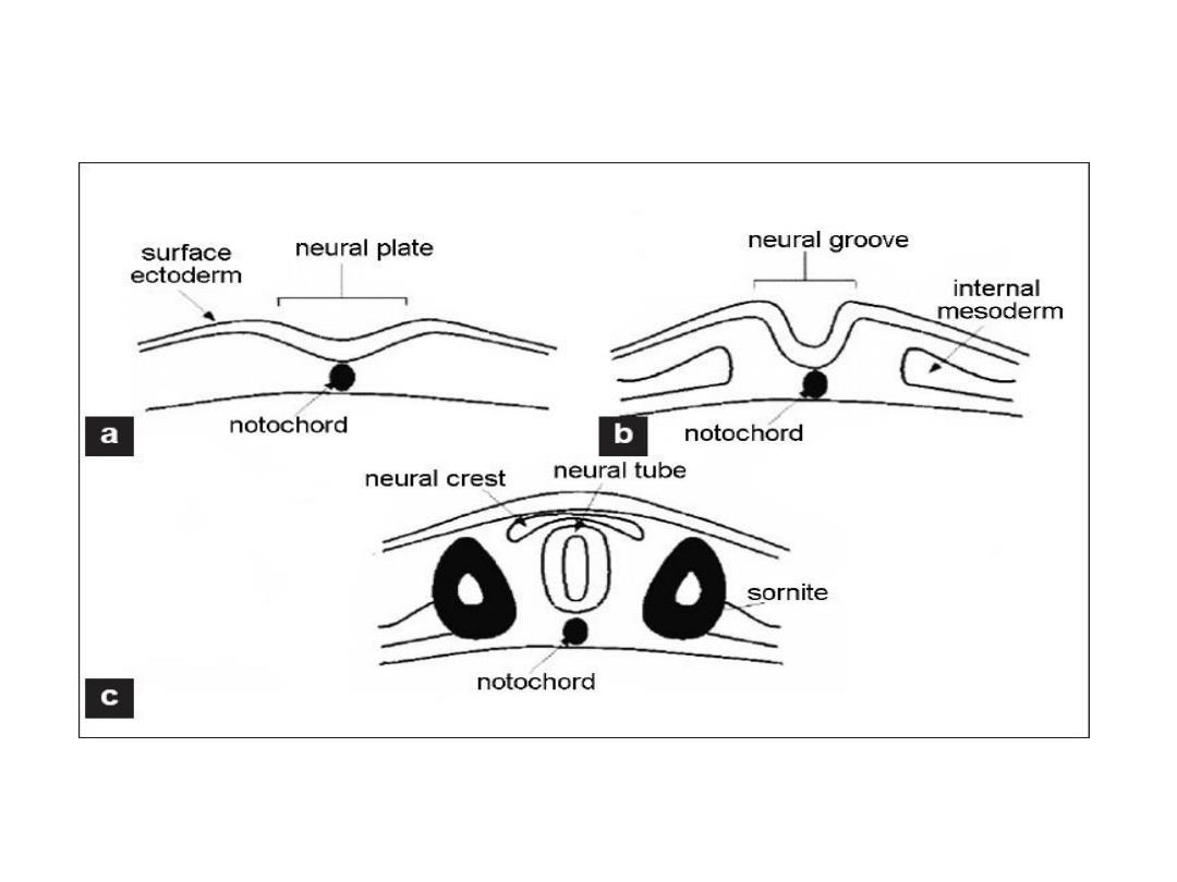
Appearance of ^ notochord & prechordal
mesoderm induces ^ overlying ectodermal to
thicken & form ^
neural plate.
Cells of ^ plate
make up ^
neuroectoderm,
& their induction
represents ^ initial event in ^ process of
neurulation.

Neurulation
• Is ^ process whereby ^ neural plate forms ^ neural tube.
• By ^ end of 3
rd
week, ^ lateral edges of ^ neural plate become
elevated to form
neural folds,
& ^ depressed midregion forms
^ neural
neural groove.
• Gradually, ^ neural folds approach each other in ^ midline,
where they fuse.
• Fusion begins in ^ cervical region & proceeds cranially &
caudally, as a result , neural tube is formed.
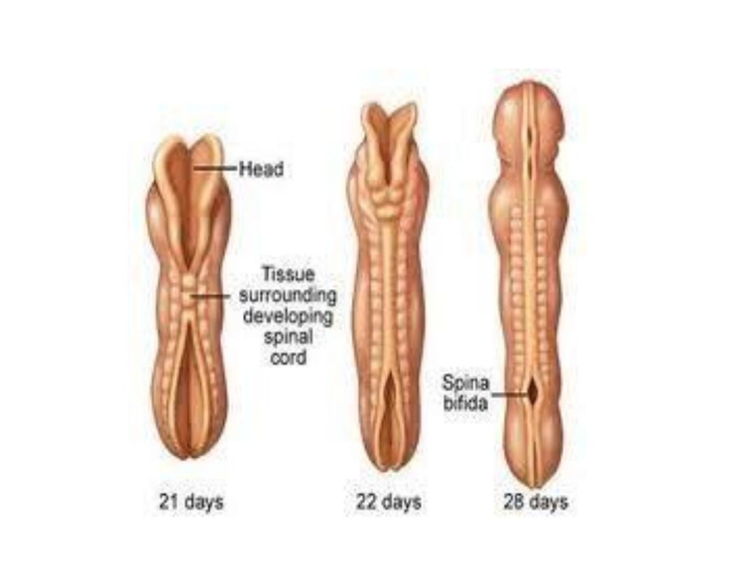
• Until fusion is completed, ^ cephalic & caudal ends of neural
tube communicate with ^ amniotic cavity by way of ^
anterior
(cranial) & posterior (caudal) neuropores.
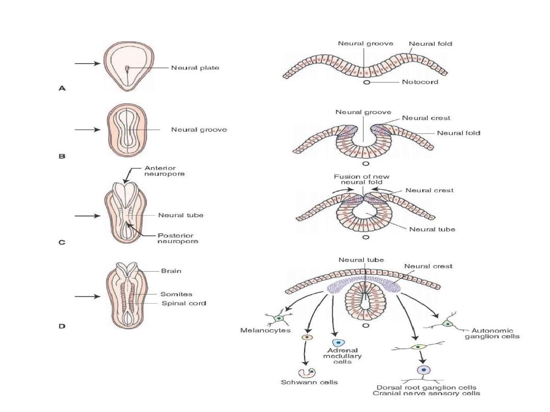

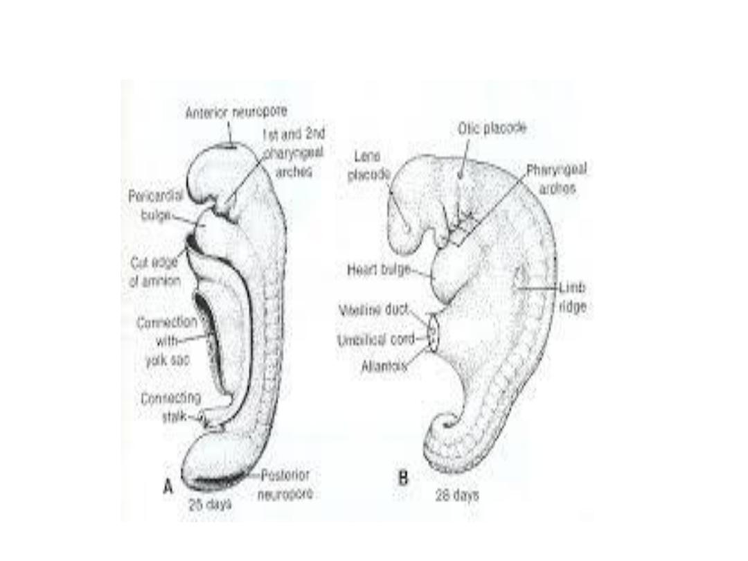
• Closure of ^ cranial neuropore occurs approximately at day
25.
• Closure of ^ caudal neuropore occurs approximately at day
28.
• After closure of ^ ends, neurulation is then complete, & ^
central nervous system is represented by a closed tubular
structure with a narrow caudal portion (spinal cord), & a
much boarder cephalic portion characterized by a no. of
dilations, (brain vesicles).

Neural crest cells
• As ^ neural folds elevate & fuse, cells at ^ lateral border of ^
neuroectoderm begin to dissociate from their neighbors, this
cell population,
neural crest,
will undergo an
epithelial-
mesenchymal transition.
• Neural crest leaves ^ ectoderm & enter ^ mesoderm.
• The neural crest leave ^ neuroectoderm & migrate in two
pathways:
• 1-Dorsal pathway: through ^ dermis, where they will enter ^
ectoderm through holes in ^ basal lamina to form
melanocytes
in skin & hair follicles.

• 2-Ventral pathway: through ^ ant. Half of each somite to
become
sensory ganglia, sympathetic , enteric neurons,
schwann cells, & cells of adrenal medulla.
• Neural crest cells also form & migrate from cranial neural
folds, leaving ^ neural tube before closure in ^ region, these
cells contribute to ^
craniofacial skeleton, neurons for cranial
ganglia, glial cells, melanocytes, & other cell types.

• By ^ time ^ neural tube is closed, two bilateral ectodermal
thickenings, ^
otic placodes & ^ lens placodes,
become visible
in ^ cephalic region of ^ embryo.
• Otic placodes develop to otic vesicles structures for
hearing & equilibrium.
• Lens placode - invaginate at 5
th
week lenses of ^ eyes.

Derivatives of ectoderm germ layer:
• ^ Central nervous system.
• ^ peripheral nervous system.
• ^ sensory epithelium of ^ ear, nose,& eye.
• ^ epidermis, including ^ hair & nails.
• Subcutaneous glands.
• ^ mammary glands.
• ^ pituitary gland.
• & enamel of teeth.

Derivatives of ^ mesoderm
• By approximately ^ 17
th
day cells of mesoderm close to ^
midline proliferate & form a thickened plate
paraxial
mesoderm,
more laterally, ^ mesoderm layer remains thin,
lateral plate,
& that connects these two mesoderm, is
intermediate mesoderm.
• A layer continuous with mesoderm covering ^ amnion, known
as ^
somatic or parietal mesoderm layer.
• A layer continuous with mesoderm covering ^ yolk sac,
^
splanchnic or visceral mesoderm layer.
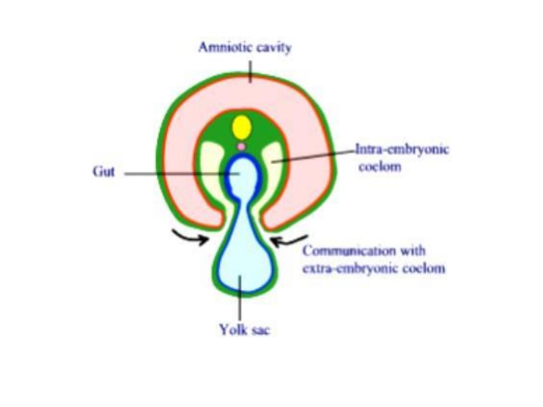
• Together, these layers line a newly formed cavity,
intraembryonic cavity,
which is continuous with ^
extraembryonic cavity on each side.

Paraxial mesoderm
• By ^ beginning of 3rd week, paraxial mesoderm begins to be
organized into segments ,
somitomeres,
1
st
appear in cephalic
region of ^ embryo, & their formation proceeds
cephalocaudally.
• In ^ head region, somitomeres in association with segments
of neural plate form
neuromeres
& contribute to
mesenchyme in ^ head.
• From ^ occipital region & caudally, somitomeres further
organize into somites.
• ^ 1
st
pair of somites arises in ^ occipital region at
approximately 20th day of development.

• ^ somite formation proceeds craniocaudally at a rate of 3
pairs per day, until , at ^ end of ^ 5
th
week, 42-44 pairs are
present.
• There are 4 occipital, 8 cervical, 12 thoracic, 5 lumber, 5
sacral, & 8-10 coccygeal pairs ( ^ 1
st
occipital & ^ last 5-7
coccygeal pairs disappear.
• By counting somites, ^ age of an embryo can be detected.

Somite differentiation
• ^ Somites at ^ beginning appears as a ball, then undergo a process
of
epithelization
& arrange themselves in donut shape around a
small lumen.
• By ^ beginning of 4
th
week, cells of ^medial & ventral wall of ^
somites loose their epithelial characteristics & become
mesenchymal again, & surround ^ neural tube & ^ notochord,
called
sclerotome,
that will differentiate into ribs & vertebrae.
• Cells at ^ dorsomedial & ventrolateral edges of ^ upper region of
somites form precursors for muscle cells, called
myotome,
which
form muscle components of each segment.
• ^ Remaining cells between these 2 groups form
dermatome,
which
forms ^ dermis of ^ back. Each dermatome & myotome has its own
segmental nerve component, no matter where ^ cells migrate.

Intermediate mesoderm
• Differentiate into urogenital structures.
• In cervical & upper thoracic regions, it forms segmental cell
clusters (future
nephrotomes
)
• More caudally, it forms an unsegmented mass of tissue, ^
nephrogenic cord.
• Excretory units of ^ urinary system & ^ gonads develop from
this partly segmented, partly unsegmented intermediate
mesoderm.

Lateral plate mesoderm
• This part splits into parietal (somatic) & visceral( splanchnic)
layers, which line ^ intraembryonic cavity & surround ^
organs, respectively.
• ^ parietal layer of lateral mesoderm forms
^ dermis of ^ skin
of ^ body wall & limbs, ^ bones & connective tissue of ^ limbs
& ^ sternum.
• Sclerotome & muscle precursor cells, that migrate into
parietal layer form ^
costal cartilages, limb muscles, & most of
body wall muscles.
• ^ visceral layer of lateral plate mesoderm, together with
endoderm forms ^ wall of ^ gut tube.

• Mesoderm cells of ^ parietal layer surrounding ^ intra
embryonic cavity form thin membranes , ^
mesothelial
membrane (serous)
which will line ^ peritoneal, pleural &
pericardial cavities.
• Mesoderm cells of ^ visceral layer form a thin serous
membrane around each organ.

Derivatives of endoderm layer
• ^ GIT tract is ^ main tract derived from endoderm.
• This layer covers ^ ventral part of embryo, & forms ^ roof of
yolk sac.
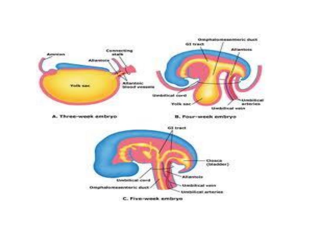
• During this period ^ embryo grow cephalocaudally & bulge in
^ amniotic cavity, forming
head fold & tail fold, also lateral
folds
form & assist in body wall closure.
• As a result of^ cephalocaudal foldings, ^ major part of
endoderm forms ^ gut tube, which consist of ( foregut, midgut
& hindgut)
• ^ midgut connects with ^ yolk sac by ^
vitelline duct.

• At ^ cephalic end ^ foregut is temporarily bounded by an
ectodermal-endodermal membrane (oropharangeal
membrane), thus, this membrane seperates ^
stomadeum,
(ectodermal origin), from ^ pharynx (endodermal origin).
• At ^ 4
th
week of development this memb. ruptures &
establishes a connection between oral cavity & foregut.
• At ^ caudal region, ^ hindgut also terminates temporarily at
an ectodermal-endodermal membrane ,
cloacal memb.,
which separates ^ upper part of anal canal (endoderm) from
lower part of anal canal(ectoderm), this membr. Ruptures
during 7
th
week of development.

• As a result of folding from head, tail & lateral folds, ^ ventral
body is closed except for a small part in ^ umbilical region
where yolk sac & connecting stalk are attached.
• Another important result of cephalocaudal & lateral foldings is
partial incorporation of allantois into ^ body of embryo, forms
cloaca.
• By ^ 5
th
week , yolk sac duct, allantois & umbilical vessels are
restricted to ^ umbilical region.
• Later, ^ endoderm layer gives rise to:
• 1- epithelial lining of respiratory tract.

• 2-^ parenchyma of thyroid, parathyroid, liver & pancrease.
• 3-reticular stroma of ^ tonsils & thymus.
• 4- epith.lining of urinary bladder & urethra.
• 5-epith.lining of tympanic cavity & auditory tube.