Genitourinary system
Part 2
Genital system

• POSITION OF THE KIDNEY:
• The kidney, initially in the pelvic region, later shifts to a
more cranial position in the abdomen. This ascent of
the kidney is caused by diminution of body curvature
and by growth of the body in the lumbar and sacral
regions.
• FUNCTION OF THE KIDNEY:
• The defi iti e kid ey for ed fro the metanephros
becomes functional near the 12th week. Urine is
passed into the amniotic cavity and mixes with the
a
ioti fluid. The fluid is s allo ed y the fetus a d
recycles through the kidneys. During fetal life, the
kidneys are not responsible for excretion of waste
products, since the placenta serves this function.

•
BLADDER AND URETHRA
• During the fourth to seventh weeks of development the Cloaca
divides into the urogenital sinus anteriorly and the anal canal
posteriorly.
• The urorectal septum is a layer of mesoderm between the primitive
anal canal and the urogenital sinus
• The tip of the septum will form the
perineal body
• Three portions of the urogenital sinus can be distinguished:
(1)-
• The upper and largest part is the urinary bladder. Initially the
bladder is continuous with the allantois, but when the lumen of the
alla tois is o literated, a thi k fi rous ord, the
urachus
, remains
and connects the apex of the bladder with the umbilicus in the
adults, it is known as the median umbilical ligament.
• (2)_
The next part is a rather narrow canal, the pelvic part of the
urogenital sinus, which in the male gives rise to the prostatic and
membranous parts of the urethra.
• (3)_
The last part is the phallic part of the urogenital
sinus.(Development of the phallic part of the urogenital sinus differs
from male to female)

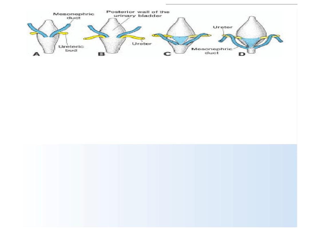
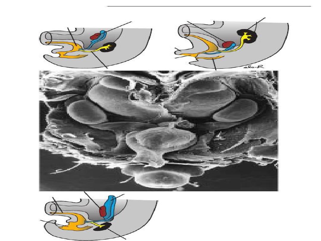
During differentiation of the cloaca, the caudal
portions of the mesonephric ducts are absorbed into the
wall of the urinary bladder. Consequently, the ureters,
initially outgrowths from the mesonephric ducts, enter
the bladder separately.
As a result of as e t of the kid eys, the orifi es of the
ureters move farther cranially; those of the mesonephric
ducts move close together to enter the prostatic urethra
and in the male become the ejaculatory ducts.
Since both the mesonephric ducts and ureters
originate in the mesoderm, the mucosa of the bladder
formed by incorporation of the ducts(the trigone of the
bladder) is also mesodermal. With time the mesodermal
lining of the trigone is replaced by endodermal
epitheliu , so that fi ally the i side of the ladder is
completely lined with endodermal epithelium.

URETHRA
The epithelium of the urethra in both sexes
originates in the endoderm; the surrounding
connective and smooth muscle tissue is derived
from splanchnic mesoderm. At the end of the third
month, epithelium of the prostatic urethra begins
to proliferate and forms a number of outgrowths
that penetrate the surrounding mesenchyme. In the
male, these buds form the
prostate gland
.
In the female, the cranial part of the urethra gives
rise to the
Urethral
and
paraurethral glands
.

Genital system
• The genital system consists of (a)- gonads or
primitive sex glands,(b)- genital ducts, and (c)
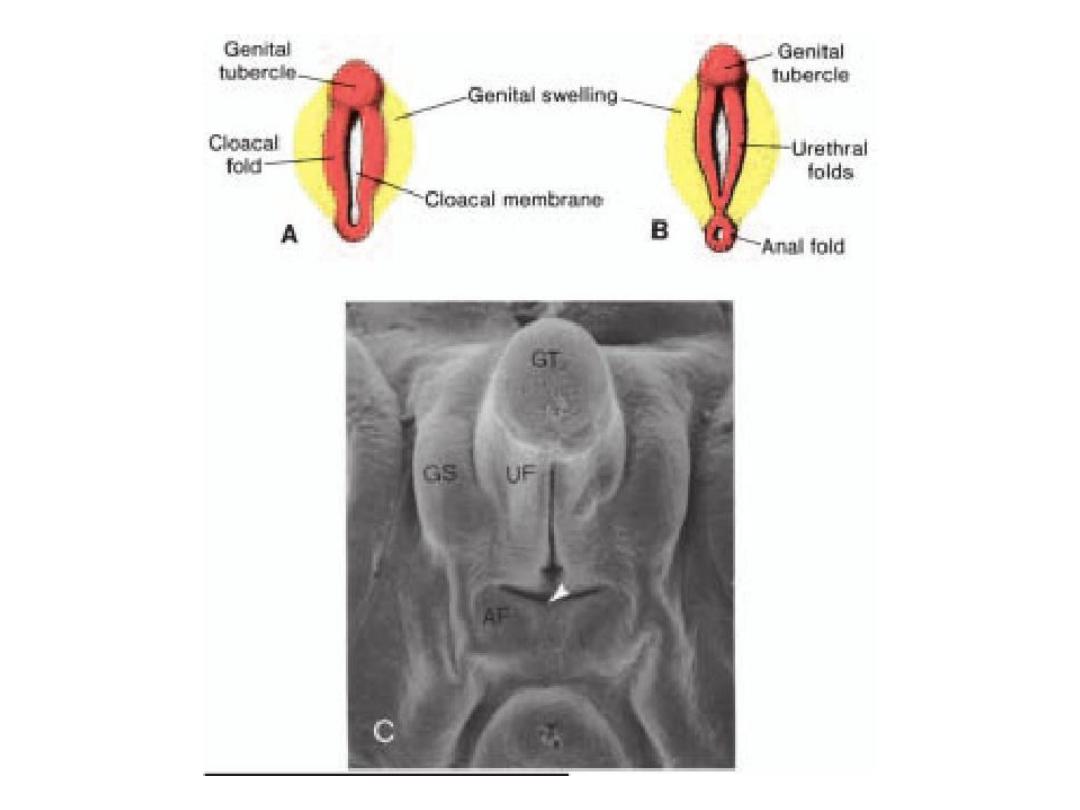
external genitalia.
• All three components go through an indifferent
stage in which they may develop into either a
male or a female. The SRY gene on the Y
chromosome produces testes-determining factor
and regulates male sexual development, that
stimulate differentiation of Sertoli and Leydig
cells in the testes. And its disappearance lead to
development of female genital system.

1-gonads
• Gonads appear initially as a pair of longitudinal ridges,
the genital or gonadal ridges.
• Germ cells do not appear in the genital ridges until the
sixth week of development.
• Primordial germ cells originate in epiblast, migrate
through primitive streak, &by 3
rd
week reside among
endoderm cells in the wall of yolk sac close to allantois.
• By 4
th
week they move by ameboid movement along
dorsal mesentry of hindgut.
• By 5
th
week arriving the primitive gonads and invading
the genital ridges by 6
th
week.

• Shortly before and during arrival of primordial
germ cells, the epithelium of genital ridge
proliferates, & penetrate the underlying
mesenchyme. Here they form irregular cords, the
primitive sex cords
.
• In both male and female embryos, these cords
are connected to surface epithelium and it is
impossible to differentiate between them. Hence
the gonad is known as the
indifferent gonads.
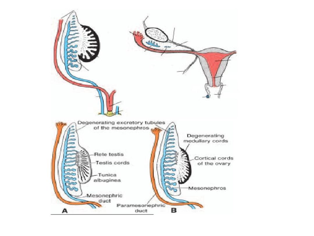
• Then after that differentiation of gonads happen
to male & female gonads i.e. ovary & testis. Each
gonad composed of medulla & cortex but

Testis ovary
• Medullary cords
develop
• No cortical cords
• Thick tunica albuginea
• Medullary cords
degenerate
• Cortical cords develop
• No tunica albuginea

Genital ducts
• Indifferent stage:
• Initially, both male and female embryos have 2
pairs of genital ducts: mesonephric (wolffian)
ducts & paramesonephric (mullerian) ducts.
• The paramesonephric duct arises as a longitudinal
invagination of the epithelium on the anterolateral
surface of the urogenital ridge. Cranially the duct
opens into the abdominal cavity with a funnel-like
stru ture. Caudally it first ru s lateral to the
mesonephric duct, then crosses it ventrally to
grow caudomedially.

• the 2 ducts are initially separated by a septum
but later fuse to form the
uterine canal
.
• The caudal tip of the combined ducts projects
into the posterior wall of the urogenital sinus,
where it causes a small swelling, the
paramesonephric or mullerian tubercle
. The
mesonephric ducts open into the urogenital
sinus on either side of the mullerian tubercle.

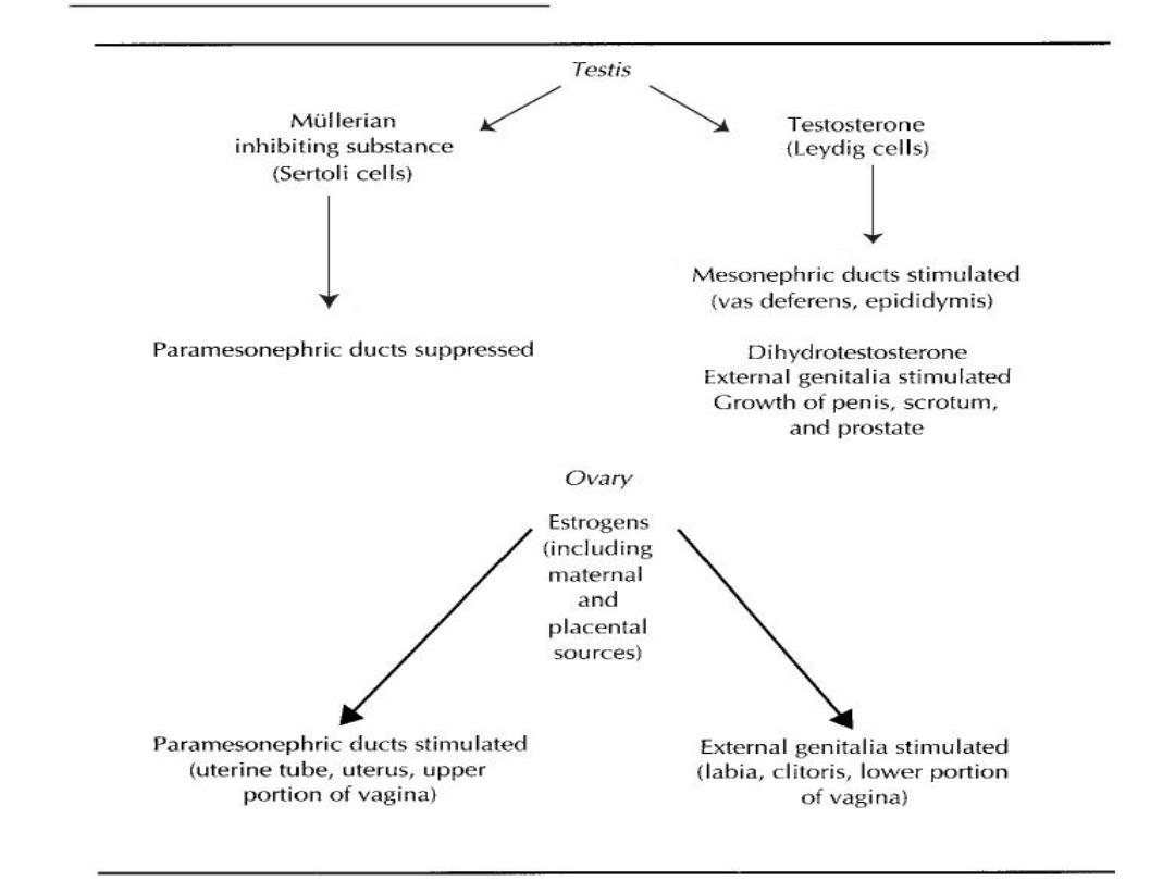
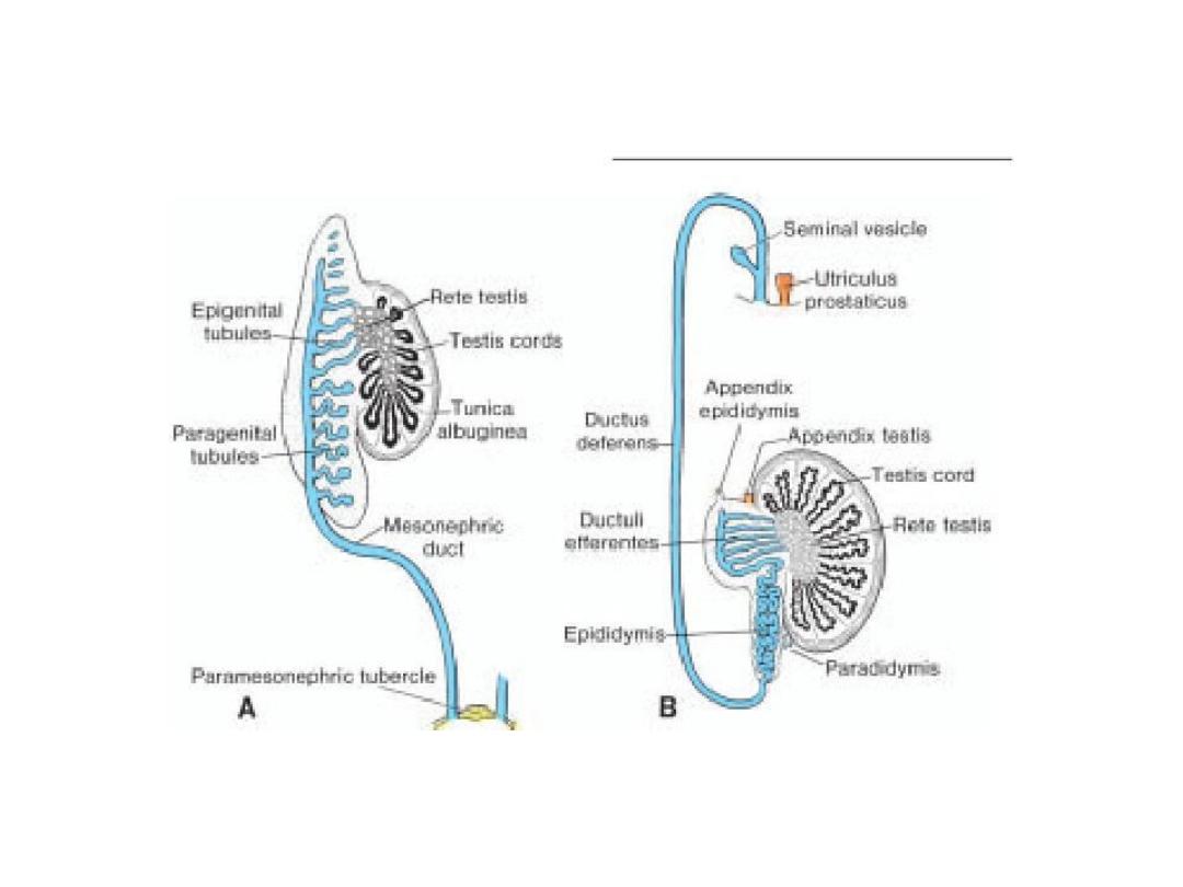
Differentiation of duct system and
external genitalia
• The indifferent duct system and external genitalia develop under
the i flue e of hor o es.
Testosterone
produced by Leydig cells in
the testes stimulates development of the mesonephric ducts (vas
deferens epididymis),while
MIS
produced by Sertoli cells in the
testes causes regression of the paramesonephric ducts (female duct
system).
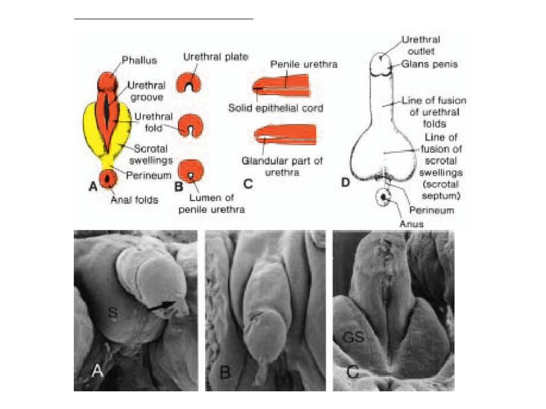
• Dihydrotestosteron
e stimulates development of the external
genitalia, penis, scrotum, and prostate.
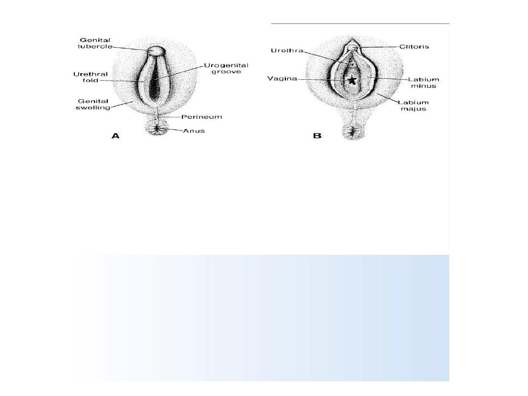
• Estrogens
i flue e de elop e t of the paramesonephric female
system, including the uterine tube, uterus, cervix, and upper
portion of the vagina. They also stimulate differentiation of the
external genitalia, including the clitoris, labia, and lower portion of
the vagina. Errors in production of or sensitivity to hormones of the
testes lead to a predominance of female characteristics under
i flue e of the ater al a d pla e tal estroge s