Gastrointestinal system

• The epithelium of the digestive system and the
parenchyma of its derivatives originate in the
endoderm; connective tissue, muscular components,
and peritoneal components originate in the
mesoderm.
• Differentiation of the gut and its derivatives depends
upon reciprocal interactions between the gut
endoderm (epithelium) and its surrounding mesoderm.
• The gut system extends from the buccopharyngeal
membrane to the cloacal membrane with craniocaudal
organization and is divided into the pharyngeal gut,
foregut, midgut, and hindgut. The pharyngeal gut gives
rise to the pharynx and related glands .

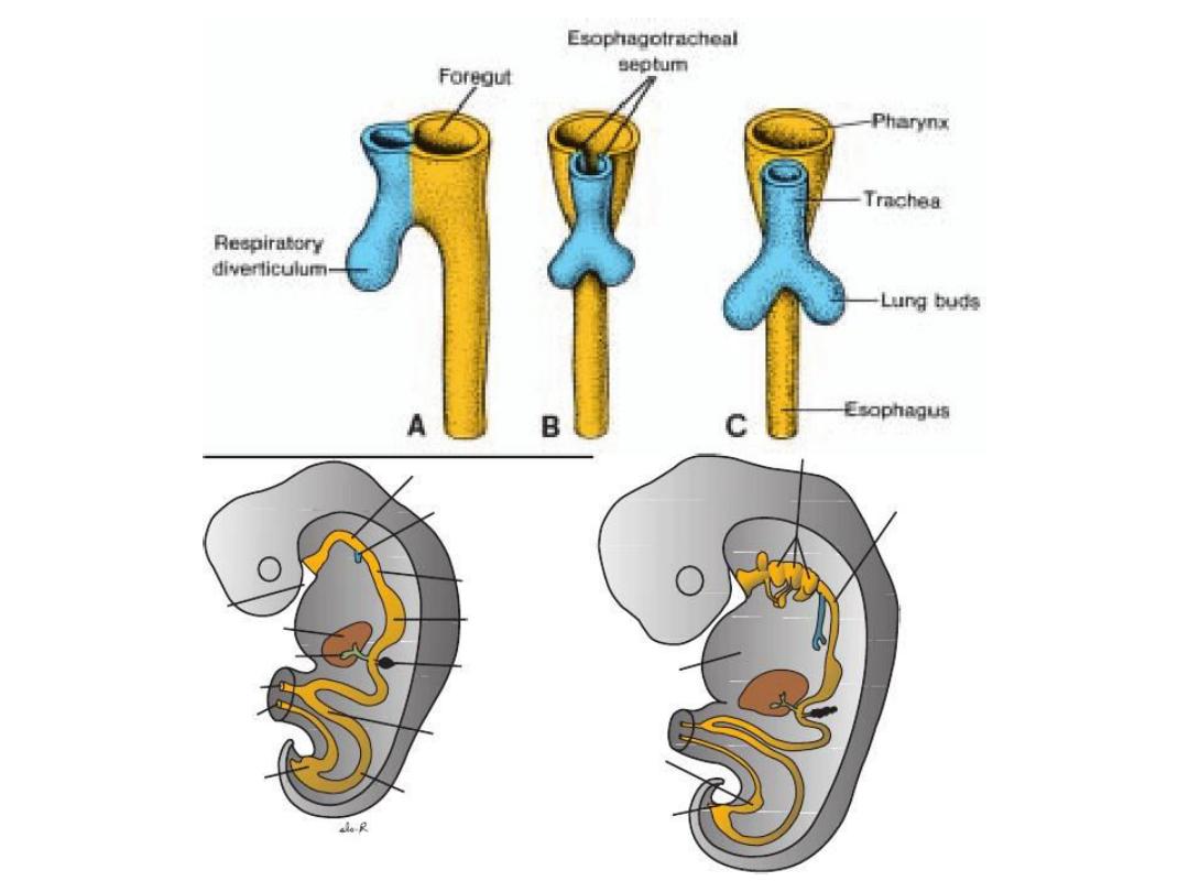
The foregut
• gives rise to the esophagus, the trachea and lung buds, the
stomach, and the duodenum proximal to the entrance of the bile
duct. In addition, the liver, pancreas, and biliary apparatus develop
as outgrowths of the endodermal epithelium of the upper part of
the duodenum.
• Since the upper part of the foregut is divided by a septum (the
tracheoesophageal septum) into the esophagus posteriorly and the
trachea and lung buds anteriorly.
• Deviation of the septum may result in abnormal openings between
the trachea and esophagus. The epithelial liver cords and biliary
system growing out into the septum transversum, differentiate into
parenchyma.
• Hematopoietic cells (present in the liver in greater numbers before
birth than afterward), the Kupffer cells, and connective tissue cells
originate in mesoderm. The pancreas develops from a ventral bud
a d a dorsal ud that later fuse to for the defi itive pa reas.
Sometimes, the two parts surround the duodenum (annular
pancreas), causing constriction of the gut

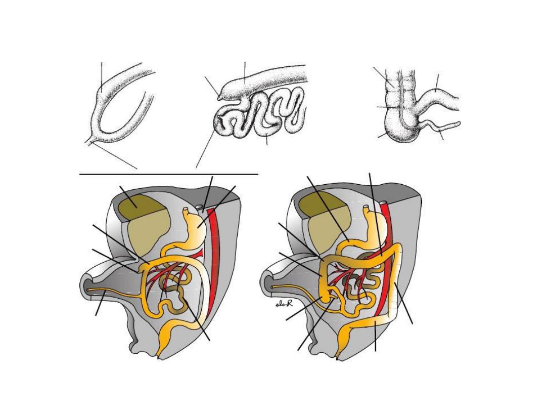
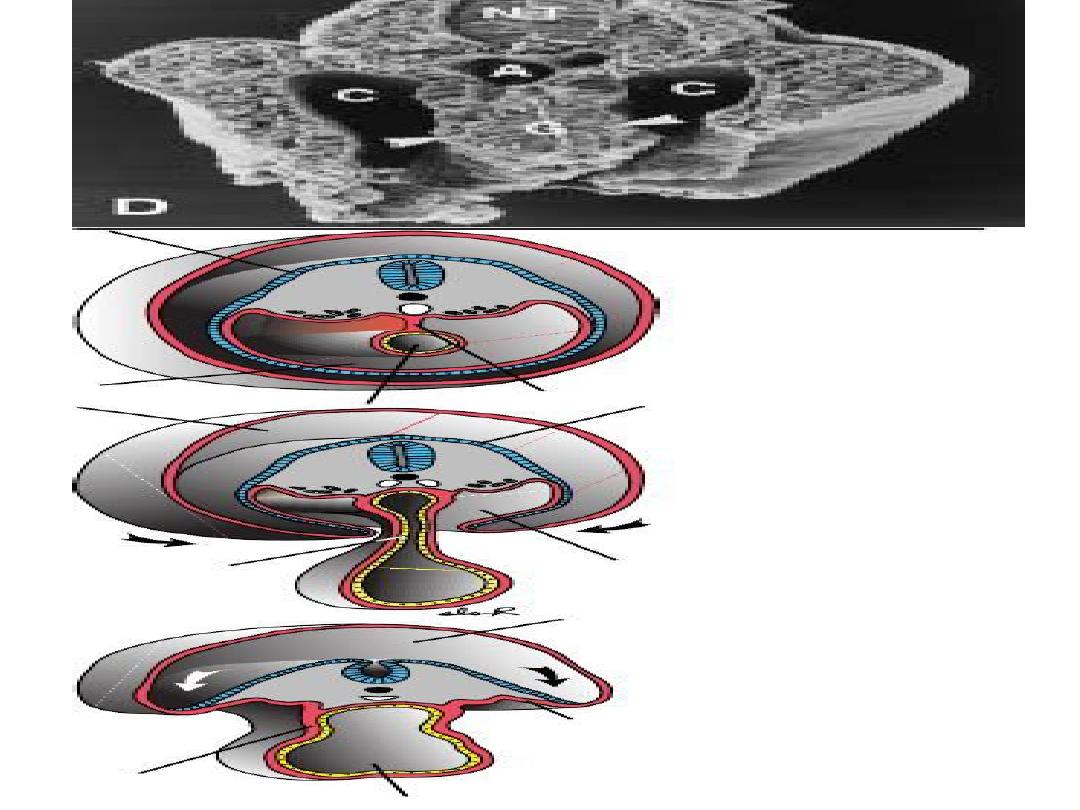
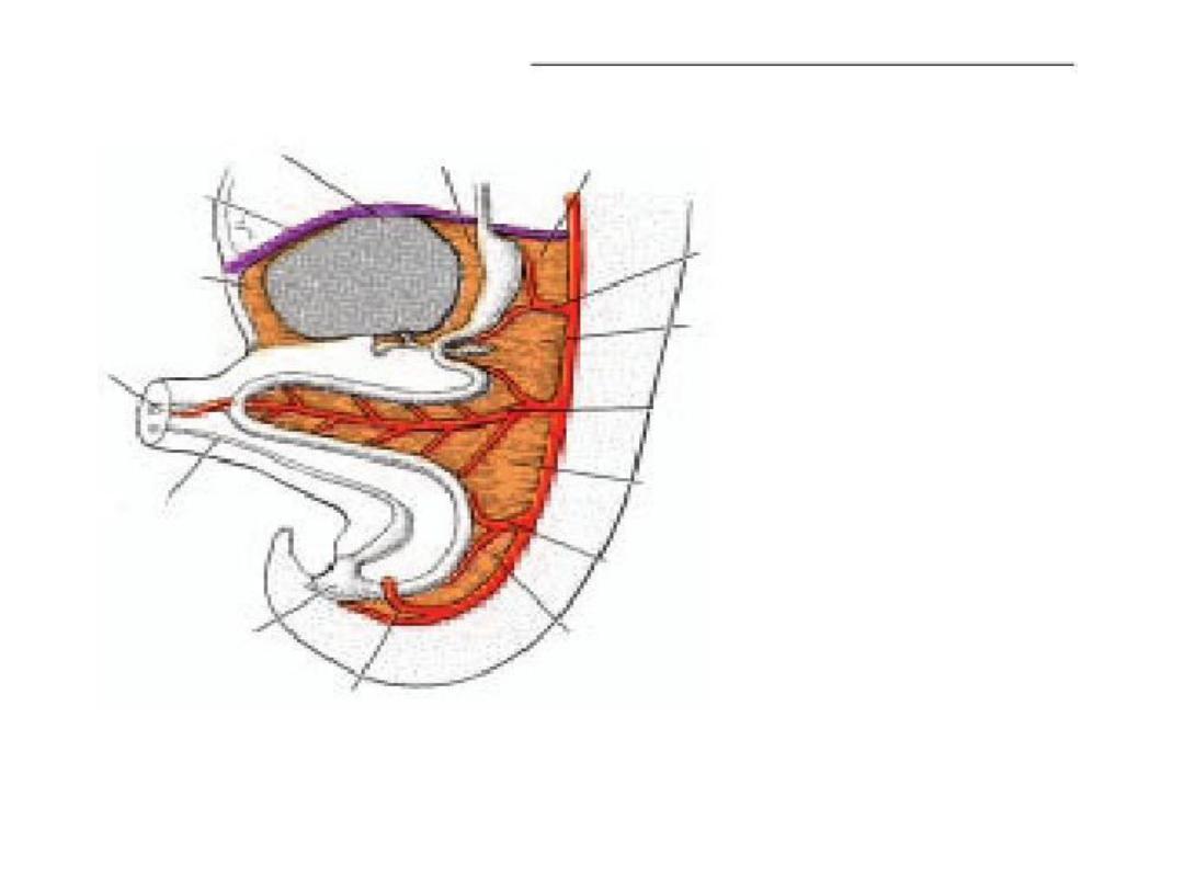
The midgut
• forms the primary intestinal loop, gives rise to the duodenum distal
to the entrance of the bile duct, and continues to the junction of
the proximal two-thirds of the transverse colon with the distal third.
• At its apex the primary loop remains temporarily in open
connection with the yolk sac through the vitelline duct. During the
sixth week, the loop grows so rapidly that it protrudes into the
umbilical cord (physiological herniation. During the 10th week, it
returns into the abdominal cavity. While these processes are
occurring, the midgut loop rotates 270
◦ counterclockwise.
• Remnants of the vitelline duct, failure of the midgut to return to
the abdominal cavity, malrotation, stenosis, and duplications of
parts of the gut are common abnormalities.

The hindgut
• gives rise to the region from the distal third of the
transverse colon to the upper part of the anal canal; the
distal part of the anal canal originates from ectoderm.
• The hindgut enters the posterior region of the cloaca(future
anorectal canal), and the allantois enters the anterior
region (future urogenital sinus).
• Breakdown of the cloacal membrane covering this area
provides communication to the exterior for the anus and
urogenital sinus. Abnormalities in the size of the posterior
region of the cloaca shift the entrance of the anus