By
Dr. Suhair Majeed

Lymphoid system :
The
lymphoid system collects excess
interstitial fluid into lymphatic capillaries
,
transports absorbed lipids from the small
intestine, and responds immunologically to
invading foreign substances.

Lymphatic System:
Lymphatic System consists of:
A. Cells
1.
Lymphocytes (B,T, natural killer)
2.
Antigen-presenting cells (dendritic cells,
Langerhans
’ cells & macrophages
B. Lymphatic tissue –diffuse and nodular
C. Lymphatic organs lymph nodes, spleen,
thymus)
D. Lymphatic vessels that carry the cells and fluid

Cont.
The main function of the lymphoid organs is
to protect the organism against invading
pathogens or antigens (bacteria, parasites, and
viruses). The immune response occurs when the
organism detects the pathogens, which can enter
the organism at any point. For this reason,
lymphatic cells, tissues, and organs have wide
distribution in the body.

Cont.
The major lymphoid organs are the
- lymph nodes,
- tonsils,
- thymus, and
- spleen.
Because bone marrow produces lymphocytes, it
is considered a lymphoid organ and part of the
lymphoid system.

Cont.
lymphatic tissue divided into:
1-
diffuse and
2- nodular
depends on the arrangement and concentration
of the cells, not on differences in fiber types.

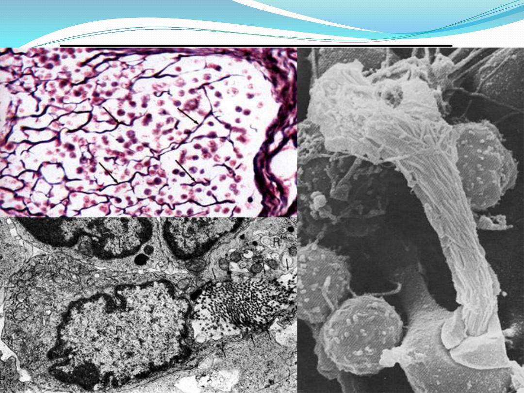
Cells of lymphatic tissue :
The cells of lymphatic tissue are present as
fixed and free cells.
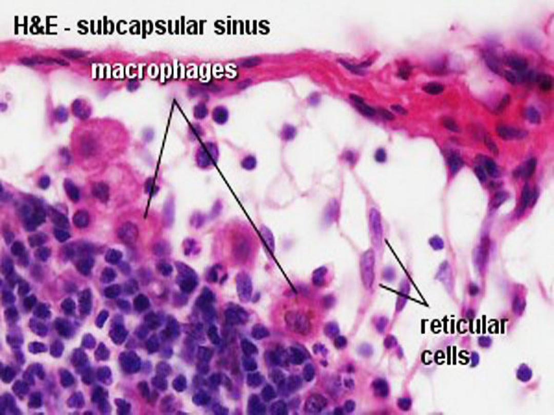
Fixed cells are the reticular
cells responsible for the formation and
maintenance of reticular fibers.
reticular cells appear as elongated or stellate
elements with round or oval, palely stained nuclei
and, lightly basophilic cytoplasm.
Reticular Fibers (type III collagen
)

Cont.
The remaining cells of lymphatic tissue are
contained within the spaces of the reticular
network and constitute the
free cells.

lymphocyte:
Lymphocytes are the cells that carry out
immune responses.
Morphologically, all types of lymphocytes
appear very similar, but functionally, they are very
different.When lymphocytes are properly
stimulated,
B lymphocytes and T lymphocytes
are produced
.

Cont.
These two subclasses of lymphocytes are
distinguished on the basis of where they
differentiate and mature into immunocompetent
cells, and on the types of surface receptors
present on their cell membranes.

T- lymphocytes or T- cells:
T cells arise from lymphocytes that are carried
from the bone marrow to the
thymus gland.
They mature, differentiate, and acquire surface
receptors and
immunocompetence before
migrating to peripheral lymphoid tissues and
organs. The thymus gland produces mature T
cells early in life.

Cont.
T cells carry out immune responses when
stimulated. There are four main types of
differentiated T cells:
-helper T cells,
- cytotoxic T cells,
-memory T cells,
- suppressor T cells

1-T- helper cells
When encountering an antigen,
helper T cells
assist other lymphocytes by secreting immune
chemicals called
cytokines( interleukins).
Cytokines are protein hormones that stimulate
proliferation, secretion, differentiation, and
maturation of B cells into
plasma cells, which
then produce immune proteins called
antibodies ( immunoglobulins).

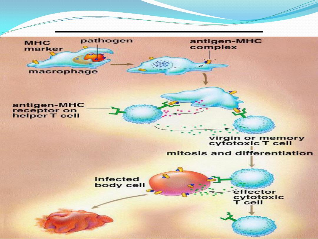
2- cytotoxic T-cells
:
Cytotoxic T cells specifically recognize
antigenically different cells such as virus-infected
cells, foreign cells, or malignant cells and destroy
them. These lymphocytes become activated
when they combine with antigens that react with
their receptors.

3- Memory T- cells :
Memory T cells are the long-living progeny of
T cells. They respond rapidly to the same
antigens in the body and stimulate immediate
production of cytotoxic T cells.
Memory T cells are the counterparts of
memory B cells.

4- suppressor T- cells
Suppressor T cells may decrease or
inhibit the functions of helper T cells and
cytotoxic T cells, and thus modulate the
immune response.

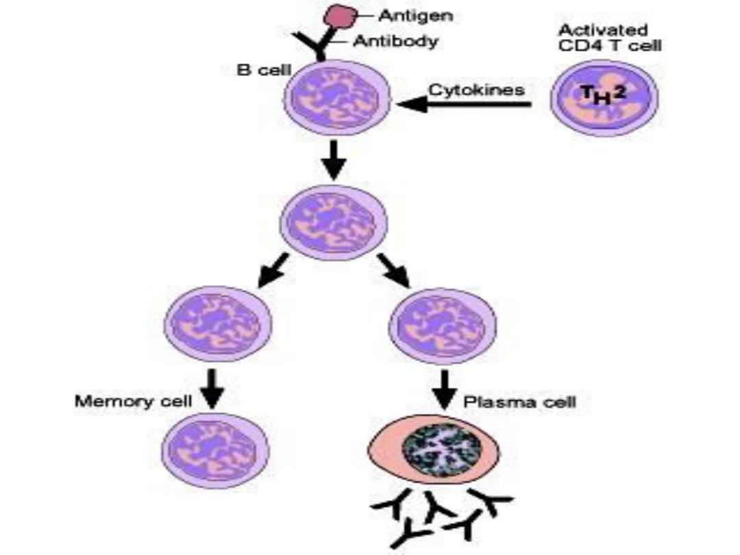
B
–lymphocyte or B-cells :
B cells mature and become immunocompetent
in bone marrow
. After maturation, blood carries
B cells to the nonthymic lymphoid tissues such as
lymph nodes, spleen, and connective tissue.
B cells are able to recognize a particular type of
antigen owing to the presence of
antigen
receptors on the surface of their cell membrane
.

Cont.
The response of B cells to an antigen, is more
intense when antigen-presenting cells, such as
helper T cells
,
present the antigen to the B cells.
Helper T cells secrete a cytokine(
interleukin 2)
that
induces increased proliferation and
differentiation of antigen-activated
B cells.
Numerous progeny of activated B cells
enlarge, divide, proliferate, and differentiate
into
plasma cells.

Cont.
Plasma cells then secrete large amounts of
antibodies specific to the antigen that triggered
plasma cell formation.
Antibodies react with the antigens and initiate
a complex process that eventually destroys the
foreign substance that activated the immune
response.
(antibody or humoral immune response ).

Cont.
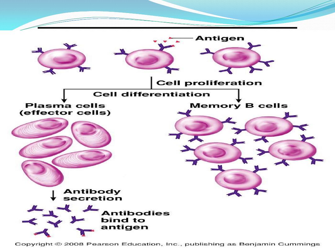
Other activated B cells do not become plasma
cells. Instead, they persist in lymphoid organs as
memory B cells. These memory cells produce a
more rapid immunologic response should the
same antigen reappear.

Cont.
In addition to T cells and B cells, cells
called :
- macrophages,
- natural killer cells, and
- antigen presenting cells, perform
important functions in immune responses.

Cont.
Natural killer cells attack virally infected
cells and cancer cells.
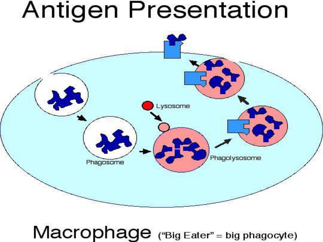
Antigen-presenting cells are found in most
tissues
. These cells phagocytose and process
antigens, and then present the antigen to T cells,
inducing their activation.

Cont.
Included in this group are the
connective
tissue macrophages, A type of wbc that engulfs
and digests cellular debries , microbes and cancer
cells in aprocess called
phagocytosis.
They play a critical role in non- specific
defense (innate immunity ),and also help intiate
specific defense mechanisms (adaptive
immunity ) by recruiting other immune cells
such as lymphocytes
.

Antigen presenting cells :
Are found in most tissues. These cells
phagocytose and process antigens, and then
present the antigen to T- cells, inducing their
activation .
Most antigen- presenting cells belong to
mononuclear phagocytic system .Included in this
group are (connective tissue macrophages
,perisinusoidal macrophages in liver (kupffer cells
), Langerhans cells in skin ,and macrophages
with in lymphoid organs.

Cont.
Follicular dendritic cells in lymphoid
nodules retain antigen molecules on their
surfaces where they remain available to B
–
lymphocytes .

Basic Types of Immune Responses
The presence of foreign cells or antigens in the
body stimulates a highly complex series of
reactions.These result in either production of
antibodies, which bind to the antigens, or
stimulation of cells that destroy foreign cells. B
cells and T cells respond to antigens by different
means. Two types of closely related immune
responses take place in the body, both of which
are triggered by antigens.

1- cell-mediated immune response
In the
cell-mediated immune response,
T
cells are stimulated by the presence of antigens
on the surface of antigen-presenting cells. The T
cells proliferate and secrete cytokines. These
chemical signals stimulate other T cells, B cells,
and cytotoxic T cells. On activation and binding
to target cells, cytoxic T cells produce protein
molecules called
perforin, which perforate the
target cell membranes, causing cell death.

Cont.
Cytotoxic T cells also destroy foreign cells by
attaching to them and inducing
apoptosis or
programmed cell death. The activated
lymphocytes then destroy foreign
microorganisms, parasites, tumor cells, or virus-
infected cells. T cells may also attack indirectly by
activating B cells or macrophages of the immune
system. T cells provide specific immune
protection without secreting antibodies.
Cell
–mediated immunity

2- humoral immune response
In the humoral immune response, exposure of
B cells to an antigen induces proliferation and
transformation of some of the B cells into plasma
cells. These secrete specific antibodies into blood
and lymph that bind to, inactivate, and destroy
the specific foreign substance or antigens.

Cont.
The activation and proliferation of B cells
against most antigens require the cooperation of
helper T cells that respond to the same antigen
and the production of certain cytokines. The
presence of the B cells, plasma cells, and
antibodies in the blood and lymph are the basis
of the humoral immune response
.
humoral immune response

Diffuse Lymphatic Tissue
is a constituent of lymphatic organs . It
appears as a loose aggregate of cells with no
distinct demarcation from surrounding tissue .
Diffuse lymphatic tissue is prominent in the
connective tissue that underlies the epithelium of
the intestine. Any antigen that does penetrate the
epithelial lining induces an immune response in
the lymphatic tissue .
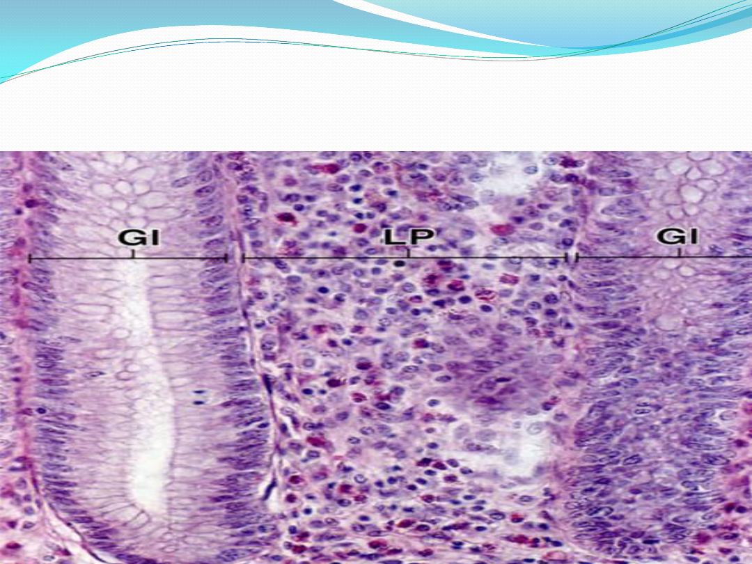
Diffuse lymphoid tissue
Lamina propria (LP) of gut shown here, but can be found
associated with mucosae anywhere in the gut, respiratory,
and genitourinary tracts.

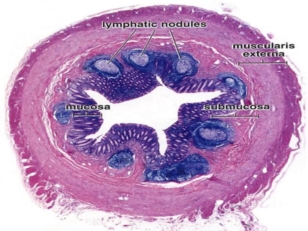
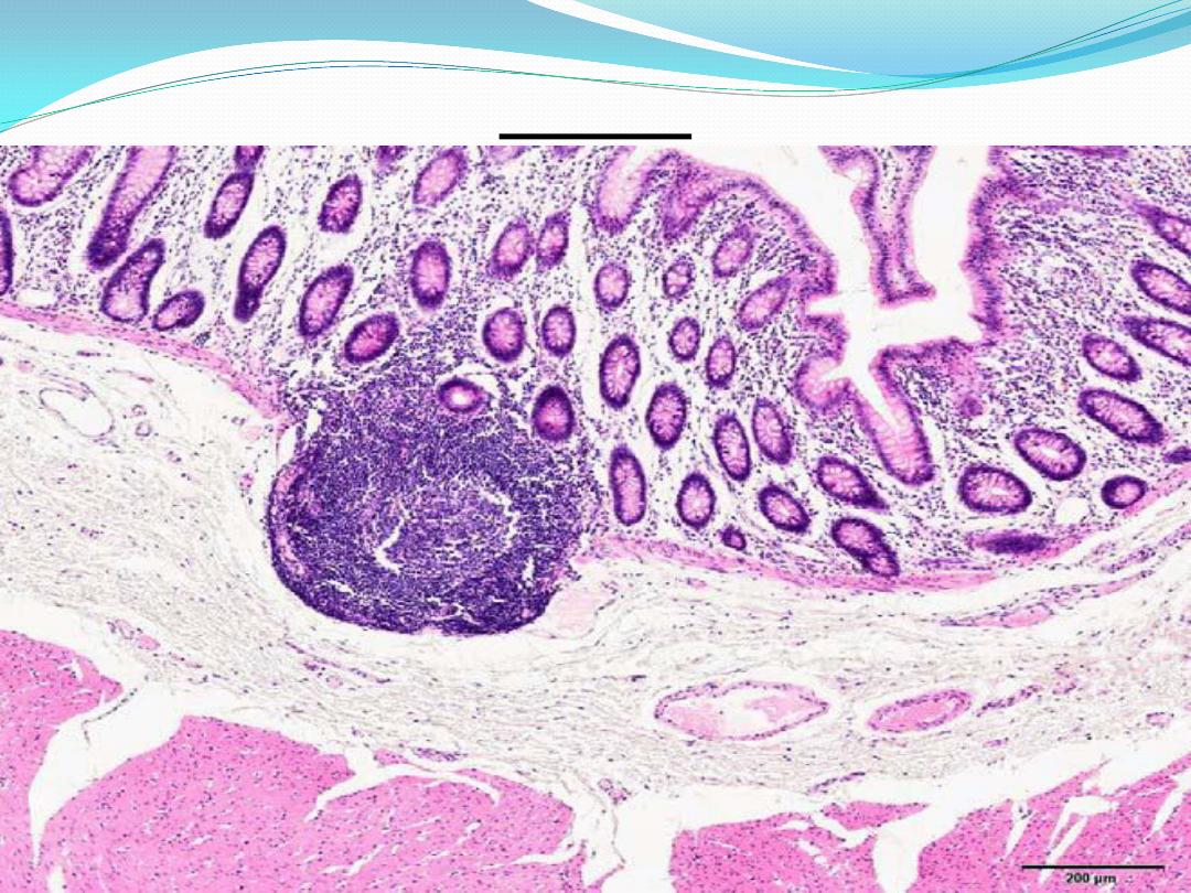
Nodular Lymphatic Tissue
Nodular lymphatic tissue contains the same
structural elements as diffuse lymphatic tissue,
differing only in that the components are
organized into compact, circumscribed
structures. Lymphatic nodules (also called
follicles) may be present as solitary nodules ,
as occur in the appendix and the Peyer's patches
of the ileum.

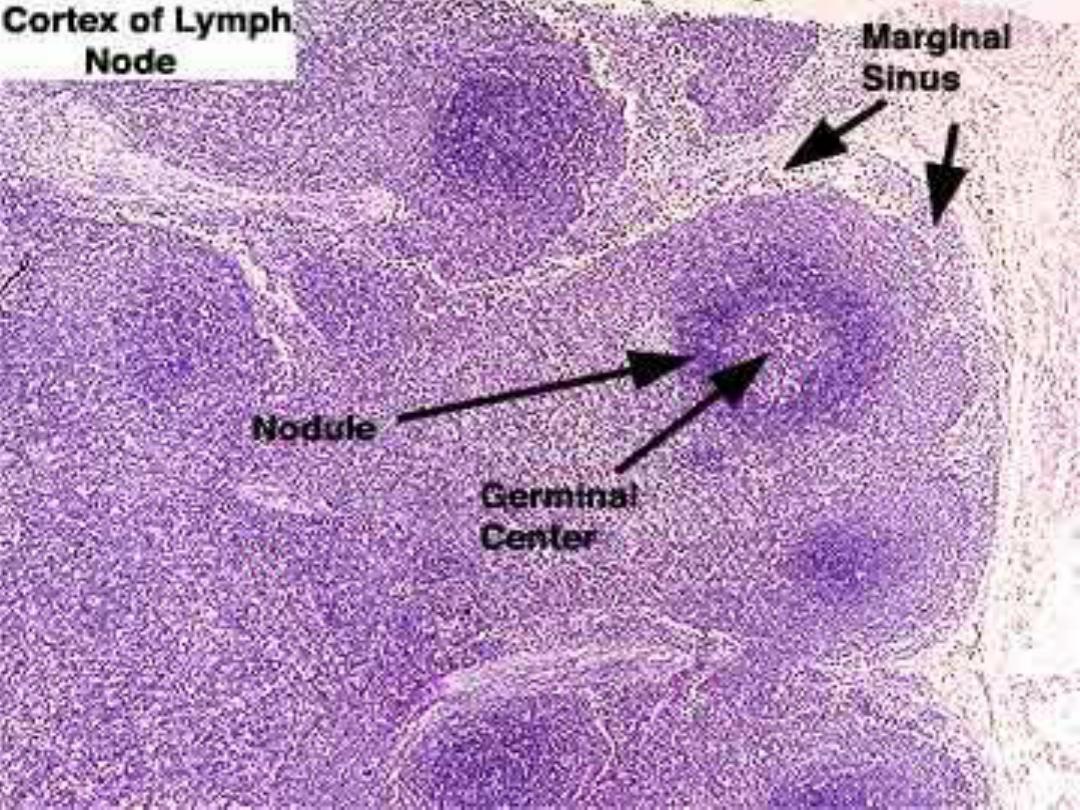
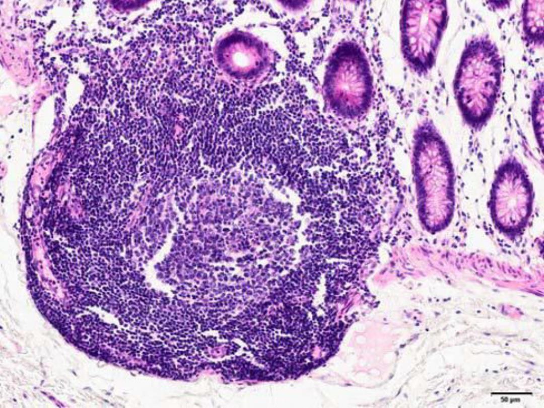
Cont.
Histologically, some lymphatic nodules
appear as rounded collections of densely packed
small lymphocytes; this nodule is called a
primary nodule.
Other lymphatic nodules contain a lightly
staining central area surrounded by a deeply
stained cap of closely packed small lymphocytes.
The pale region has been called a
germinal
center and the whole structure a secondary
nodule.

Cont.
Lymphatic nodules are prominent in organs
such as the tonsils, lymph nodes, and spleen but
are absent from the thymus . Germinal centers
produce B-
cells that can migrate through the cap
to leave the center and eventually pass to other
lymphatic tissues.
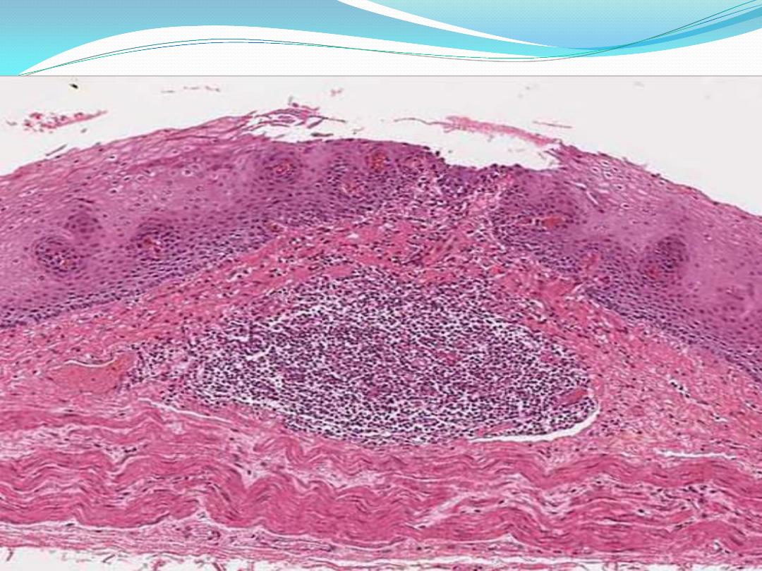
Primary lymphatic nodule/follicle (LN)
Aggregation of lymphocytes in lamina propria or submucosa

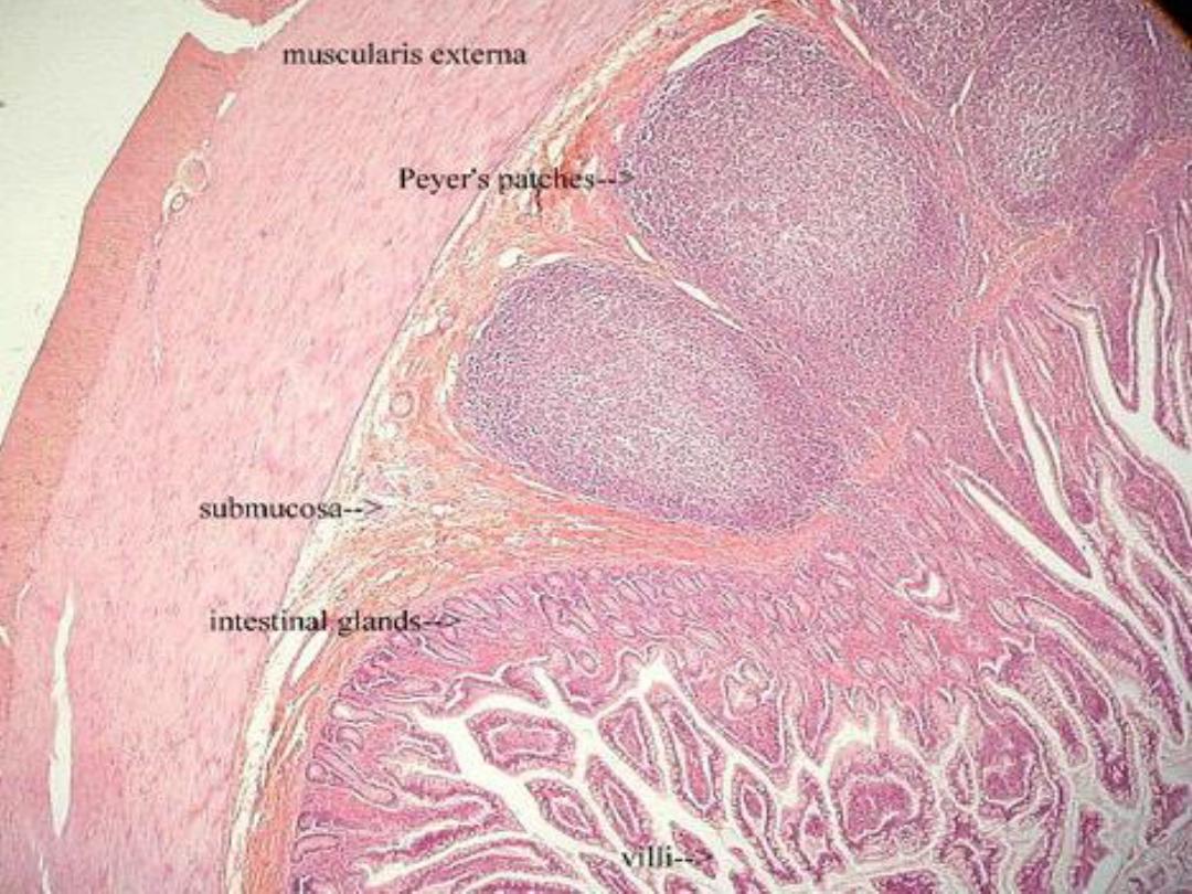
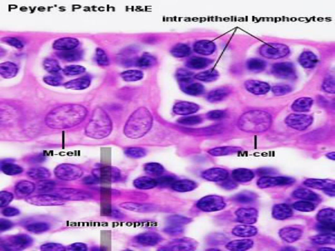
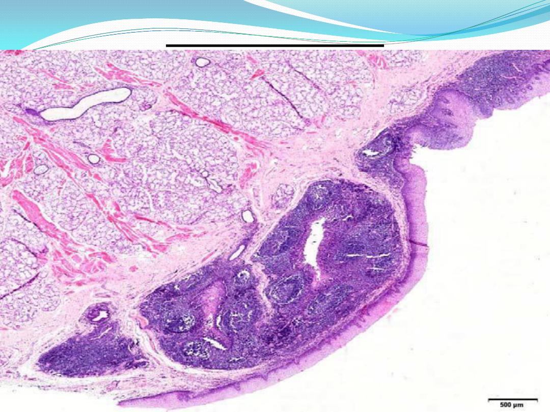
Peyer’s patches :
Peyer’s patches occur in the wall of the ileum.
Consist of very large spherical aggregates
(nodules ) of dense lymphoid tissue which may
show germinal centers .
most of the mass of each nodule is located in
the submucosa ,but the nodule extends into
lamina propria (Subepithelial loose FECT ) and
bulges into the lumen of ileum.

Cont.
Their function in screening of the lumen of
small intestine, probably to prevent colon
bacteria from migrating up into small intestine .
IgA antibodies secreted by plasma cells generated
by
peyer’s Patches seriously impair bacterial
motility and inhibit attachment of bacteria to
intestinal walls .

appendix
The lymphoid nodules in the appendix are
simillar in structure and basic function to
peyer’s patches .
Nodules in appendix probably prime the
immune system against microorganisms which
inhabit the colon ,so that if these organisms
penetrate into the colon wall or the peritoneal
cavity they are efficiently destroyed
.

Lymphatic Organs :
Lymphatic organs are divided into
1- primary (central) and
2- secondary (peripheral) organs.
Primary lymphatic organs are the first to
develop and include the thymus and the bone
marrow .
The secondary lymphatic organs are the
lymph nodes, spleen, tonsils.

Thymus
The thymus is a bilobed, encapsulated lymphatic
organ located in the upper anterior mediastinum
and lower part of the neck. . The thymus is the
only primary lymphatic organ and is the first
organ of the embryo to become lymphoid . Unlike
the spleen and lymph nodes, it is well developed
and relatively large at birth, after which the organ
undergoes progressive involution and is partially
replaced by fat and connective tissue.
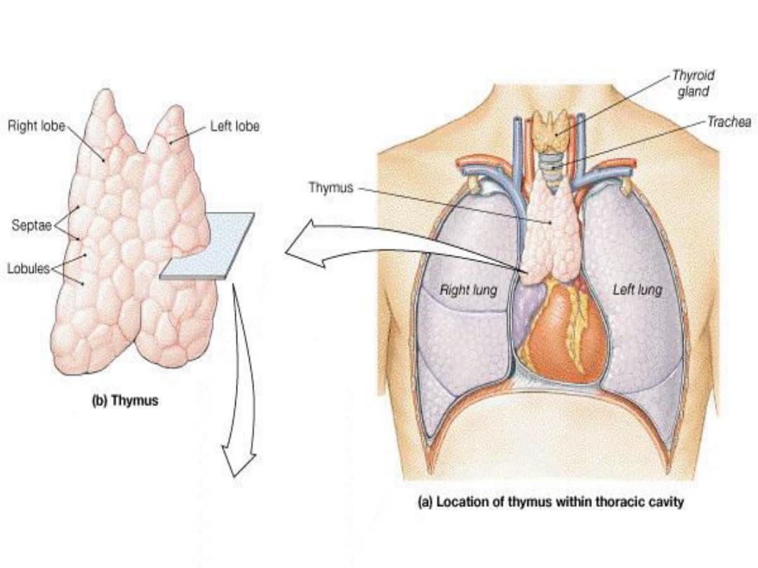

Structure:
The thymus consists of two
lobes joined by
connective tissue. A thin capsule of loosely
connective tissue surrounds each lobe and
provides
septa that extend into the thymus,
subdividing each lobe into a number of irregular
lobules.
Each lobule consists of :
-cortex ,
- medulla.
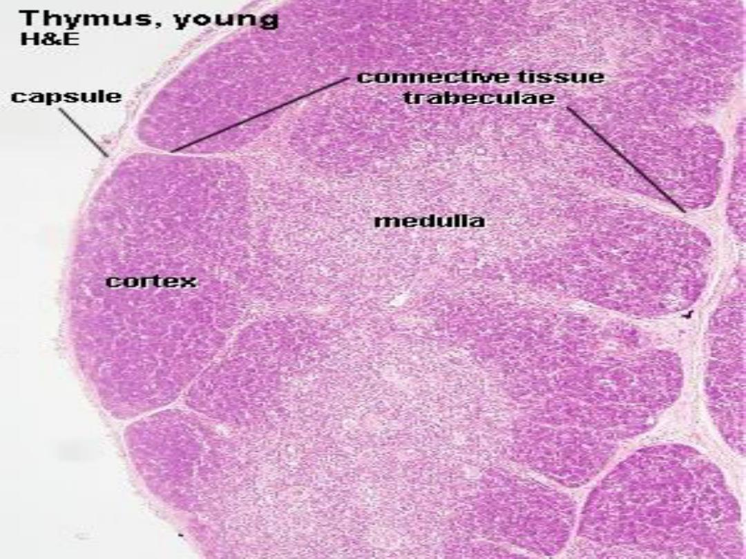
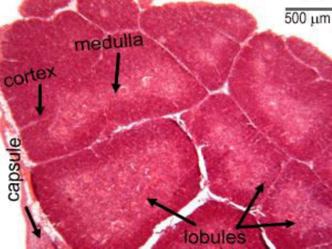

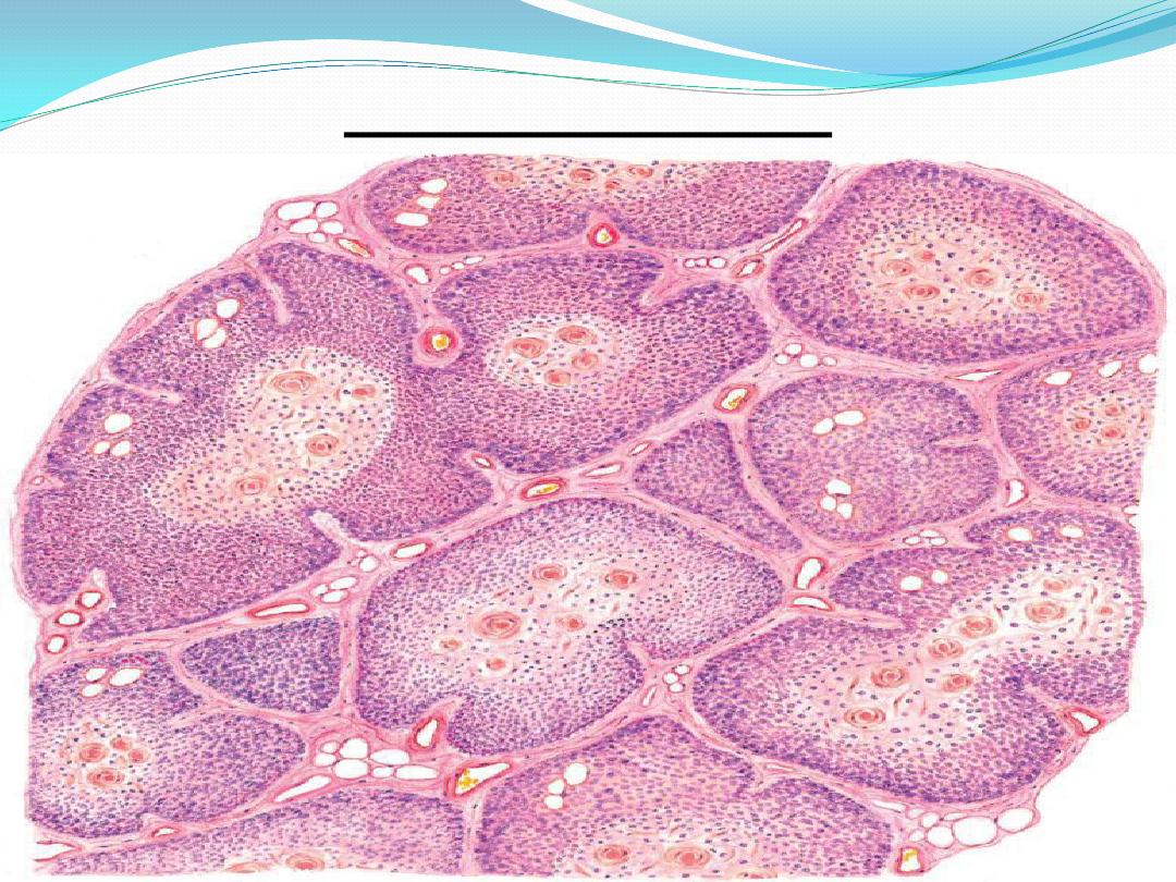
Cortex :
under the capsule is a dark-staining
cortex
with a network of interconnecting spaces. These
spaces become colonized by
immature
lymphocytes that migrate from hemopoietic
tissues to undergo maturation and
differentiation . The epithelial cells of the thymus
gland provide structural support for the increased
lymphocyte population.

Medulla :
it appears to be isolated within a lobule,
surrounded by a complete layer of cortex.
Lymphocytes are less numerous than in the
cortex, it appear lighter-staining
.
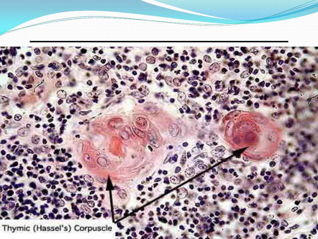
The epithelial cells form a coarser framework
that contains fewer lymphocytes and whorls of
epithelial cells that combine to form
thymic
(assall’s corpuscles, which are the
charecteristic feature of medulla of thymus
gland.

Hassall’s corpuscles
The
thymic (assall’s corpuscles are oval
structures consisting of round or spherical
aggregations (whorls) of flattened epithelial cells.
The thymic corpuscles also exhibit calcification
or degeneration centers
that stain pink or
eosinophilic
. The functional significance of these
corpuscles remains unknown.
Blood vessels and adipose cells are present in
both the thymic lobules and in trabecula .
Thymus gland
Thymic
Hassel’s corpuscle

Functional correlation
The thymus gland performs an important role
early in childhood in immune system
development
. Undifferentiated lymphocytes are
carried from bone marrow by the bloodstream
to
the thymus gland. In much of the thymic cortex,
the epithelial reticular cells, also called
thymic nurse cells, surround the lymphocytes
and promote their differentiation, proliferation
,
and maturation.

CONT.
As maturation progresses in the cortex,
the cells are presented by antigen-presenting cells
with self and foreign antigens.Lymphocytes that
are unable to recognize or that recognize self-
antigens die and are eliminated by macrophages
(negative selection), which is about 95% of the
total. Those lymphocytes that recognize the
foreign antigens (positive selection) reach
maturity, enter the medulla from the cortex, and
are then distributed in the bloodstream.

Cont.
the lymphocytes mature into
immunocompetent T cells, helperT cells, and
cytotoxic T cells, they acquire their surface
receptors for recognition of antigens.
After maturation, the T cells leave the thymus
gland via the bloodstream and populate the
lymph nodes, spleen, and other thymus-
dependent lymphatic tissues in the organism.

Lymph nodes
Lymph nodes are small encapsulated
lymphatic organs set in the course of lymphatic
vessels.
They are prominent in the neck, axilla, groin,
and mesenteries and along the course of large
blood vessels in the thorax and abdomen.
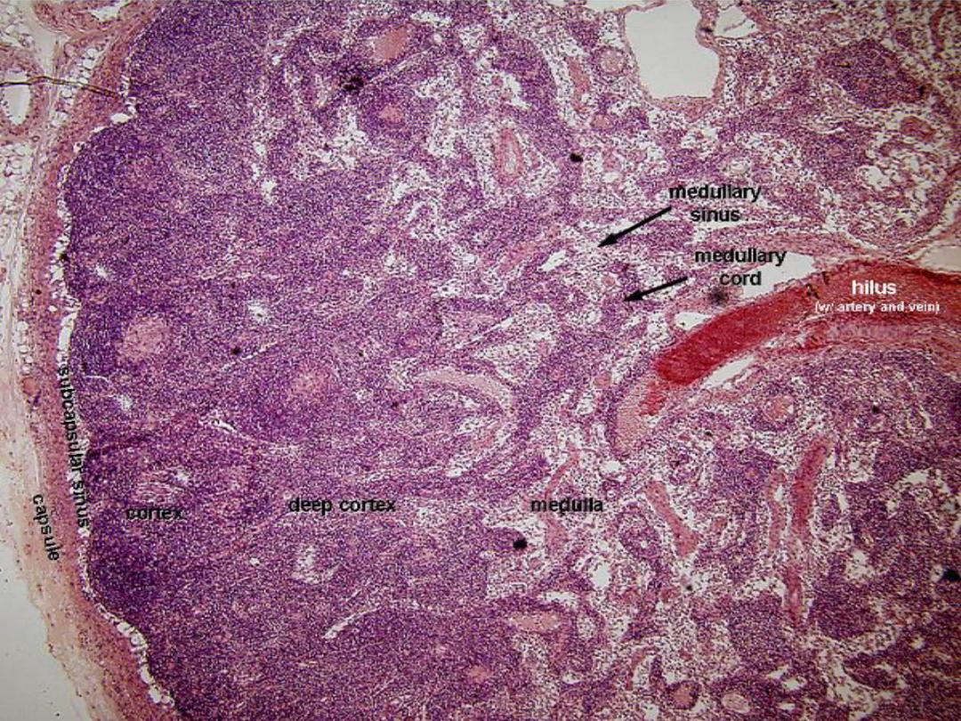
They appear as flattened, ovoid or bean-
shaped structures with a slight indentation at
one side, the
hilus, through which blood and
lymphatic vessels enter or leave.
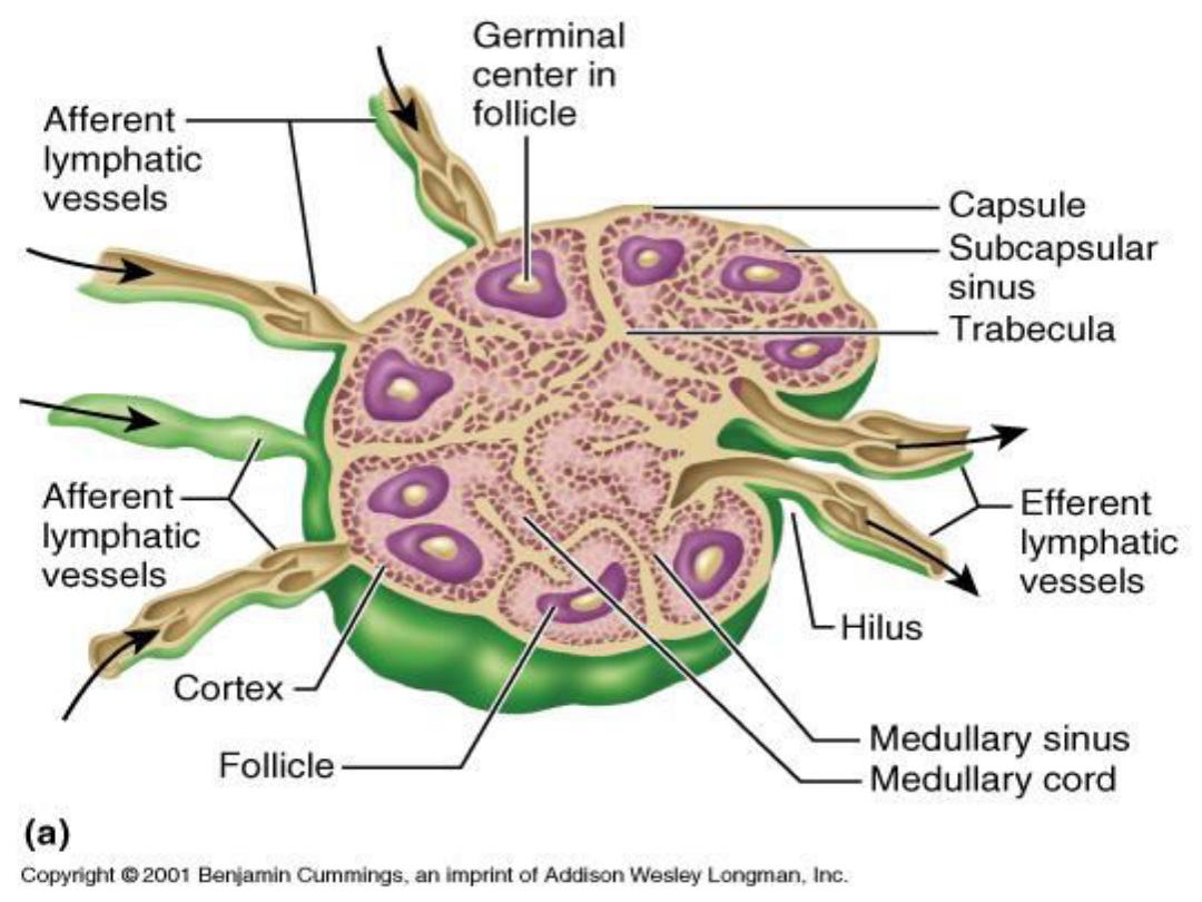

Cont.
Lymph nodes are the only lymphatic
structures that are set into the lymphatic
circulation and thus are the only lymphatic
organs to have afferent and efferent lymphatics.
Afferent lymphatics enter the node at multiple
sites
, anywhere over the convex surface; efferent
lymphatics leave the node at the hilus. Both sets
of
vessels have valves that allow unidirectional
flow of lymph through a node.
Lymph node
Lymph node

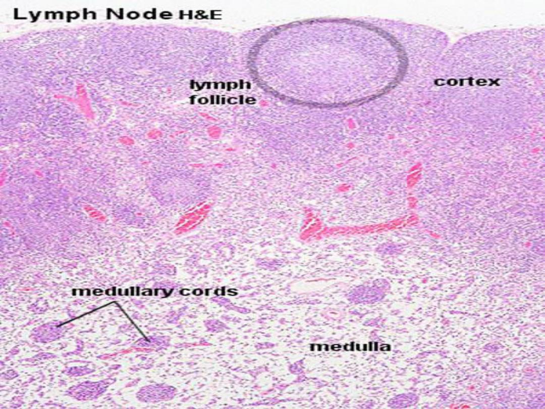
Structure:
lymph nodes consist of
diffuse and
nodular lymphatic tissue enclosed
in a capsule that is thick at the hilus. The
capsule consists of closely packed collagen
fibers, with few elastic fibers.
From the inner surface of the capsule,
trabeculae of dense connective tissue
extend into the node. Trabeculae
subdividing the cortex into several irregular
"compartments.

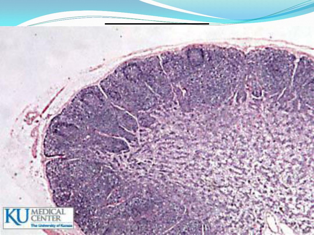
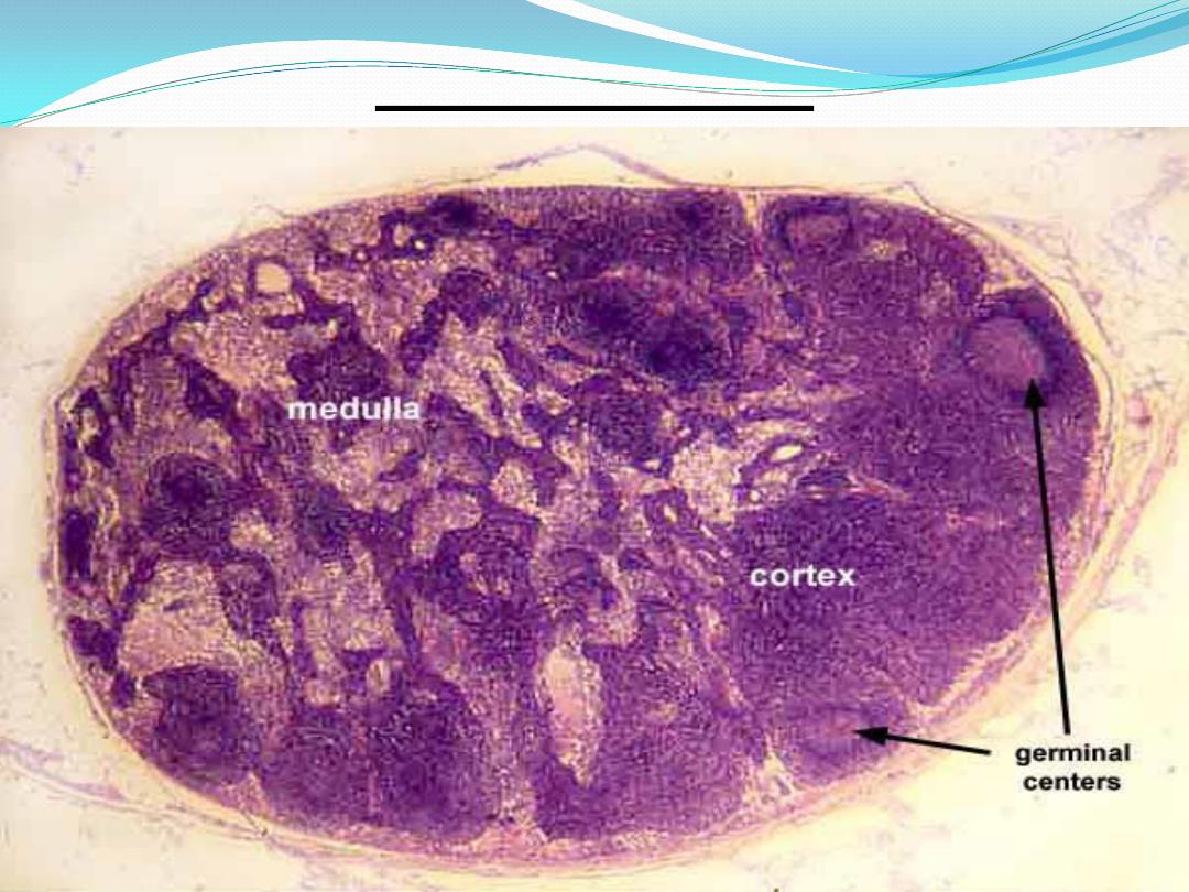
cortex
The cortex forms a layer under the capsule.
The cortex is divided into an
outer cortex
that lies under the capsule and contains nodular
and diffuse lymphatic tissue, and a
deep (inner)
cortex that consists of diffuse lymphatic tissue
only.
A network of reticular fibers and spherical,
aggregations of lymphocytes called
lymphoid
nodules characterize the cortex. Some of them
exhibit
germinal centers.

medulla
The medulla appears as a paler area of variable
width, surrounding the hilus . It consists of
diffuse lymphatic tissue arranged as irregular
medullary cords.
Medullary cords are networks of reticular fibers
filled with plasma cells, macrophages, and
lymphocytes separated by capillary-like channels
called
medullary sinuses.

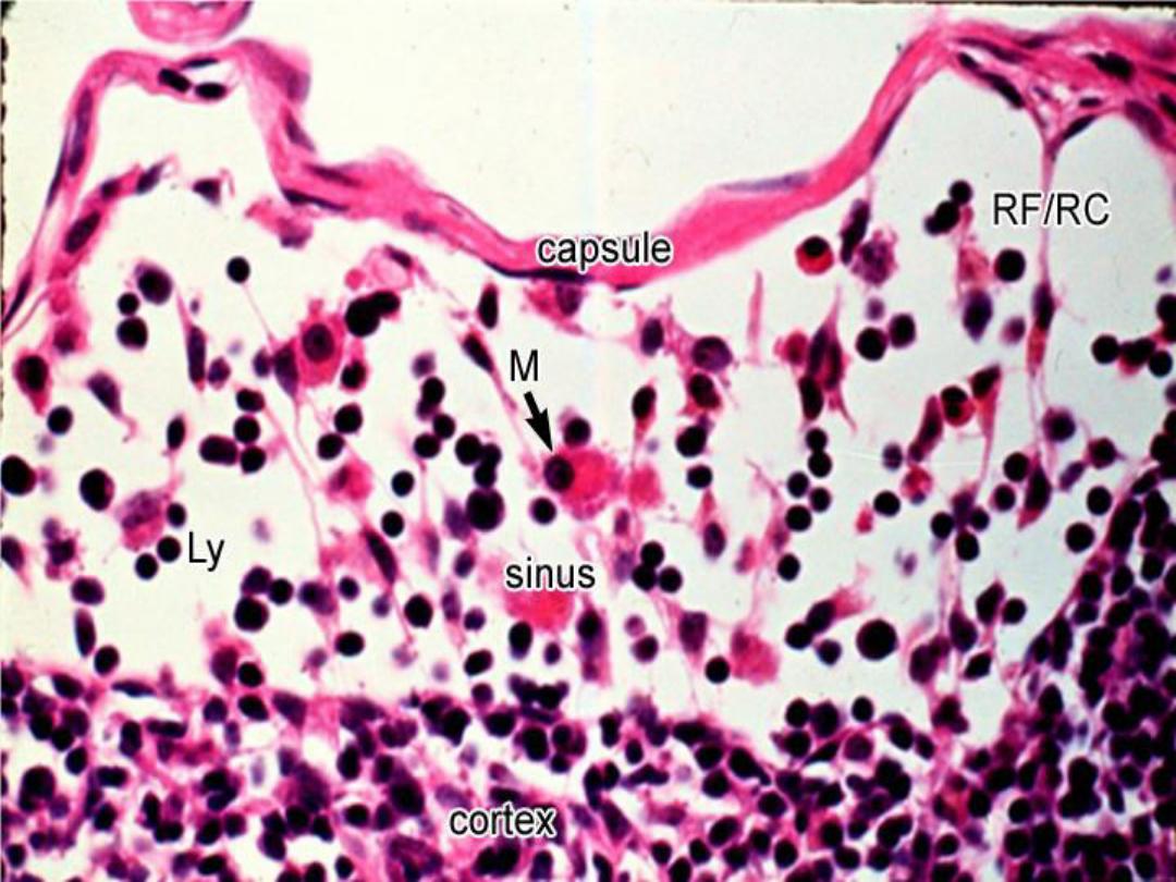
Lymph Sinuses
Within the lymph node is a system of channel-
like spaces, the lymph sinuses, through which
lymph percolates. Lymph enters the node
through afferent lymphatic vessels and empty
into the
subcapsular (marginal sinus) which
separates the cortex from the capsule. present as
a wide space extending beneath the capsule. It is
continuous with the
cortical (trabecular)
sinus. which extend into the cortex, usually
along the trabeculae.

Cont.
These become continuous with
medullary
sinuses that run between the medullary cords
and trabeculae of the medulla.
Sinuses in the cortex are less numerous than
in the medulla and narrow . They run in the
medullary parenchyma as irregular cordlike
arrangement .
Medullary sinus

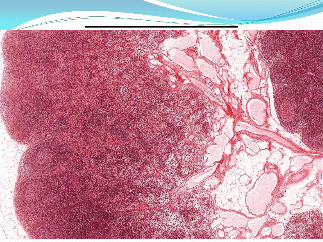
Spleen
The spleen is a large lymphoid organ with a
rich blood supply
.
The spleen is enclosed by a
capsule of dense connective tissue( fibro-elastic
connective tissue, some smooth muscle, and an
outer covering mesothelium . )
.
On the medial
surface of the spleen, the capsule is form a cleft-
like
hilus through which blood vessels, nerves,
and lymphatics enter or leave the spleen.
spleen

Cont.
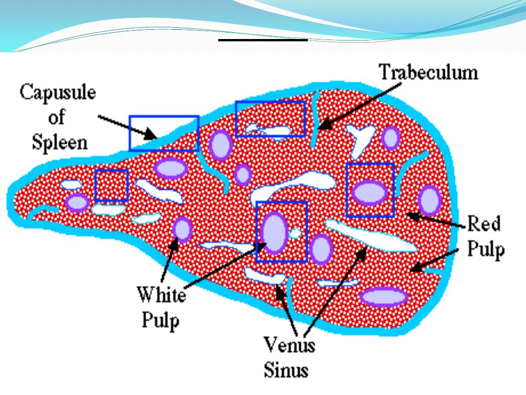
Broad bands of connective tissue, the
trabeculae, extend from the inner surface of the
capsule, the trabeculae subdivide the organ into
compartments. The main trabeculae enter the
spleen at the hilus and extend throughout the
organ. Located within the trabeculae are
trabecular arteries and trabecular veins .The
spaces between trabeculae are filled by a reticular
network of fibers and associated reticular cells.
The substance of the spleen is called the
splenic
pulp .

Cont.
the splenic pulp is consist of :
- the light areas form the
white pulp and
consist of diffuse and nodular lymphatic tissue.
- The dark red tissue is the
red pulp and consists
of diffuse lymphatic tissue that is suffused with
blood.
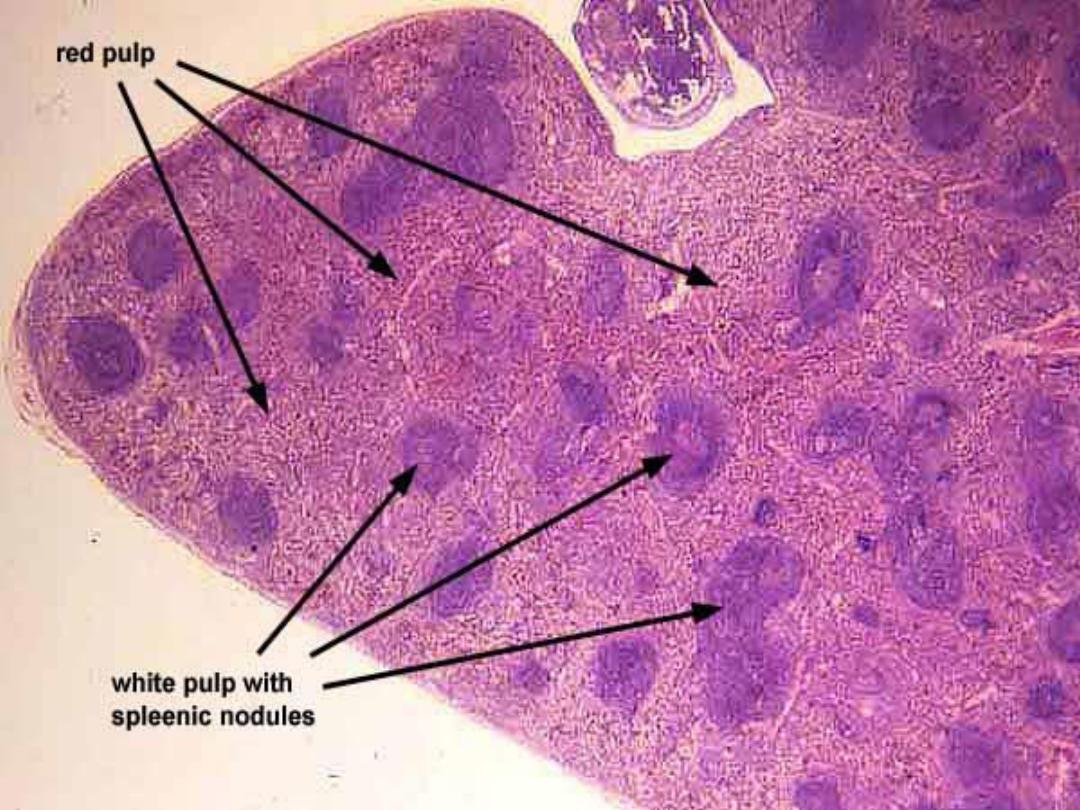

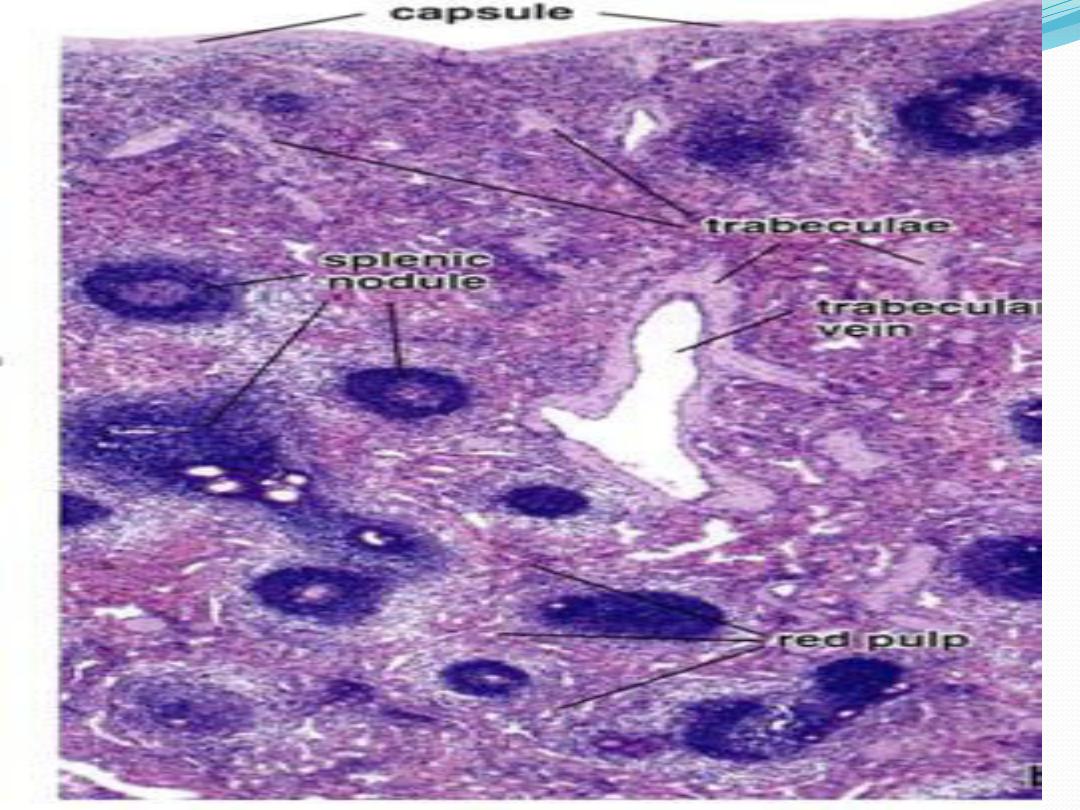
White pulp :
The spleen is characterized by numerous
aggregations of
lymphatic nodules , they
contain mainly
B cells. The lymphatic nodules
also contain
germinal centers that decrease in
number with age
. Passing through each
lymphatic nodule is a blood vessel called a
central artery that is located in the periphery of
the lymphatic nodules .

Cont.
Central arteries are branches of trabecular
arteries that become ensheathed with lymphatic
tissue as they leave the connective tissue
trabeculae .
The cells found in the periarterial lymphatic
sheath are mainly T cells.

Cont.
Antigen-presenting cells and
macrophages reside within the white pulp.
These cells detect trapped bacteria and antigens
and initiate immune responses against them.
As a result, T cells and B cells interact,
become activated, proliferate, and perform their
immune response.

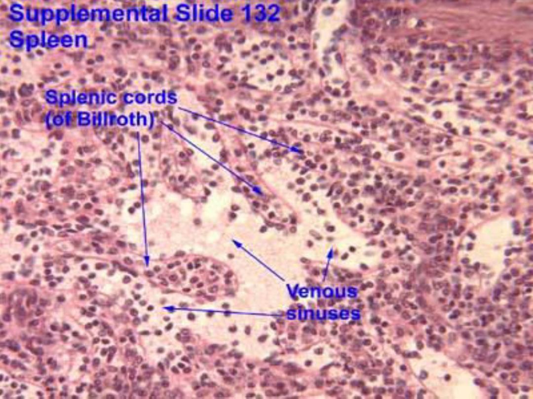
Red pulp :
red pulp is red because of its extensive vascular
tissue. The red pulp also contains
-
pulp arteries ,
- venous sinuses , and
- splenic cords (of Billroth) .
The splenic cords appear as diffuse strands of
lymphatic tissue between the venous sinuses and
form a spongy meshwork of reticular connective
tissue.

Cont.
They are thin aggregations of lymphatic tissue
containing small lymphocytes, associated cells,
and various blood cells.
Venous sinuses are dilated vessels lined with
modified endothelium of elongated cells .
pulp arteries represent the branches of the
central artery after it leaves the lymphatic nodule
. Capillaries and pulp (venules) are also present.
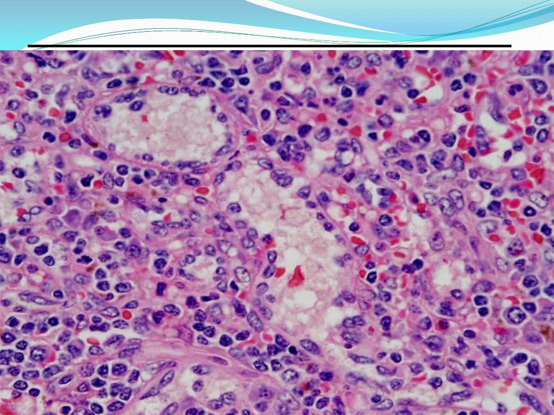
Venous sinuses &splenic cords

Cont.
The main function of the red pulp is to filter
the blood. It removes antigens, microorganisms,
platelets, and aged or abnormal erythrocytes
from the blood
.
The spleen does not exhibit a distinct cortex
and a medulla, as seen in lymph nodes. However,
lymphatic nodules are found throughout the
spleen
.
In addition, the spleen contains venous
sinuses , in contrast to lymphatic sinuses that are
found in the lymph nodes.

Cont.
The spleen also does not exhibit subcapsular
or trabecular sinuses. The capsule and trabeculae
in the spleen are thicker than those around the
lymph nodes .

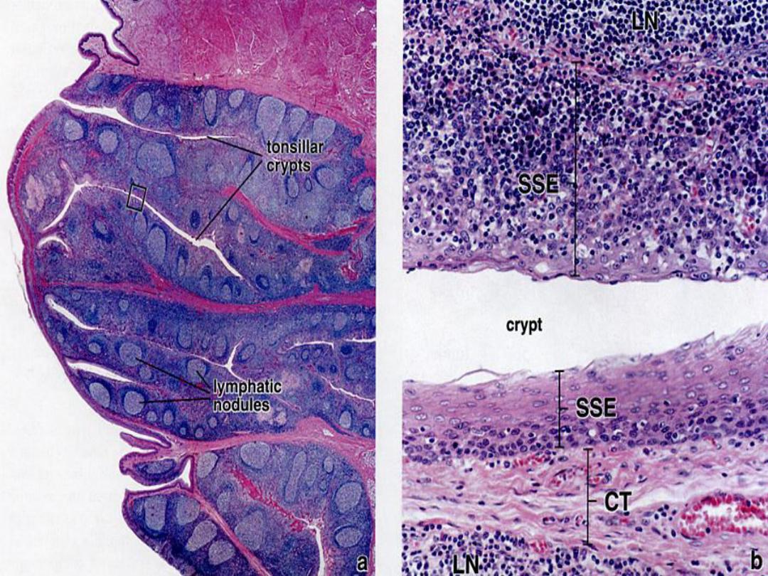
Tonsils :
Tonsils are aggregates of lymphatic nodules
associated with the pharynx and oropharynx.
These structures are spread through different
areas - oropharynx, nasopharynx, and tongue -
and form the
1- palatine,
2- pharyngeal, and
3-
lingual tonsils .

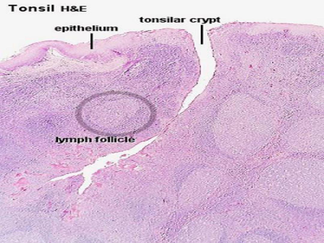
palatine tonsils
The palatine tonsils are paired, oval lymphatic
organs located laterally at the junction of the oral
cavity and oropharynx.
A
stratified squamous epithelium covers
the free surface of the tonsil and is very closely
associated with the lymphatic tissue. Deep
invaginations of the epithelium form the tonsillar
crypts that reach almost to the base of the tonsil.

Cont.
Lymphatic nodules, many with germinal
centers, usually are arranged in a single layer
beneath the epithelium, embedded in a mass of
diffuse lymphatic tissue. A
partial capsule
beneath the basal surface of the tonsil separates it
from surrounding structures . Septa of loose
collagen fibers extend from the capsule
into the tonsillar tissue and partially divide the
crypts and their lymphatic tissue from one
another.

Cont.
The connective tissue is infiltrated by
lymphocytes , plasma cells, and mast cells.
Neutrophil may be present and are numerous
during inflammation of the tonsils
.

Lingual Tonsils:
The lingual tonsils form nodular bulges in
the root of the tongue, and their general structure
is similar to that of the palatine tonsil.
Crypts
are deep, and are lined by
stratified squamous
epithelium that invaginates from the surface.
The associated lymphatic tissue consists of
diffuse and nodular types.
Lingual tonsil

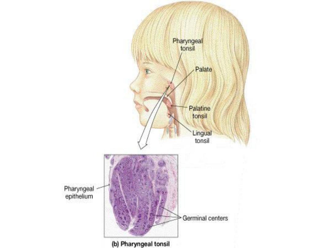
Pharyngeal Tonsil :
The pharyngeal tonsil is located on the posterior
wall of the nasopharynx. Its surface epithelium is
a
ciliated pseudostratified columnar
epithelium that contains goblet cells. Patches of
stratified squamous epithelium may be present .
crypts are not as deep as in the palatine tonsils . A
thin capsule separates the pharyngeal tonsil from
underlying tissues and provides fine septa that
extend into the substance of the tonsil.

Mucosa associated lymphoid
tissue (MALT) :
can be found in various locations of digestive
or respiratory systems. Lymphatic nodules are
always located within the connective tissue
under the lining epithelium (lamina propria
).
The
nodules are prominent by their size and
deep blue staining of nuclei of lymphocytes, the
germinal centers of these nodules contain
some
developing plasma cells and supporting c.t.
cells.
MALT

Thank you