
LIVER,
GALLBLADDER, AND
PANCREAS
by
Dr. Suhair Majeed

The liver, gallbladder, and pancreas are
accessory organs of the digestive tract that deliver
their secretory products to the small intestine by
excretory ducts.
The common bile duct from the liver and the
main pancreatic duct from the pancreas join in
the duodenal loop to form a single duct common
to both organs.

CONT.
This duct then penetrates the duodenal wall and
enters the lumen of the small intestine. The
gallbladder joins the common bile duct via the
cystic duct. Thus, bile from the gallbladder and
digestive enzymes from the pancreas enter the
duodenum via a common duct.

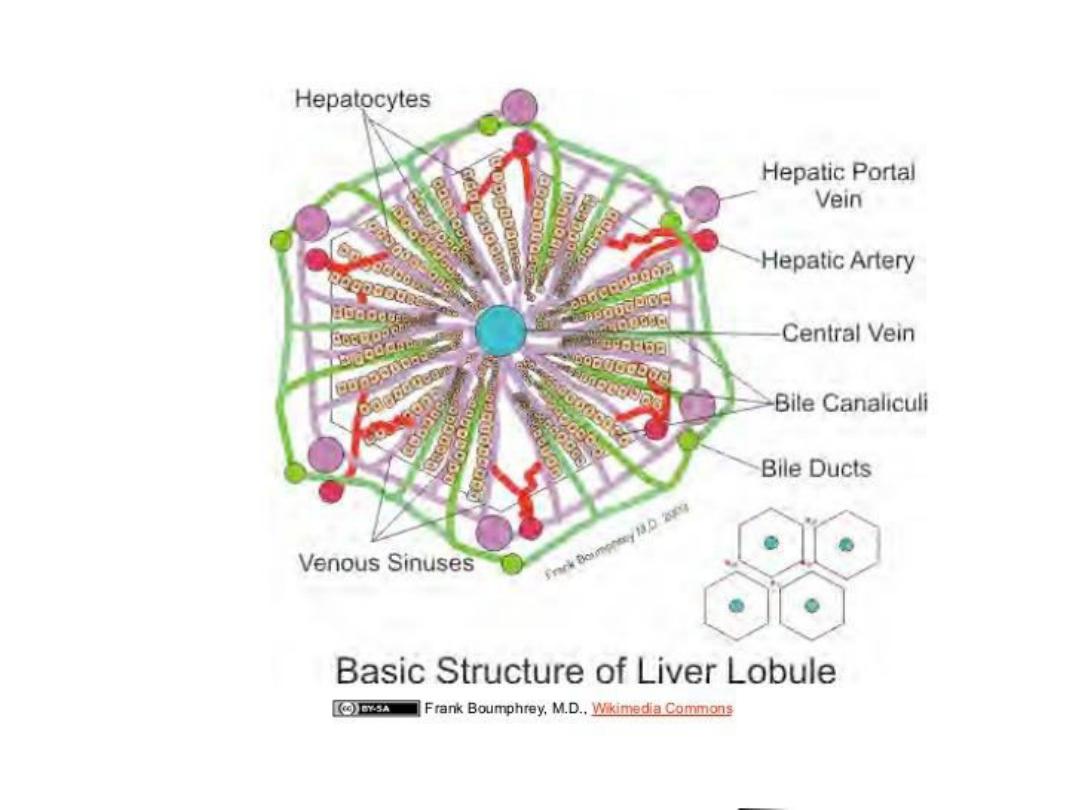
1- LIVER
The liver is invested by a delicate connective
tissue capsule . The capsule contains numerous
elastic fibers and is covered by a mesothelium .
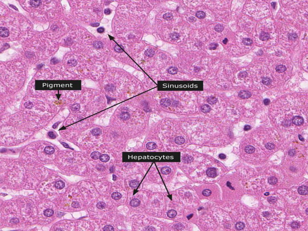
The liver is composed of epithelial cells, the
hepatocytes, arranged in branching and
anastomosing plates separated by blood
sinusoids. Both form a radial pattern about a
central vein.
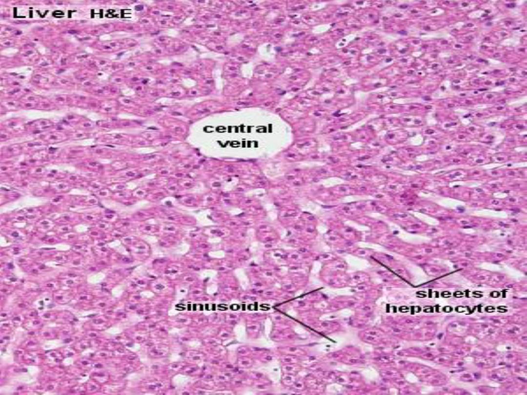

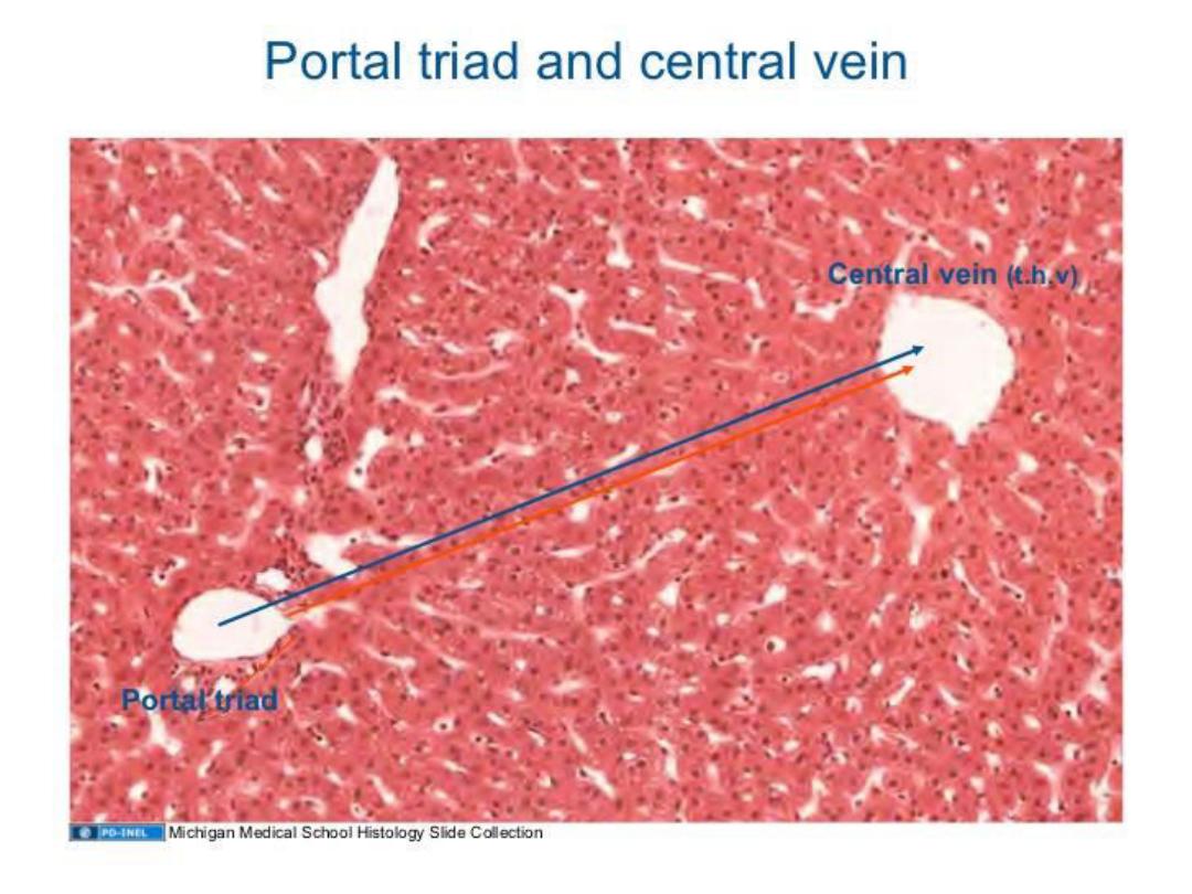
CONT.
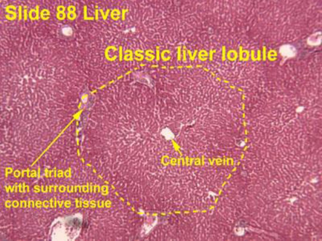
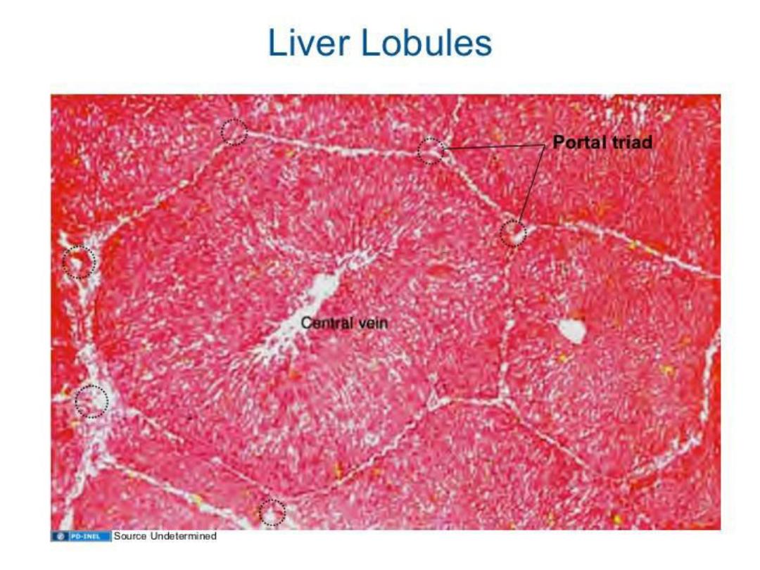
The spokelike arrangement of hepatic
plates about a central vein constitutes the basis
of the classic hepatic lobule, which appears
hexagonal in cross section, with a central vein at
the center and portal areas at the corners.

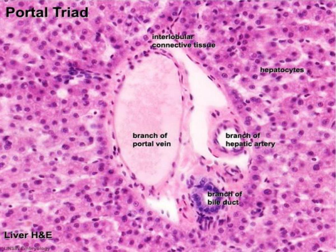
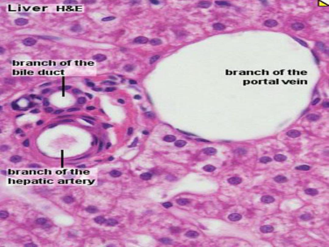
CONT.
A portal area contains :
- a branch of the portal vein,
-a branch of the hepatic artery,
-a bile duct,
- a lymphatic channel.
All are enclosed in a common investment of
connective tissue .
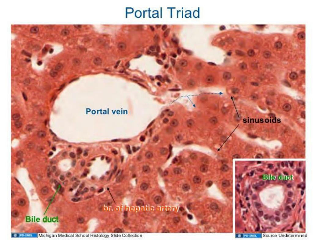

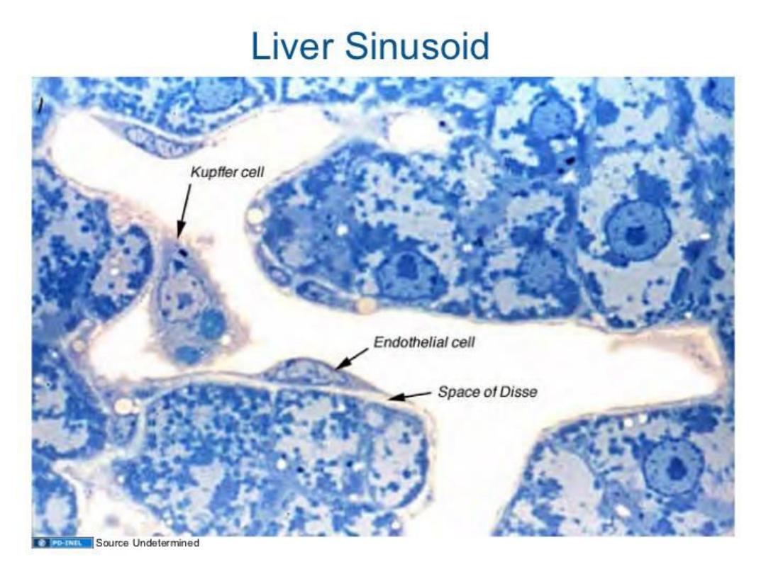
HEPATIC SINUSOIDS
Hepatic sinusoids are larger and more
irregular in shape than ordinary capillaries. The
sinusoidal lining consists of a simple layer of
squamous epithelium supported by connective
tissue.
Three types of cells are associated with the
sinusoidal lining: endothelial cells, stellate cells
(Kupffer cells or hepatic macrophages), and fat-
storing cells (lipocytes).

CONT.
The sinusoidal lining is separated from the liver
cells by a narrow perisinusoidal space (of Disse) .
the perisinusoidal space has considerable
significance in the exchange of materials between
the liver and plasma.

HEPATOCYTES
The parenchyma of the liver consists of large
polyhedral hepatocytes arranged in plates that
radiate from the region of the central vein. The
surfaces of an individual hepatocyte either
contact an adjacent liver cell or border on a
perisinusoidal space. This latter surface bears
numerous well developed microvilli. The nuclei of
hepatocytes are large and round and occupy the
center of the cell.

CONT.
Inclusions such as glycogen and lipid are
common in the cytoplasm.
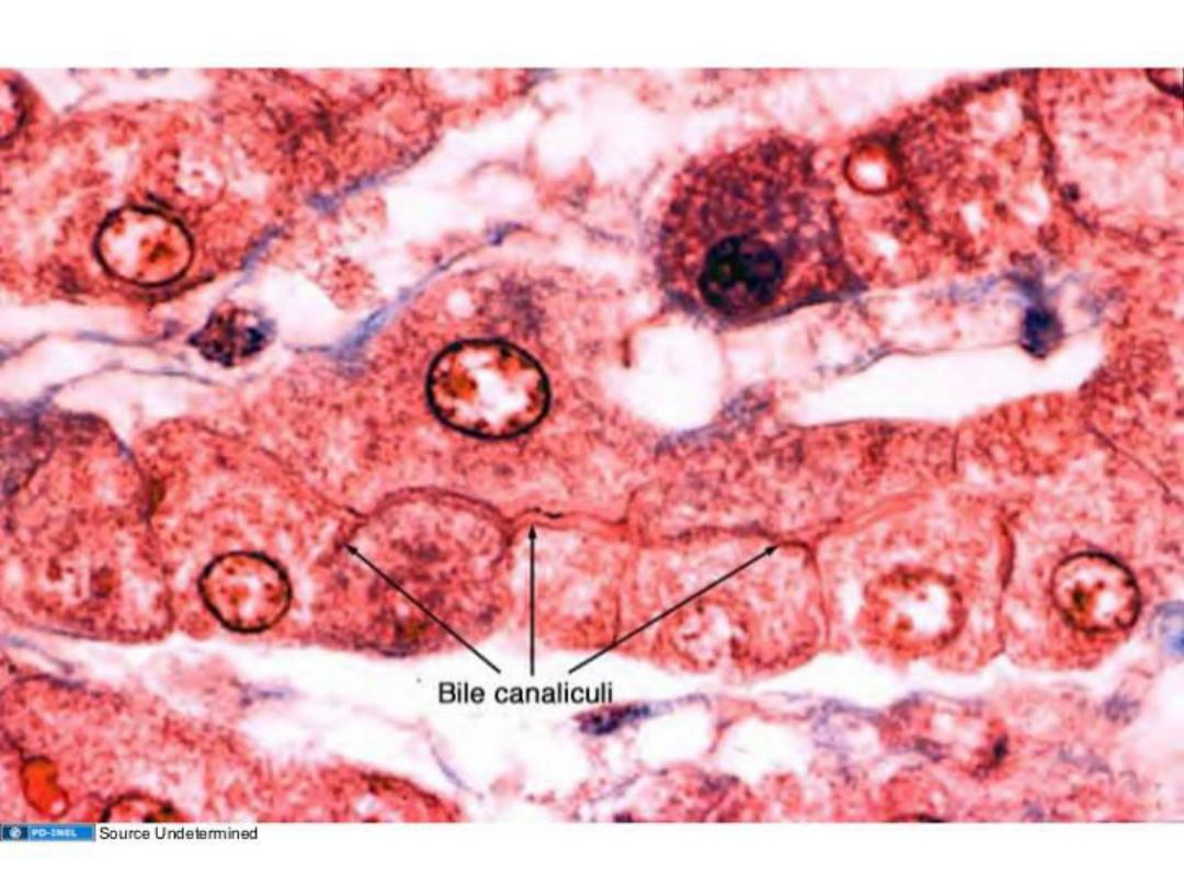
Hepatocytes secrete bile into tiny channels
called bile canaliculi located between individual
hepatocytes.

BILE DUCTS
Bile canaliculi unite with bile ducts in the portal
canals by small, interconnecting channels called
bile ductules. They are small and have thin walls,
lined by a low cuboidal epithelium.
The terminal ductules empty into interlobular
bile ducts of the portal areas. Interlobular ducts
unite to form the extrahepatic ducts.

CONT.
Two large extrahepatic ducts, the left and right
hepatic ducts, exit the lobes of the liver and unite
to form the major excretory duct of the liver, the
common hepatic duct. It is joined by the cystic
duct from the gallbladder to form the common bile
duct, which empties into the duodenum.
The major extrahepatic ducts are lined by a tall
columnar, mucus-secreting epithelium.

FUNCTIONAL CORRELATIONS:
LIVER :
The liver performs hundreds of functions.
Hepatocytes perform more functions than any
other cell in the body, and perform both endocrine
and exocrine roles.
- Exocrine Functions
-Endocrine Functions

1- EXOCRINE FUNCTIONS:
One major exocrine function of hepatocytes is
to synthesize and release 500 to 1,200 mL of
bile into the bile canaliculi per day. From these
canaliculi, bile flows through a system of ductules
and ducts to enter the gallbladder, where it is
stored and concentrated by removal of water.
Release of bile from the liver and gall bladder is
primarily regulated by hormones.

CONT.
Bile flow is increased when a hormone such as
cholecystokinin is released by the mucosal
enteroendocrine cells, stimulated when dietary
fats in the chyme enter the duodenum. This
hormone causes contraction of smooth muscles
in the gallbladder wall and relaxation of the
sphincter, allowing the bile to enter the
duodenum.

CONT.
Bile salts in the bile emulsify fats in the
duodenum. This process allows for more efficient
digestion of fats by the fat-digesting pancreatic
lipases produced by the pancreas.
The digested fats are subsequently absorbed
by cells in the small intestine and enter the blind-
ending lymphatic lacteal channels located in
individual villi. From the lacteals, fats are carried
into larger lymphatic ducts that drain into the
major veins.

CONT.
Hepatocytes also excrete bilirubin, a toxic
chemical formed in the body after degradation of
worn-out erythrocytes by liver macrophages,
(Kupffer cells ). Bilirubin is taken up by
hepatocytes from the blood and excreted into bile.

CONT.
Hepatocytes also have an important role in
the immune system.
Antibodies produced by plasma cells in the
intestinal lamina propria are taken from blood
by hepatocytes and transported into bile
canaliculi and bile. From here, antibodies enter
the intestinal lumen, where they control the
intestinal bacterial flora.

2- ENDOCRINE FUNCTIONS :
Hepatocytes are also endocrine cells. The
arrangement of hepatocytes in a liver lobule
allows them to take up, metabolize, accumulate,
and store numerous products from the blood.
Hepatocytes then release many of the
metabolized or secreted products back into the
bloodstream, as the blood flows through the
sinusoids and comes in direct contact with
individual hepatocytes.

CONT.
The endocrine functions of the liver hepatocytes
involve synthesis of numerous plasma proteins,
including albumin and the blood-clotting factors
prothrombin and fibrinogen. The liver also stores
fats, various vitamins, and carbohydrates as
glycogen.
When the cells of the body need glucose,
glycogen that is stored in the liver is converted
back into glucose and released into the
bloodstream.

CONT.
Hepatocytes also detoxify the blood of drugs
and harmful substances as it percolates through
the sinusoids. Kupffer cells in the sinusoids are
specialized liver phagocytes derived from blood
monocytes. These large, branching cells filter and
phagocytose particulate material, cellular debris,
and worn-out or damaged erythrocytes that flow
through the sinusoids.
The liver also performs vital functions early in
life. In the fetus, the liver is the site of
hemopoiesis.

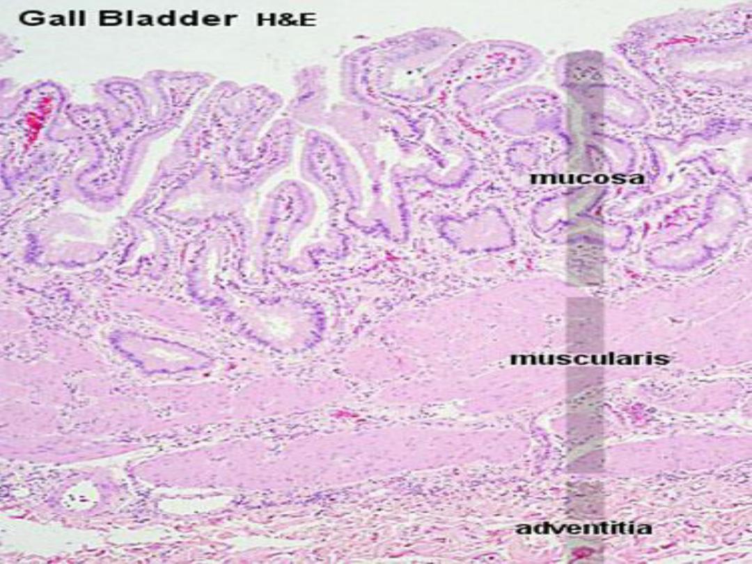
2- GALLBLADDER
The gallbladder is a saclike structure on the
inferior surface of the liver, It is joined to the
common hepatic duct by the cystic duct, whose
mucous membrane forms prominent spiraling
folds that contain bundles of smooth muscle.
These folds make up the spiral valve that prevents
the collapse or distention of the cystic duct during
sudden changes in pressure.

CONT.
The wall of the gallbladder consists of a mucous
membrane, a muscularis, and a serosa or
adventitia.
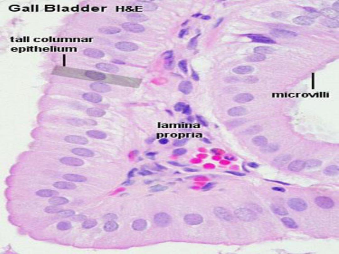
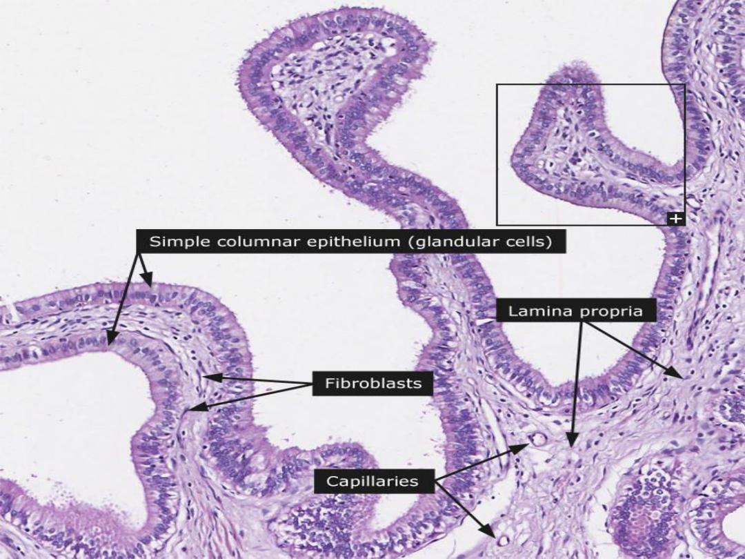
The mucous membrane of the gallbladder wall
consists of a simple columnar epithelium and an
underlying lamina propria. The oval nuclei are
located basally in the cells and the luminal
surfaces show numerous short microvilli .

CONT.
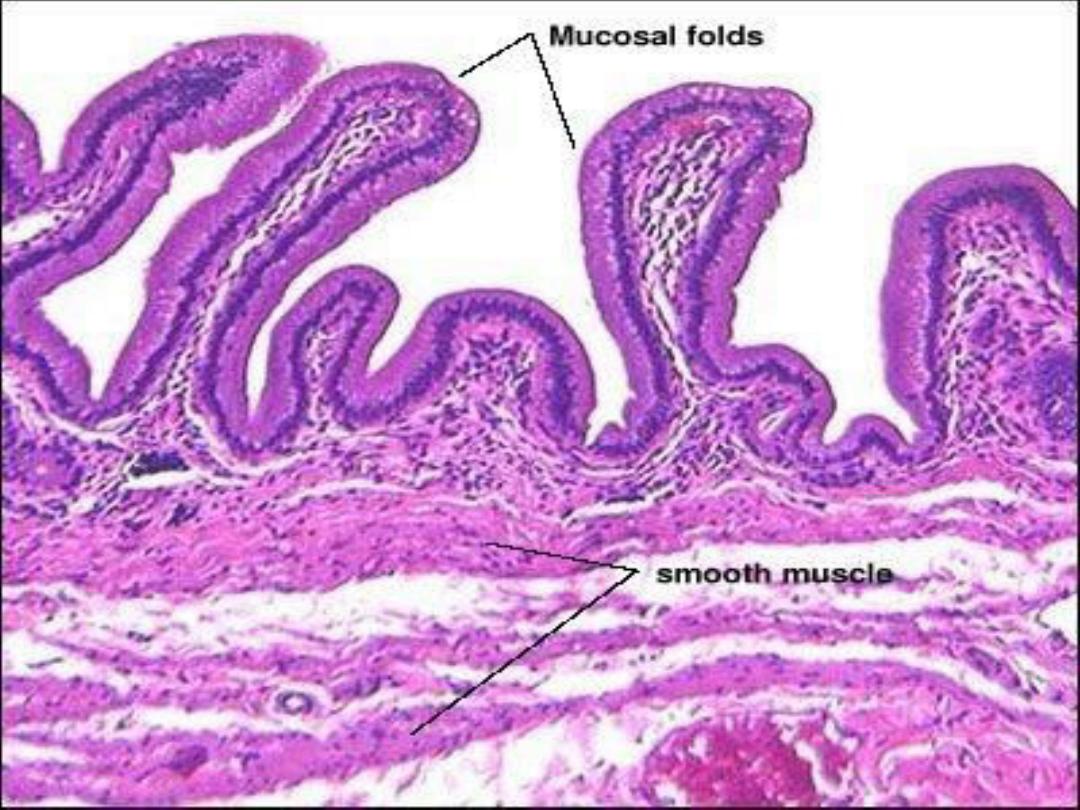
The mucosa of the nondistended gallbladder
forms large irregular folds called rugae, which
flatten out as the gallbladder fills with bile.
There is no submucosa in the gallbladder. The
muscularis consists of bundles of smooth muscle
that spiral around the lumen of the gallbladder.
The smooth muscle cells contain numerous
receptors for cholecystokinin.
.

CONT.
Gaps between the smooth muscle bundles
are filled with reticular, and elastic fibers.
Because of the musculoelastic wall and the
rugae, gallbladder has the capacity for
distention

CONT.
The surrounding fibroconnective tissue of the
adventitia is dense and is continuous with the
connective tissue of the liver capsule.
The gallbladder stores and concentrates bile,
which is elaborated continuously by the liver.
On stimulation by cholecystokinin, gallbladder
wall contracts and the sphincters of the common
bile duct and ampulla relax, allowing bile to be
released into the duodenum.

FUNCTIONAL CORRELATIONS:
THE GALLBLADDER :
The primary functions of the gallbladder are to
collect, store, concentrate, and expel bile when
it is needed for emulsification of fat. Bile is
continually produced by liver hepatocytes and
transported via the excretory ducts to the
gallbladder for storage. Here, sodium is actively
transported through the simple columnar
epithelium of the gallbladder into the extracellular
connective tissue, creating a strong osmotic
pressure.

CONT.
Release of bile into the duodenum is under
hormonal control. In response to the entrance of
dietary fats into the proximal duodenum, the
hormone cholecystokinin (CCK) is released into
the bloodstream by enteroendocrine cells located
in the intestinal mucosa. CCK is carried in the
bloodstream to the gallbladder, where it causes
strong rhythmic contractions of the smooth
muscle in its wall.

CONT.
Water and chloride ions passively follow,
producing concentrated bile.
At the same time, the smooth sphincter
muscles around the neck of gallbladder relax.
The combination of these two actions forces
the bile into the duodenum via the common
bile duct.

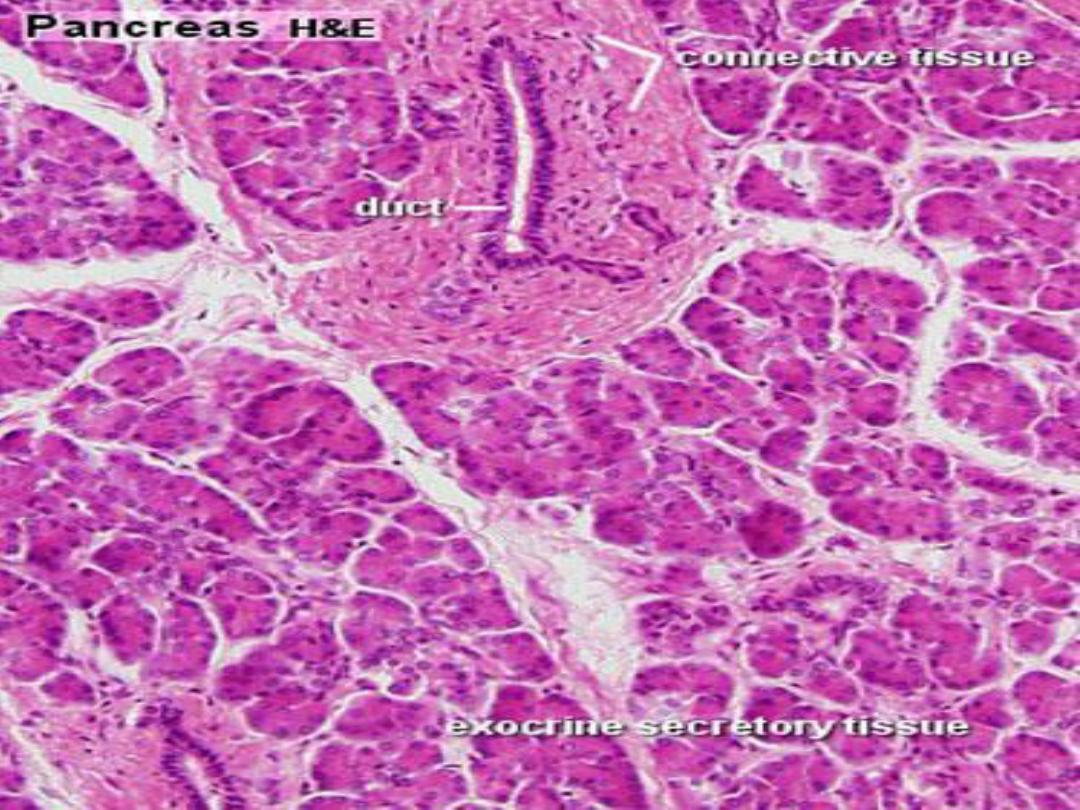
3- PANCREAS
The pancreas is a soft, elongated organ
located posterior to the stomach. The head of the
pancreas lies in the duodenal loop and the tail
extends across the abdominal cavity to the
spleen.
It lacks a definite capsule but is covered by a
thin layer of connective tissue that extends
delicate septa into the substance of the pancreas
and subdivides it into numerous small lobules.
Blood vessels, nerves, lymphatics, and excretory
ducts course through the septa.
.

CONT.
The pancreas consists of an exocrine portion,
which elaborates numerous digestive enzymes
and bicarbonate, and an endocrine portion,
whose secretions are important in carbohydrate
metabolism. Unlike the liver, the exocrine and
endocrine functions of the pancreas are
performed by different groups of cells.
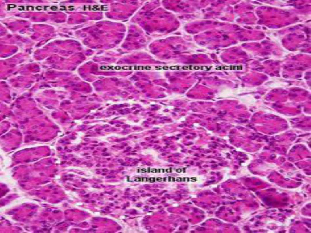

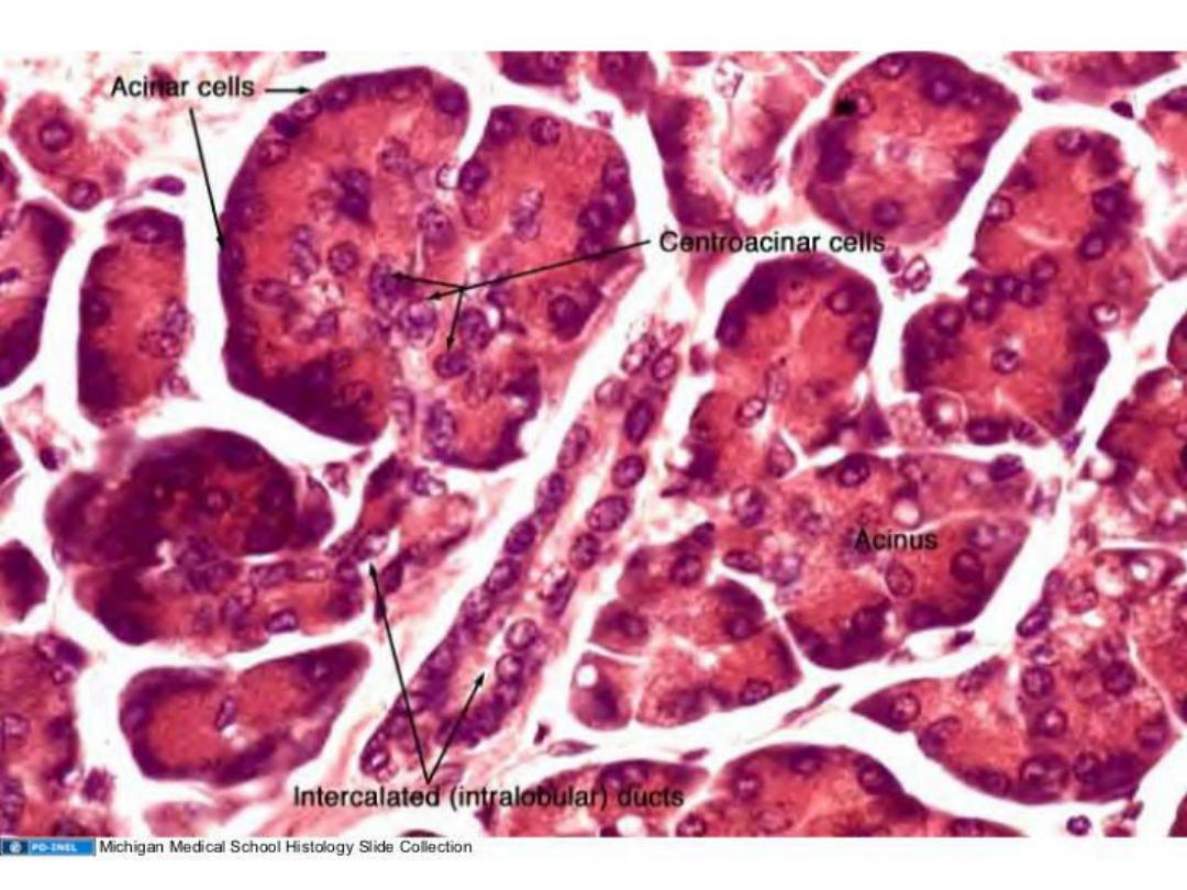
1- EXOCRINE PANCREAS :
Most of the pancreas is an exocrine gland. The
exocrine secretory units or acini , each acinus
consists of a single pyramidal cells whose narrow
apices border on a lumen, while their broad
bases lie on a thin basement membrane. whose
apices are filled with secretory granules.
These granules contain the precursors of
several pancreatic digestive enzymes that are
secreted into the excretory ducts in an inactive
form.
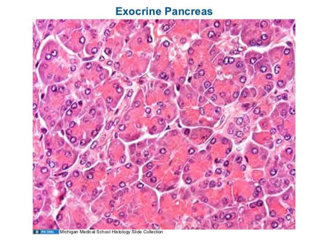

CONT.
The secretory acini are subdivided into
lobules and bound together by loose connective
tissue.
The excretory ducts in the exocrine pancreas
start from within the center of individual acini as
pale-staining centroacinar cells, which continue
into the short intercalated ducts.


DUCT SYSTEM
An extensive duct system permeates the
pancreas. At their beginnings, the ducts extend
into the acini and are interposed between the
acinar cells and the lumen.
Ductal cells within the pancreatic acini are
called centroacinar cells and appear as flattened,
cells. The wall of the duct system formed by
centroacinar cells is continuous outside the
secretory unit with intercalated and
intralobular ducts
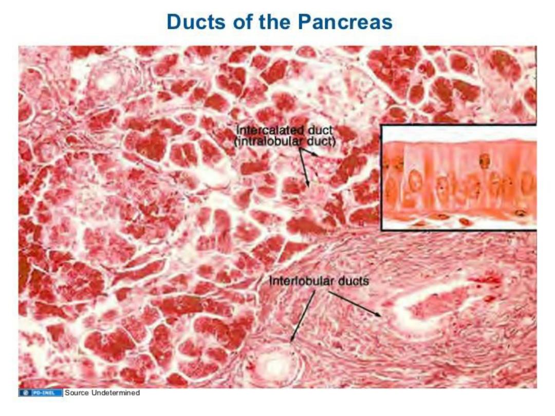

CONT.
These ducts are tributaries of the interlobular
ducts found in the loose connective tissue
between lobules; the transition between ducts is
gradual. The epithelial lining is simple squamous
in the intercalated ducts, to cuboidal in the
intralobular ducts, and columnar in the
interlobular ducts.
The interlobular ducts drain into the primary
and accessory pancreatic ducts.

CONT.
The primary duct runs the length of the
pancreas, increasing in size near the duodenum,
where it runs parallel to the common bile duct,
with which it often shares a common opening at
the greater duodenal papilla .
The intercalated ducts merge to form
intralobular ducts in the connective tissue, which,
in turn, join to form larger interlobular ducts that
empty into the main pancreatic duct.
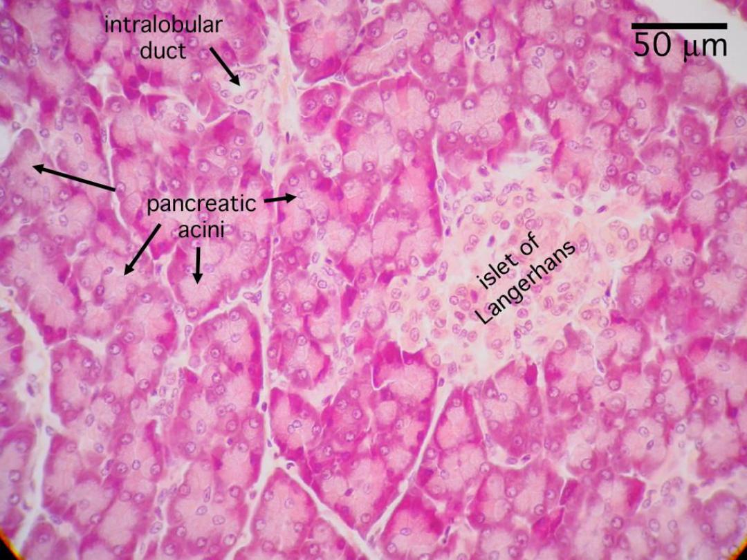

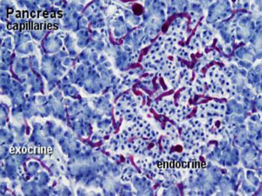
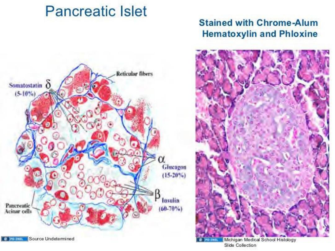
2- ENDOCRINE PANCREAS :
The endocrine units of the pancreas are
scattered among the exocrine acini as isolated,
pale-staining vascularized units called pancreatic
islets (of Langerhans). Each islet is surrounded by
fine fibers of reticular connective tissue, four cell
types can be identified in each pancreatic islet:
alpha, beta, delta, and pancreatic polypeptide
(PP) cells.
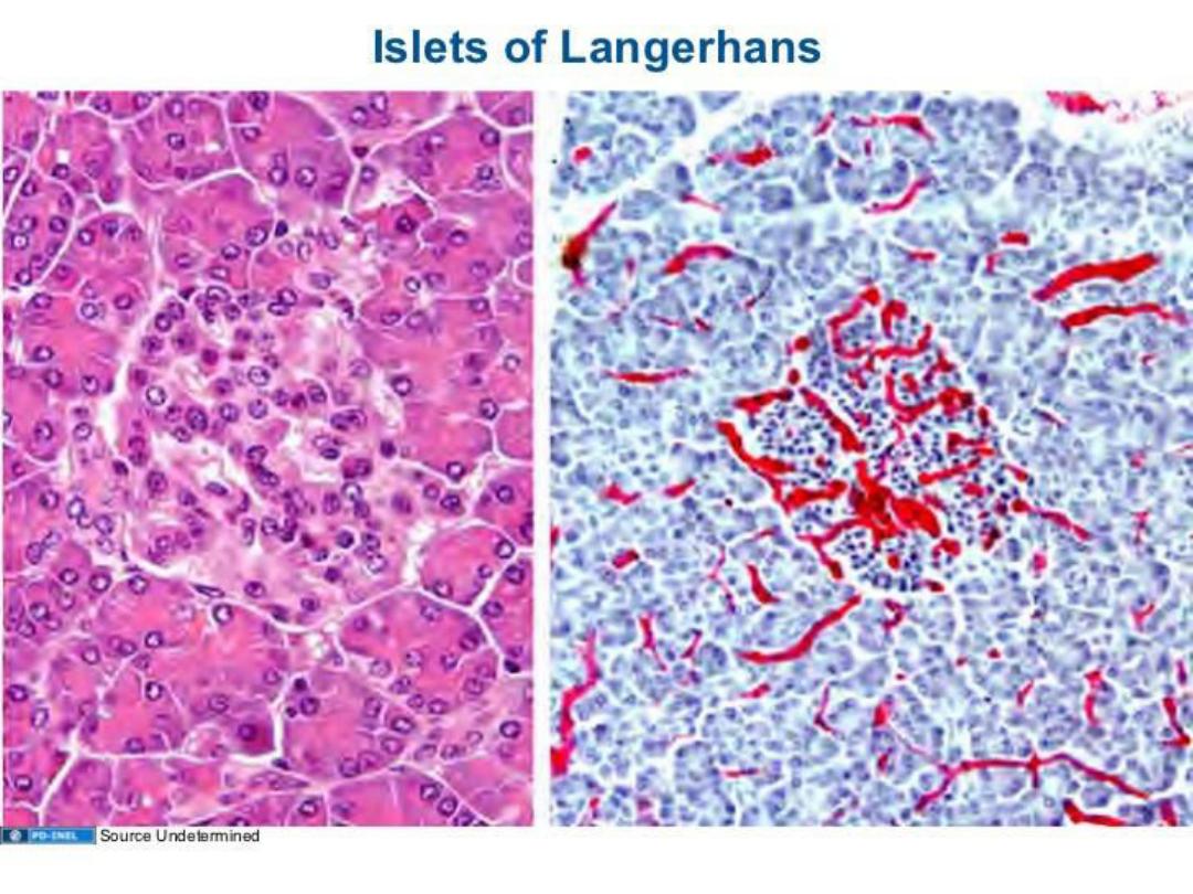

CONT.
Alpha cells constitute about 20% of the islets
and are located primarily around the islet
periphery.
beta cells are most numerous, constituting
about 70% of the islet cells, and are primarily
concentrated in the center of the islet. The
remaining cell types are few in number and are
located in various places throughout the islets.

FUNCTIONAL CORRELATIONS:
EXOCRINE PANCREAS
The exocrine and endocrine functions of the
pancreas are performed by separate exocrine and
endocrine cells. The pancreas produces
numerous digestive enzymes that exit the gland
through a major excretory duct, whereas the
different hormones are transported via blood
vessels. Both hormones and vagal stimulation
regulate pancreatic exocrine secretions

CONT.
Two intestinal hormones, secretin and
cholecystokinin (CCK), secreted by the
enteroendocrine cells in the duodenal mucosa
into the bloodstream, regulate pancreatic
secretions.
The release of the hormone secretin
stimulates exocrine pancreatic cells to produce
large amounts of a watery fluid rich in sodium
bicarbonate ions.

CONT.
This fluid, which has little or no enzymatic
activity, is primarily produced by centroacinar cells
in the acini and by cells that line the smaller
intercalated ducts. The main function of this
bicarbonate fluid is to neutralize the acidic chyme,
stop the action of pepsin from the stomach, and
create a neutral pH in the duodenum for the
action of the digestive pancreatic enzymes.

CONT.
In response to the presence of fats and
proteins in the small intestine, CCK is released
into the bloodstream. CCK stimulates the acinar
cells in the pancreas to secrete large amounts of
digestive enzymes: pancreatic amylase for
carbohydrate digestion, pancreatic lipase for lipid
digestion, deoxyribonuclease and ribonuclease for
digestion of nucleic acids, and the proteolytic
enzymes trypsinogen, chymotrypsinogen, and
procarboxypeptidase.

CONT.
Pancreatic enzymes are first produced in the
acinar cells in an inactive form and are only
activated in the duodenum by the hormone
enterokinase secreted by the intestinal mucosa.
This hormone converts trypsinogen to trypsin,
which then converts all other pancreatic
enzymes into active digestive enzymes.

FUNCTIONAL CORRELATIONS:
ENDOCRINE PANCREAS
Pancreatic islets secrete two major hormones
that regulate blood glucose levels and glucose
metabolism.
Alpha cells in the pancreatic islets produce the
hormone glucagon, which is released in response
to low levels of glucose in the blood.

CONT.
Glucagon elevates blood glucose levels by
accelerating the conversion of glycogen, amino
acids, and fatty acids in the liver cells into
glucose.

CONT.
Beta cells in pancreatic islets produce the
hormone insulin, whose release is stimulated
by elevated blood glucose levels after a meal.
Insulin lowers blood glucose levels by accelerating
membrane transport of glucose into liver
cells,muscle cells, and adipose cells. Insulin also
accelerates the conversion of glucose into
glycogen in liver cells. The effects of insulin on
blood glucose levels are opposite to that of
glucagon.

CONT.
Delta cells secrete the hormone somatostatin.
This hormone decreases and inhibits secretory
activities of both alpha (glucagon-secreting) and
beta (insulin-secreting) cells through local action
within the pancreatic islets.
Pancreatic polypeptide cells (PP) produce the
hormone pancreatic polypeptide, which inhibits
production of pancreatic enzymes and alkaline
secretions.

THANK YOU