1
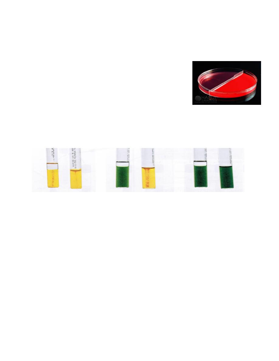
Non hemolytic
Hemolytic
Spore Staining method:
Red
bacilli arranged in short chains with oval central & non bulging
green
endospore.
Albert Stain :
light green bacilli
with bluish
black metachromatic granules.
non spore forming
pleomorphic with club expansion
(metachromatic=
Volutin
granules) & arranged in Chinese
letter form.
Quick Review of Microbiology 1
st
Semester
Microscopic Appearance
Staphylococci: gram +ve cocci arranged in clusters.
Non motile non spore forming
non capsulated
.
(3non)
Streptococci: gram +ve cocci arranged in chains.
Non motile non spore forming,
capsule depends on species.
Streptococcus pnemoniae : gram +ve lancet shaped diplococci or short chains.
Non motile non spore forming
,
capsulated
.
Neisseria: Gram –ve kidney shaped diplococcic.
Non motile non spore forming.
Pathogenic N. are intracellular or extracelluar, non-Pathogenic are extracellular.
~
Bacillus anthrcis: Gram +ve large rectangular rods that form
long
chains.
Capsulated in body tissues & fluids
, and non motile
.
Spore is oval central and non bulging (forms spore in culture) .
Bacillus subtilis & cereus (saprophytic bacilli): Gram +ve rods that form
short
chains.
non capsulated motile.
Spore is oval central , terminal or subterminal and non bulging.
~
Clostridium Tetani: Gram +ve bacilli with strait or slightly curved ends.
non capsulated motile.
Spore is spherical terminal & bulging (drum stick appearance)
Clostridium others: Gram +ve bacilli with slightly curved ends.
non capsulated motile
. (
except Cl.perfrengins > capsulated non motile
)
Spore is oval central or subterminal & bulging.
~
Corynebacterium diphtheriae : Gram +ve bacilli
non motile non spore forming and non capsulated
.
(3non)
pleomorphic with club expansion (metachromatic=Volutin granules)
& arranged in Chinese letter form.
-Slide of N. from culture ( could be patho. or non-patho)
-Slide of N. prepared directly from clinical specimen: CSF (Meningitidis) or
Vaginal/Urethral discharge (Gonorrheae), showing Gram –ve diplococcic inside pus cells.
2
You should complete colonies
morphologies by: S S S C C C O O E M
Non-pathogenic are not fastidious.
Nutrient agar > non pathogenic
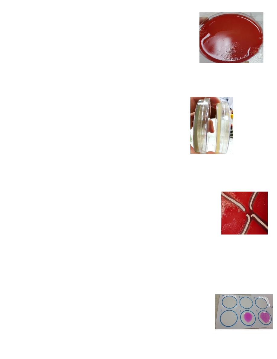
Blood agar> pathogenic & non pathogenic
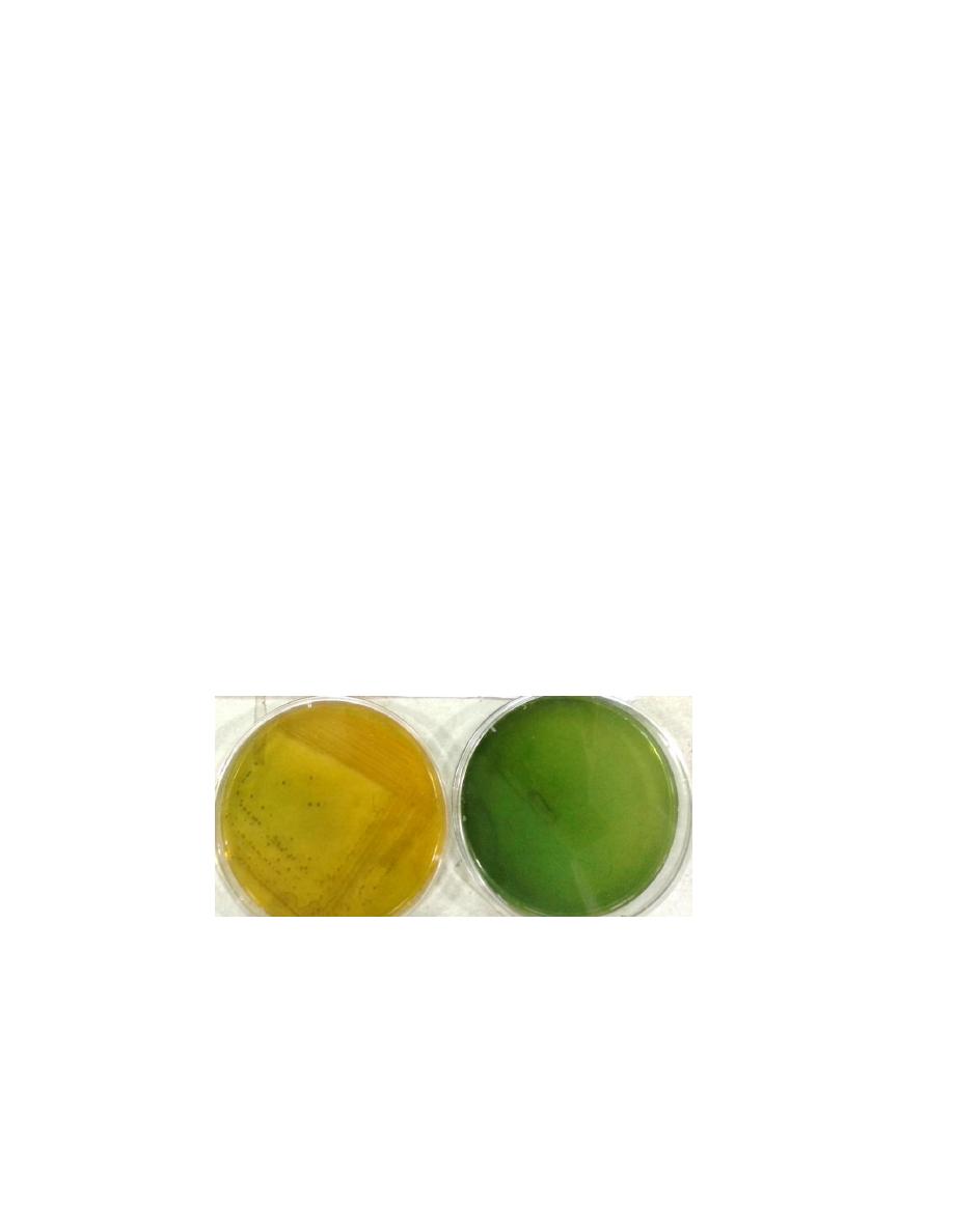
Macroscopic Appearance ( Cultural Characteristic )
Staphylococcus:
facultative anaerobes .
can tolerate 7.5-10% NaCl
, incubated in 37 for 24h.
S.aureus are B hemolytic
.
Streptococcus:
most
of them are
facultative anaerobes.
fastidious
, growth is enhanced by
5-10% CO2
, incubated in 37 for 24h.
Streptococcus pneumoniae :
facultative anaerobes
fastidious
, growth is enhanced by
5-10% CO2
, incubated in 37 for 24h.
Alpha hemolytic.
Neisseria :
Aerobic
Pathogenic are
fastidious
and they require
5-10% CO2 for primary isolation
, incubated
in 37 for 24h.
Non hemolytic
Translucent colonies in young > opaque in adult.
Bacillus:
Aerobic
.
(saprophytic bacilli ) Large , round or irregular, dull surface , may
become creamy, moist, (
hemolytic on blood agar
), opaque , convex with
undulating margin.
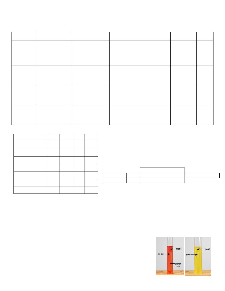
Clostridium: Anaerobic ,
Fastidious
B hemolytic
with diaphanous growth(except Cl.perfrengins that produce
target haemolysis & no diaphanous growth)
Corynebacterium:
Aerobic
B-hemolytic

3
Bacitracin sensitivity +
Short chains
Optochin sensitivity +
Bile solubility +
Grow in 6.5 NaCl
Biochemical Activities
Staphylococci: (catalase +)
-Nutrient agar: Aureus (golden yellow) epidermidis ( white) saprophyticus (Lemon
yellow)
-Blood agar: Aureus is B-hemolytic
-Coagulase + (clot or agglutination) : Aureus
-Mannitol Salt agar: selective for staphylococcus , differential between Aureus that
ferment mannitol forming yellow colones & epidermidis (pink or red)
-Gelatinase + (liquify gelatin): Aureus
Sterptococci (catalase –ve):
oxidase –ve
B- hemolytic:
Pyogens >>>>>
Agalactiae
Alpha Hemolytic:
Viridans
Pnemoniae >>>>
Non ( Y) hemolytic:
Bovis
enterococcus fecalis >>>>>
Neisseria:
Oxidase
+ve
Sugar utilization
( +ve is yellow “due to
change of color of
phenol red”
-ve is red )
Thayer Martin medium is selective for pathogenic Neisseria
Sucrose
Maltose
Glucose
Species
-
+
+
N.meningitides
-
-
+
N.gonorrhoeae
+
+
+
N.Mucosa

4
Bacillus:
-Catalase +ve
-liquify gelatin +ve
-Hemolysis on blood agar
-Hydrolyze starch: clear area (of hydrolyzed starch) around colonies in
(blue)
starch
medium (with poured iodine solution)
Clostriduim:
-Anaerobic blood agar
-Brucella blood agar
-Colistinnalidixic acid
-Sodium thyoglycolate
-Cooked meat broth :
1-Saccharrolytic group
(All gas gangrene group except C. histolyticum) ferments muscle
glycogen & produce of acid and gas.
2-Proteolytic group (C. histolyticum ,C. botulinum, C. tetani slightly proteolytic) digests
meat particles & forms black, foul smelling due to sulfur compounds.
-Litmus Milk: Saccharolytic group produce
pink stormy result
.
Corynebacterium:
1-Selective Medium: Potassium Tellurite (with added blood or serum) > black colonies
because of reduction of tellurite in bacterial wall into telluride ( 3 colonies types can be
demonstrated in such medium : Gravis , intermedius , & mitis ).
2-Enriched Medium: Lofflers medium
Blood agar
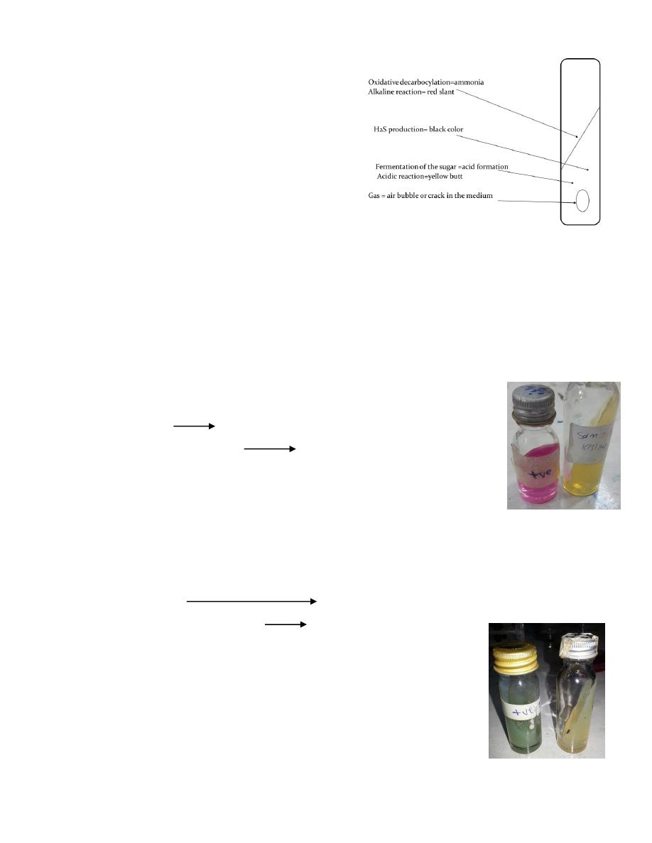
Toxigenicity tests:
- In vivo : Animal inoculation for isolation or demonstration of exotoxin erythema & necrosis
-In vitro: Gel diffusion technique “ Eliks Test” (Toxin/Antitoxin reaction)
-PCR : detects toxin genes
-ELISA: detects toxin
-Tissue culture : bacteria are inoculated into cell culture , the toxin diffuses & kills the cells.

5
Not stained by Gram stain.
Enterobacteriaceae
Quick Review of Microbiology 2
nd
Semester
Microscopic Appearance
Mycobacterium:
Microscopical appearance: (Acid Fast Bacilli) It is long, slender rods , tapered at the
ends, straight or slightly curved or bent (curved) , stain
red
by Ziehl-Neelsen stain
method with blue counter stained background . Staining may be irregular with
granules appearing darker than other parts of bacterial cells.
Enterobacteriaceae
:
G-ve ,motile (except Shigella, Klebsiella) , non spore forming.
E. coli
:
G-ve rods with rounded edges , motile , non spore forming
Some species are capsulated with K antigen
Klebsiella
: capsulated .
(Colonies are high convex , mucoid , tends to coalse)
Salmonella
:
G-ve , motile , non spore forming.
Shigella
: G-ve non motile non capsulated.
Proteus
:
very
pleomorphic & highly motile
Pseudomonas :
G-ve strait or slightly curved rods , motile by one or more polar
flagella , & non spore forming.
Cocobacilli:
Gram –ve cocobacilli with intermediate shape between cocci & bacilli.
H. influenzae :
G-ve cocobacilli (
sometimes pleomorphic
) non motile . >
Brucella :
gram negative small coccobacilli arranged in pairs or small chains
Bord
etella
:
They are minute gram negative coccobacilli showing
bip
olar
stain they are
motile
or
non motile
V. Cholera
:
G-ve comma shaped or rarely filamentous , with marked
pleomorphism
.
Motile with flagellae . Stain with dilute Carbol Fuchsin.
6
Media
Mycobacterium :
Culture : Aerobic
,
CO2 increase the growth
, Incubated at 37
C for up to 8weeks.
1-Agar base media (oleic acid albumin media) e.g. Dubos medium and Middle brook 7H10,
these media contain defined salts, vitamins, co-factor, oleic acid, albumin, glucose,
glycerol, these media used for the observation of the colonies morphology ,& used for
susceptibility test.
2-Egg-base media: Lowenstein Jensen
s medium(L-J medium) & Stonebrink
s
medium, contain defined salt, glycerol & complex organic material in form of
fresh egg, egg yolk, potato flour & other ingredient in various combinations.
Malachite green is included in the media to inhibit other bacteria .
Used for primary isolation of Mycobacteria because they need small inoculum size.
3-Liquid media:These media enriched with either ascitic fluid or bovine albumin fraction
V. e.g. Sula medium.
E. coli
&
Klebsiella: (LF)
Macconkey’s agar (Selective & Differential ): contains lactose , both bile salts & crystal
violet (to inhibit the growth of G+ bacteria) and Neutral red.
NLF (
Pale colonies = Yellow
)
LF (
Pink
< 6.8 pH)
NLF
E.coli
Klebsiella
7
Salmonella & Shigella : (Grow on ordinary Media)
1-
Macconkey’s agar :
NLF ( Pale colonies = Yellow)
2-
Bismuth Sulfite Agar BSA (Selective for Salmonella): black colonies with metallic sheen
Main Composition:
1.Bismuth sulfite
2.Brilliant green
3.Ferrous sulfate
Bismuth sulfite and brilliant green (selective agent) inhibit the growth of
gram positive and coliform bacteria and allow the growth of Salmonella.
Sulpher compound H2S Black colonies( sulphate act as substrate to H2S)
Bismuth indicator Metallic Bismuth Metallic Sheen.
Metallic salts in media stain the colony and the surrounding medium black or brown.
3- Salmonella-Shigella Agar SSA (Selective & Differential) : (1ry isolation)
-Salmonella : Pale colonies with black center (NLF)
-Shigella : Pale colonies without black center (NLF)
-Coliform : Pink/Red colonies (LF)
It highly selective formulate to inhibit the growth of most coliform bacteria
and permit the growth of Salmonella and Shigella from enviromental and
clinical specimens.
Main Composition:
-Bile salt inhibit the G+ and other G- bacteria.
-Sodium thiosulfate source of sulfer.
-Neurtral red indicator.
High conc. of bile salt to make it more selective because salmonella and shigella can
tolerate high conc. of bile
Salmonella doesn`t produce black colony on MacConkey because the agar lack sulphate.
4-Selenite (Selective for Salmonella & Enriched) : 1ry isolation
It increases the number of Salmonella & Inhibit the growth of coliform and Shigella
(temp and/or PH dependent)
8
Proteus : (Grow on ordinary Media)
Fishy bad odour,
very pleomorphic, & highly motile;
swarming
phenomenon occurs on a non inhibitory media as nutrient & blood
agar , but inhibited with increase in agar content as Macconkey’s
agar.
Pseudomonas : (Grow on ordinary Media)
-Can tolerate up to 42 C & 8.5 pH (pH is similar to V. cholera)
-Two important characterstics : sweetish aromatic (grape-like) odour & green coloration
diffuse in medium.
-Pigments on nutrient agar: (Part of pathogenisis)
Pyocyan: blue
(Fluorescein) Pyoverdin: green or yellow
Pyorubin: red
Pyomelanin: balck or dark brown
H. influenzae :
Green Yellow
Fastidious
(needing X=hemin & V=NAD factors)
&
5% CO2
incubated for 18h in :
-Blood agar (X)
-Choclate agar (X ,V) (Larger coloneis)
-Haemophilus isolation agar
-Fildes digest agar (X,V)
Satellite test: The NAD diffuses into the surrounding medium and stimulates
the growth of haemophilus in the vicinity of the staphylococcus.
Brucella:
Specimens (blood and bone marrow) are inoculated in an aerobic bottle at 35C° for 4-6
weeks.
Media:
Liver broth & Liver agar
Serum Dextrose agar
Charcoal yeast extract agar
BACTEC 9240 and Septi-check system
Serology :
-Slide method (Rose Bengal screening test)
>>>
-Tube method (Titration method) Agglutination titer ≥ 1/160 significant.
-2ME (2mercaptoethanol) which inactivate IgM producing +ve result for IgG.
9
Bord
etella
:
Mercury droplet
or
pearl appearance
Some Species need X,V factors for 1ry isolation.
Media:
-Bordet Gengon : 1ry isolation for 2-4 days at 36C
-Regan Lowe (superior to BG)
V. Cholera:
(Grow on ordinary Media)
Growth is enhanced by
1% NaCl
. The optimum pH 7.6-8 (can tolerate up to 8.5) it grows
in 40C°.
Media:
1-Nutrient agar: Darting “shooting” stars motility with colonies are smooth yellow with
opaque centers & transparent periphery.
2-Alkaline Peptone Water (APW) : 1ry isolation
3-Thiosulphate Citrate Bile Salts Sucrose TCBS (Selective) ( Subcultivation )
-Sodium thiosulfate provides a source of sulfer & acts in combination with ferric citrate
to detect the production of H2S.
-Bile salts inhibits the growth of G+ve M.O.
-Sucrose is the fermentable carbohydrate that act with the help bromothymol blue in
differentiation of vibrio spp. which utilize sucrose.
.
Yellow: Vibrio
Green: non-inoculated
10
> Mannitol fermenter
>
> Ferments Mannitol , Sucrose, Maltose with acid production
only.
Biochemical Activities
IMViC:
I
M
V
C
E.Coli
+
+
-
-
K.pneumoniae
-
-
+
+
Salmonella
-
+
-
+
Shigella
+
+
-
-
Proteus
V
+
-
V
Pseudomonas
+
Vibrio
+
-
-
Carbohydrate Utilization Test:
Detects the ability of m.o. to ferment a specific sugar.
Medium : Broth of CHO-fermentation medium (consisted of sugar , basic medium
bromothymol blue) into which small inverted durham tube is placed .
+ Yellow with gas
(Acid formation by fermenatation . H2 & CO2 production)
- Red without gas
Media
Reagent
Mechanism
+ve
-ve
I = Indol
Peptone water
Kovacs reagent
(para-dimethyl
aminobenzaldehyde,
isoamyl alcohol and
conc. HCl)
Decompose tryptophan to indol
( Colorimetric reaction)
Red ring
Yellow
M = Methyl
red
Glucose
phosphate
peptone water
(GPPW)
Methyl red reagent
Acid (pH 4.4 or less) production
due to glucose fermentation
Red
Yellow
V = Voges
Proskauer
Glucose
phosphate
peptone water
(GPPW)
5% alpha napthol in
ethanol + 40% KOH
Acetyl-methyl carbinol (acetoin) is
oxidized to butylene glycol
(di-acetyl) that is tested for, by a
Colorimetric reaction.
Pink/Crimson
Green
C = Citrate
Simmons citrate
Bromothymol blue
Utilization of citrate and use
carbon for energy and growth and
use ammonium as source of
nitrogen
Blue with
streak of
growth
Green
without
growth
Ornithine decarboxylase
P.vulgaris
++--
+
Sucrose fermenter
P.mirabilis
-+-+
-
11
TSI (Tri Sugar Iron Agar)
Corner stone in differentiation of G-ve bacilli.
This media consist of
Three sugars (glucose, lactose, sucrose).
Pepton ( as nitrogen source )
Ferrus sulphite.
Sodium thiosulphite.
Phenol red indicator
-E.coli & Klebsiella (Lactose Fermenters) : A/A ± Gas (V.cholera is A/A)
-Salmonella : K/A + H2S ± Gas > (Proteus is the same , but with more H2S production)
-Shigella : K/A - H2s – Gas > (Yersinia is the same)
Pseudomonas (Non Fermenters) : K/K
(H2S is produced in acidic medium)
Urase Test:
Urea + Water urease ammonia + carbon dioxide
Phenolphthalein (colorless) ammonia phenolphthalein (pink-red pH>8.1).
+ pink
,
- yellow
Salmonella : Urase -
Shigella : Urase -
Proteus : Urase +
Phenylalanine Deaminase (Guthrie) Test :
L-phenylalanine phenylalanine deaminase phenylpyruvic acid + ammonia
Phenylpyruvic acid +Fecl3 (10%) acid green color
+ Green
,
- Yellow
+ve for : Proteus , Morganella , Providencia
Note: Proteus is Urase +ve
Phenylalanine Deaminase +ve
Gelatinase +ve (Part of pathogenicity)
12
Enterobacteriaceae:
(Facultative anaerobes)
Oxidase – (Ferment Glucose)
Catalase +
Reduce nitrate to nitrite
Grow well on Macconkey’s agar at 37°C
Non Fermenters Gram -ve Bacilli :
(Aerobes)
Oxidase +
Lack of evidence of glucose fermentation
Lack of evidence of growth Macconkey’s agar.
Pseudomonas:
1-Oxidase + (oxidize glucose)
2-Catalase +
3-No lactose fermentation
4-ONPG –ve
5-Oxidative Fermentative Test (Hugh & Leifson medium) : This is a sensitive test for the
detection of bacteria that utelize glucose oxidatively
Fermentative : E.coli Oxidative : Pseudomonas Asaccharolytic : Morexella
6-Motility test : (inverted tree appearnace)
1-True Motility: upper 5 mm of tube show inverted tree (↓growth ,↑motility)appear as
wide base & narrow apex . Through :
Wet perapartion
Hanging drop preparaion
Semisolid media (0.5% agar)
2-Brownian movement (not true movement): due to vibration caused by molecular
bombardment.
7-Utilization of glucose: The end products of fermentation are relatively strong acid that
can be detected in a conventional fermentation test medium.The acid formed in
oxidative degradation of glucose is extremely weak so it is tested by semisolid of
medium.
The concentration of peptone is deceased 1% to 0.2% the concentration of CHO is
increased from 0.5-1%. (pepton:CHO 1:5)
The lower protein/ CHO ratio reduce the formation of alkine amines that can neutralize
the small quantities of weak acid may form from oxidative metabolism. The large
amount of CHO serve to increase the amount of acid that can potentially be formed. The
semisolid consistency of the agar permits acids that formed on the surface.
13
Vibrio:
Oxidase +
Catalase +
Reduction of nitrate +
Brucella:
Dye Sensitivity
Co2 req. H2S req.
Carbol Fuchsin
Thionin
Br. abortus
+
+
-
+
Br. melitensis
-
-
-
-
Br. suis
-
+
+
-
Some pics are taken from “ 3
rd
stage practical labs “ CD
Note: It’s better to print this in colors through: Muhadharaty.com/microbiology
Ameer Saadallah
(2011-2017) Batch No.53