
Cerebrospinal Fluid
System
By
Dr. Mufeed Akram Taha
FIBMS Neurology
Kirkuk College of Medicine

Cerebrospinal Fluid System
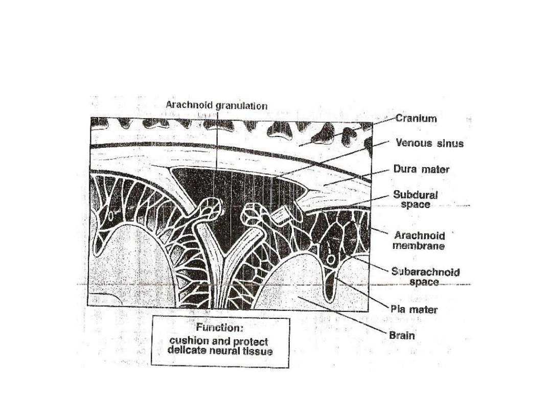
It has a volume of about 150 ml and found in
the ventricles of the brain, in the cisterns
around the brain, and in the subarachnoid
space around both the brain and the spinal
cord. All these chambers are connected
with one another and the pressure of the
fluid is regulated at a constant level. CSF
can be sampled with a lumbar puncture.
The major function of the CSF is to:

[1] Forms a protective water jacket which cushions
the brain within its solid vault. This is due to fact
that the brain actually floats in the fluid.
Therefore, a blow to the head moves the entire
brain simultaneously with the skull, causing no
one portion of the brain to be momentarily
contorted by the blow.
[2] Alteration of volume can compensate for
fluctuations in amount of blood within skull and
thus keep total volume of cranial content
constant.
[3] Low K ion concentration allows neurons to
generate very high electrical potentials.

CSF is formed at a rate of about 500 ml / day.
Two thirds or more of this fluid originates as
a secretion from choroid plexuses in the four
ventricles, mainly in the two lateral
ventricles. The CSF in the ventricles flows
through the foramens of Magendie and
Luschka to the subarachnoid space, which is
absorbed through the arachnoid villi into
veins.

The composition of CSF is essentially the same as
that of extracellular fluid of the brain. There
appears to be free communication between
the brain interstitial fluid and CSF. The surfaces
of the ventricles are lined with thin epithelial
cells called ependyma and the outer surface of
the brain is covered by a thin membrane called
the pia mater. Both of which (ependyma and
pia mater) are extremely permeable so that
almost all substances that enter the CSF can
also diffuse readily into the interstitial fluid of
the brain through these membranes, and vice
versa.

The CSF pressure normally is regulated almost
entirely by absorption of the fluid through
arachnoid villi to superior sagittal sinus. The
reason for this is that the normal rate of CSF
formation is constant. On the other hand, the
villi function like valves that allow the fluid and
its contents to flow readily into the blood of the
venous sinuses while not allowing blood to flow
backward in the opposite direction. Normally,
this valve action of the villi allows CSF to begin
to flow into the blood when its pressure is about
1.5 mm Hg greater than pressure of the blood in
the venous sinuses.

Then as the CSF pressure rises still higher, the
valves open widely, so that under normal
conditions, the pressure almost never rises
more than a few mm of Hg higher than
pressure in the venous sinuses. On the other
hand, in diseases that involved the villi, can
cause high CSF pressure.
If an obstruction occurs in the ventricular system
or foramina, the result is called
noncommnnicating hydrocephalus; if the
obstruction is at the arachnoid villi, it is called
communicating hydrocephalus.

BIood-Brain Barrier (BBB)
There are barriers between the blood and the CSF (called
blood- CSF barrier due to the epithelial cells of the
choroid plexus) and between the blood and interstitial
fluid of the brain (called blood-brain barrier due to the
endothelium of the cerebral capillaries in conjunction
with astrocytic processes). However, both barriers are
similar. general, blood-brain and -CSF barriers are highly
permeable to water, CO2, 0
2
, and most lipid soluble
substances such as alcohol and most anesthetics, slightly
permeable to the electrolytes, and almost totally
impermeable to plasma proteins, cholesterol (because
both of them have large molecular size), and most non-
lipid soluble large organic molecules.
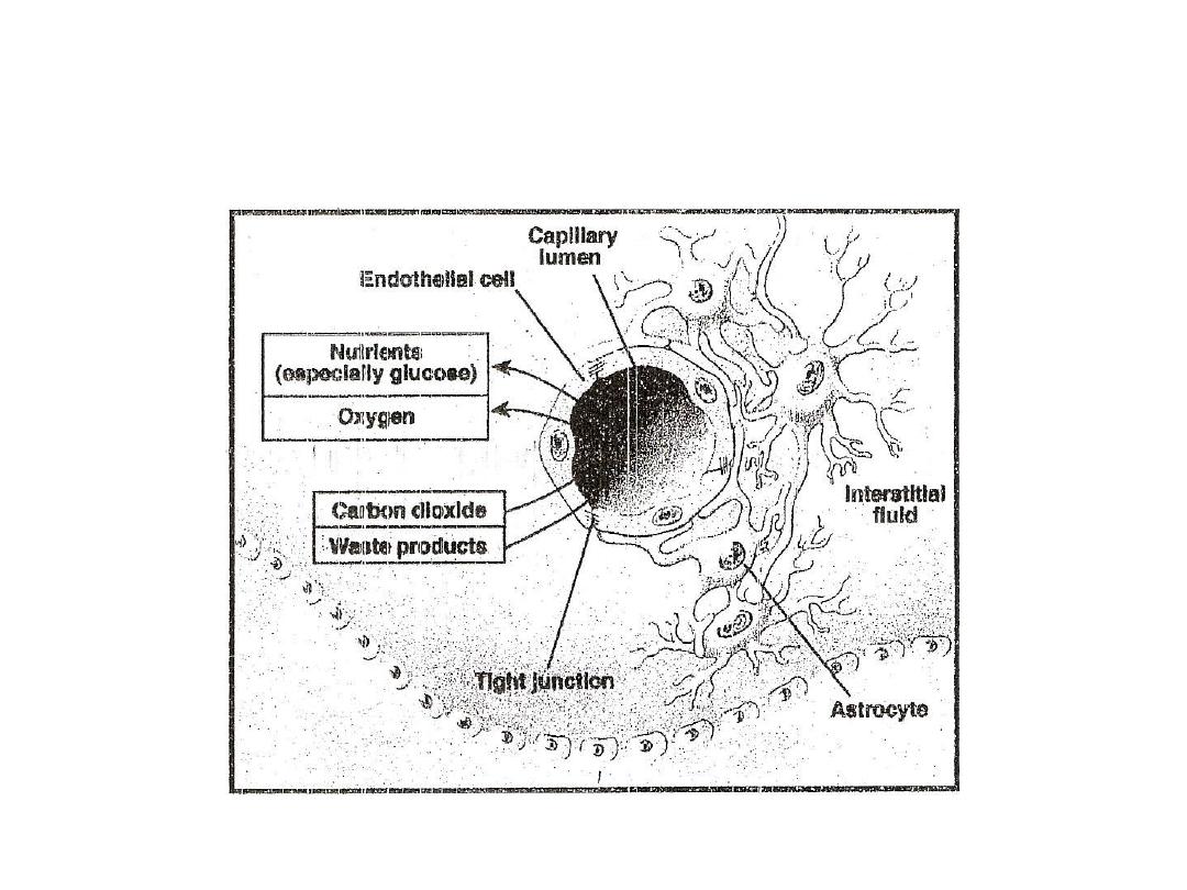

The cause of the low permeability of these barriers is
due to the tight junctions between the endothelial
cells of the capillaries and epithelial cells of the villi.
There are areas of the brain lacks the presence of
such barriers such as some areas of the
hypothalamus, pituitary gland, and pineal body.
The ease of diffusion in these areas is important
because they have sensory receptors that respond
to different changes in the body fluids and their
responses provide the signals for feedback
regulation of each of the factors. The BBB can be
disrupted temporarily by inflammation, irradiation,
tumors, sudden severe increases in blood pressure

or by intravenous injection of hypertonic fluids (which
is used clinically to extract cerebral edema fluid).
The functions of the BBB are:
1. It maintains a constant environment for neurons in
the CNS and protect the brain from endogenous
and exogenous toxins.
2. It prevents the escape of neurotransmitters from
their functional sites in the CNS into the general
circulation.
3. Drugs penetrate the BBB to varying degrees. For
example non ionized (lipid soluble) drugs cross
more readily than ionized (water soluble) drugs.
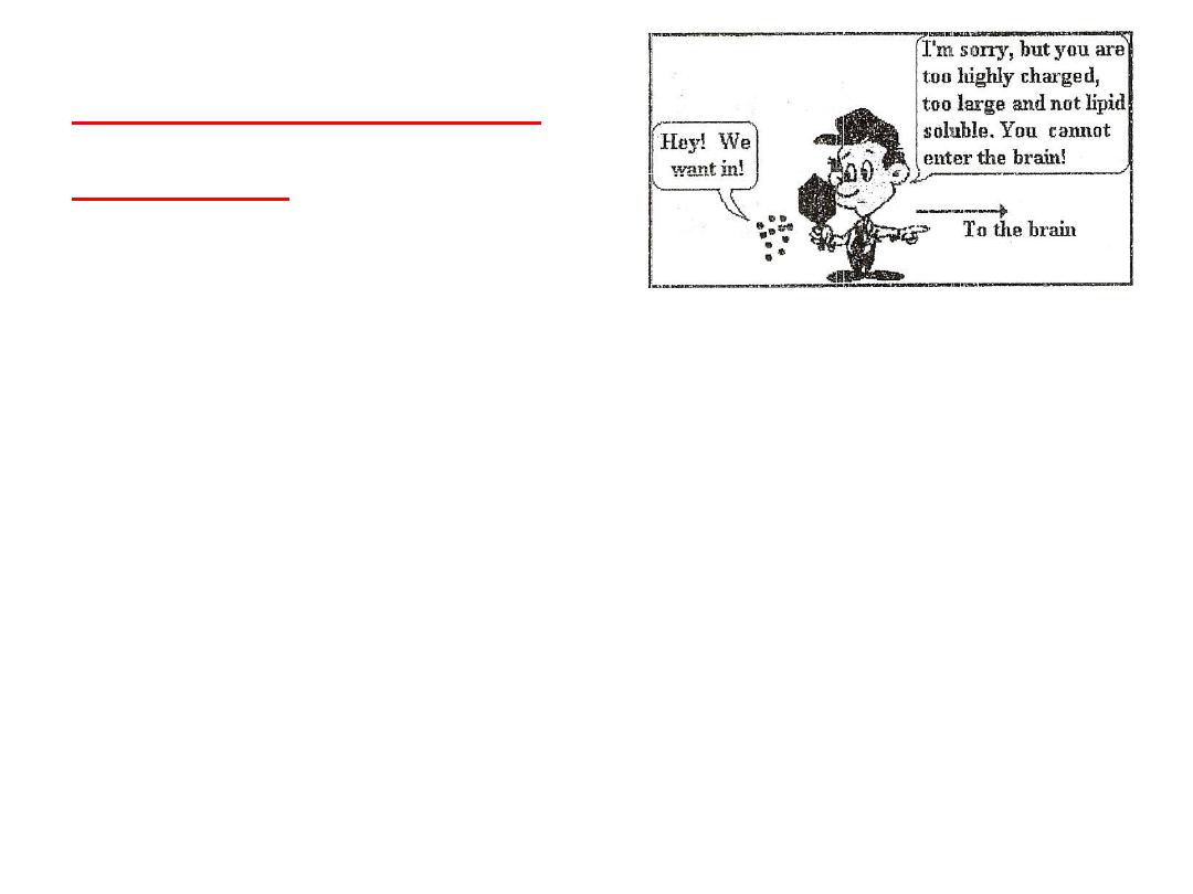
General Properties of
the BBB:-
1.
Large ole ules do ’t pass through the BBB
easily.
2.
Low lipid fat solu le ole ules do ’t
penetrate into the brain. However, lipid
soluble molecules, such as barbiturate drugs,
rapidly cross through into the brain.
3. Molecules that have a high electrical charge
to them are slowed.
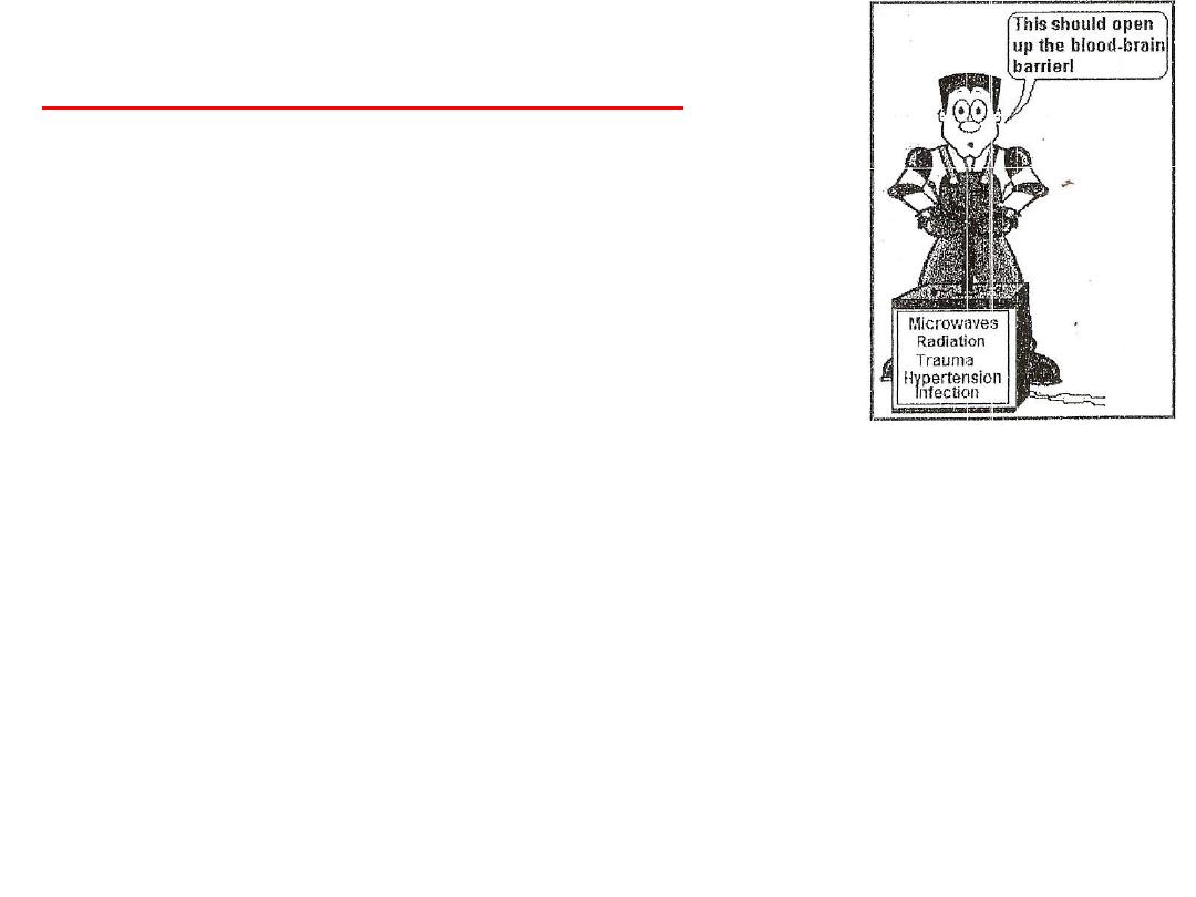
The BBB can be broken down by:-
1. Hypertension (high blood pressure):
high blood pressure opens the BBB.
2. Development: the BBB is not fully
formed at birth.
3. Hyperosmolitity: a high concentration
of a substance in the blood can open the BBB.
4. Microwaves: exposure to microwaves can open BBB.
5. Radiation: exposure to radiation can open the BBB.
6. Infection: exposure to infectious agents can open BBB.
7. Trauma, Ischemia, Inflammation, Pressure: injury to
the brain can open the BBB.

Cerebral blood flow (CBF): CBF is about 80 ml/100
g/ min in gray matter and about 20 ml/100
g/min in white matter. If CBF is deceased to less
than 10 ml/ 100 g/ min, irreversible tissue
dammage can occur at normal body
temperatures.As in the coronary circulation, CBF
is autoregulated, meaning that it remains
constant between a mean blood pressure of 50-
150 mm Hg. When the mean pressure is greater
than 150 mm Hg, the BBB may be disrupted,
The curve is shifted to a higher mean blood
pressure in patient with chronic Hypertension.

Regional metabolic activity, arterial O2 & CO2
concentration helps in determining regional CBF,
unlike the coronary circulation, cerebral
resistance vessels are more sensitive to PCO2
than PO2 , So even slight increase in PCO2 will
cause a large increase in CBF.
Many drugs affects on CBF; for example
Barbiturates constrict cerebral blood vessels,
while volatile anesthetic agents dilate them.
Constriction of the cerebral vasculature can help
decrease intracranial pressure and dilatation can
increase intracranial pressure.

Intracranial pressure (ICP):-
It’s the pressure i side the ra iu . Withi
certain limit, if one of the 3 brain
compartments (i.e, CSF, blood vessels
and brain tissue) increases in volume, its
compensted by successfully by a
decrease in volume of one or both of the
other two compartments without an
associated change in intracranial
pressure.

Thank You