
Carbohydrate Metabolism
Glucose Metabolism
● Glucose is the preferred source of energy for most tissues .
● Erythrocytes are fully reliant on glucose for energy .
● Brain , in normal conditions , uses only glucose as ATP source and metabolize it
at a constant rate ; it depends on glucose in blood ; at basal metabolic rate 60%
of glucose oxidized is by brain .
● Glucose metabolism include the following metabolic pathways :
1.
Glycolysis & Citric acid cycle
2.
Glycogenesis & Glycogenolysis
3.
Gluconeogenesis
4.
Pentose Phosphate Pathway
5.
Minor Pathways ( ex : Polyol Pathway )
● Glucose is oxidized to generate energy ( ATP ) by the pathways of Glycolysis &
Citric Acid Cycle .
● When the blood glucose level is high (after meals ) the excess is converted to
glycogen and stored (Glycogenesis ) .The body has limited capacity for storing
glycogen and the remaining excess glucose is converted to fat and stored
( Lipogenesis ) .
● During fasting , when glucose level in blood decreases , the supply of glucose
for the brain is maintained by Glycogenolysis ( glycogen breakdown ) and
Gluconeogenesis ( synthesis of glucose from non-carbohydrate sources ) .
Oxidation of Glucose
A
– Aerobic Oxidation of Glucose
● Under aerobic conditions ( oxygen in plenty ) and in the tissues containing
mitochondria , glucose is oxidized in two stage (Glycolysis & Citric acid cycle)
to generate energy ( ATP ) .
Glycolysis (
first stage
)
A series of reactions by which each molecule of glucose ( 6-carbon compound ) is
converted to two molecules of pyruvate ( 3-carbon compound ) as end product .
It is cytosolic pathway taking place in all the cells of the body . Pyruvate is then
transported into mitochondria where it is completely oxidized through Citric Acid
cycle ( second stage) to CO
2
and H
2
O .
Reactions of glycolysis :
Step 1 : phosphorylation of glucose by ATP and glucose-6-phosphate is formed . The
reaction is irreversible and catalyzed by Hexokinase (present in all tissues )
& Glucokinase ( present only in liver and pancreas ) .
Step 2 : isomerization of glucose-6-p to fructose-6-phosphate .

Step 3 : phosphorylation of fructose-6-p to give F-1,6-bisphosphate by the enzyme
phosphofructokinase (PFK-1) . Reaction is irreversible . PFK-1 is the rate-
limiting enzyme .
Step 4 : cleavage of F-1,6-bisphosphate into two 3-carbon compounds :
glyceraldehydes-3-p & dihydroxy acetone-p . These are inter-converted
to each other by an isomerase enzyme .
Step 5 : phosphorylation and simultaneous oxidation of glyceraldehydes-3-p to
1,3-bisphosphoglycerate . One molecule of NADH is produced .
Step 6 : transfer of high energy phosphate group (~P) from 1,3-bisphosphoglycerate
to ADP forming ATP ( substrate level phosphorylation) and the product
3-phosphoglycerate .
Step 7 : isomerization of 3-phosphoglycerate to give 2-phosphoglycerate .
Step 8 : dehydration of 2-phosphoglycerate to form phosphoenol pyruvate (PEP) .
Step 9 : transfer of ~P from PEP to ADP producing ATP and pyruvate .The enzyme
is pyruvate kinase (PK) and the reaction is irreversible
Note :
● NADH formed in both stages of aerobic oxidation of glucose oxidation
( glycolysis & citric acid cycle ) is oxidized back to NAD
+
through the electron
transport chain and significant number of ATP molecules are generated .
A
– Anaerobic Glycolysis :
Under anaerobic conditions ( hypoxia ; oxygen in short supply) or in the
absence of mitochondria , Pyruvate is reduced to lactate by the enzyme lactate
dehydrogenase (LDH) to regenerate NAD
+
.This is called anaerobic glycolysis
in which the end product of glycolysis is lactate .
■ Skeletal muscle tissue is highly anaerobic tissue because :
1. actively contracting skeletal muscle during intensive exercise develops hypoxic
conditions because the need for ATP formation exceeds the rate of oxygen
consumption .
2. muscle LDH (M
4
) has a low
k
m
( higher affinity ) for pyruvate .
3. Fast
–twitch white skeletal muscles lack myoglobin and contain very few
mitochondria .
*********************
■ Anaerobic glycolysis is the only source of energy in erythrocytes as they lack
mitochondria .
■ Cancer cells grow more rapidly than normal cells leading to relative degree
of hypoxia . Cancer cells metabolize glucose by anaerobic pathway and at a
higher rate and so lactate accumulates causing acidic environment in the tumor .
■ Cardiac muscle tissue is aerobic tissue ; LDH in heart muscles ( H
4
) has low
affinity for pyruvate and high affinity for lactate .
Heart muscles metabolize glucose aerobically and have low anaerobic glycolytic
activity and poor survival under hypoxic (ischemic ) conditions .
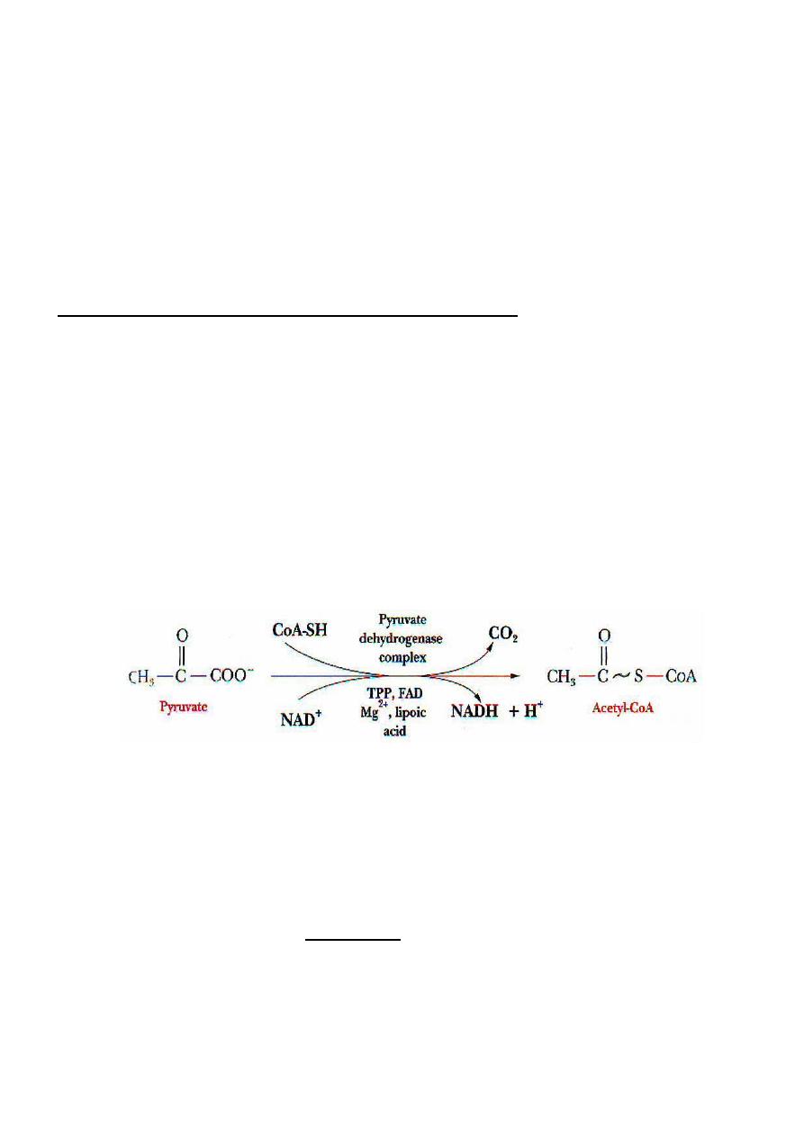
Pyruvate Dehydrogenase ( PDH ) Reaction :
● In the matrix of mitochondria , pyruvate undergo oxidative decarboxylation and
converted into 2-carbon high energy compound Acetyl-CoA .
This reaction is catalyzed by a multi-enzyme complex system called pyruvate
dehydrogenase complex (PDH system) which require five coenzymes ( derived from
five vitamins ) and also require Mg
2+
as cofactors :
■ Thiamine pyrophosphate (TPP)
■ Lipoamide
■ Coenzyme A ( CoA-SH )
■ FAD , and
■ NAD
+
● In this reaction , One molecule of NADH is formed and one molecule of CO
2
is
liberated .
● Acetyl-CoA is then enters the citric acid cycle and completely oxidized to CO
2
and
H
2
O and generate significant ATP .
● Pyruvate dehydrogenase (Oxidative decarboxylation of pyruvate to acetyl-CoA )
is the link between glycolysis and citric acid cycle .
● This reaction is completely irreversible process and there is no alternate
reaction in the body that forms pyruvate from acetyl-CoA .
*************************************

Energy Yield ( ATP ) from Glycolysis
* Number of ATP molecules generated by glycolysis per molecule of glucose under
aerobic conditions ( oxygen in plenty ) :
Step Enzyme Source Number of ATP gained
or used
───────────────────────────────────────────────────
1- Hexokinase ATP Minus one ( used )
3- Phosphofructokinase-1(PFK-1) ATP Minus one (used)
5- Glyceraldehyde-3-p dehydrogenase NADH 3 x 2 = 6 ( gained )
6- Phosphoglycerate Kinase ATP 1 x 2 = 2 ( gained )
9- Pyruvate Kinase ATP 1 x 2 = 2 ( gained )
───────────────
Total = 10
− 2 = 8 ATP
* Number of ATP molecules generated by glycolysis per molecule of glucose under
anaerobic conditions ( hypoxia ) or in the lack of mitochondria :
Step Enzyme Source Number of ATP gained or used
1- Hexokinase ATP Minus one ( used )
3- Phosphofructokinase-1 (PFK-1) ATP Minus one ( used )
6- Phosphoglycerate Kinase ATP 1 x 2 = 2 ( gained )
9- Pyruvate Kinase ATP 1 x 2 = 2 ( gained )
───────────────
Total = 4
− 2 = 2 ATP
****************************

Regulation of Glycolysis
Phosphofructokinase-1 ( PFK-1 )
is the key rate-limiting enzyme in glycolysis .
The activity of this enzyme is controlled as follows :
A - PFK-1 is inhibited by :
1 - allosterically inhibited by high ATP level .
2 - feed-back inhibition by the product Citrate ( allosteric inhibition ) .
3 - low pH ( high H
+
concentration ) due to increased Lactate .
II - PFK-1 activated by :
1.
AMP ( high levels ) .
Advantage of having both the enzymes Hexokinase & Glucokinase in liver :
1.
Hexokinase has high affinity for glucose ( K
m
≈ 0.04 mM ) . Since the resting
level for blood glucose is about 5mM , therefore hexokinase would be expected
to be fully active for all body cells at the resting level and the liver would not be
competing with other cells for glucose .
On the other hand , Glucokinase has lower affinity for glucose ( K
m
≈ 6 mM ) ;
have significant activity when blood glucose levels exceed 10 mM ( such as after
a carbohydrate
–rich diet ) , and at that concentration liver competes with other
tissue for glucose and the excess glucose preferentially taken into liver where it
can be stored as glycogen .
2.
Hexokinase is inhibited allosterically by high levels of its product glucose-6-p .
But glucokinase is not inhibited by the product glucose-6-p . Therefore , liver
can metabolize glucose preferentially over the other tissues .
Clinical aspects :
*
Hexokinase deficiency
inherited deficiency of the enzyme hexokinase cause hemolytic anemia ; less
ATP is generated in erythrocytes and are easily destroyed .
*Pyruvate Kinase deficiency in erythrocytes
There is decreased production of ATP from glycolysis . Red blood cells have
insufficient ATP for their sodium pump . The cells become dehydrated and are
phagocytosed by cells in the spleen and a hemolytic anemia result .

Intermediates of glycolysis including 2,3-Bisphosphoglycerate accumulate .
Elevated levels of 2,3-BPG decreases the affinity of hemoglobin for oxygen and
the oxygen-carrying capacity of RBC
S
decreased .
*Taruis disease
● Rare inherited deficiency in the muscle and erythrocyte PFK isoenzyme ;
there is anemia , muscle weakness and work capacity is low specially on high
carbohydrate diet which is improved on fasting and starvation because the
muscle start metabolizing fatty acids .
■ Glucose feeding to these individuals increases insulin levels and inhibits
lipolysis in adipose tissue so they have less fatty acids in the blood to serve
as fuel .
*Dietary deficiency of thiamine cause pyruvate to accumulate . Nutritionally
deprived alcoholics are thiamine-deficient and may develop potentially fatal
pyruvate and lactic acidosis .
******************************************