Basic Anatomy
The orbits are a pair of bony cavities that contain the
549
The Orbital Region
eyeballs; their associated muscles, nerves, vessels, and fat;
the eye is closed.
nea when the eye is open and rises only slightly when
gin of the cornea. The lower lid lies just below the cor
straight ahead, the upper lid just covers the upper mar
cornea of the eye. When the eye is open and looking
the eye is closed, the upper eyelid completely covers the
lids and is the entrance into the conjunctival sac. When
is the elliptical opening between the eye
bral fissure
palpe
The
lateral angles.
medial
each other at the
larger and more mobile than the lower, and they meet
light by their closure (Fig. 11.16). The upper eyelid is
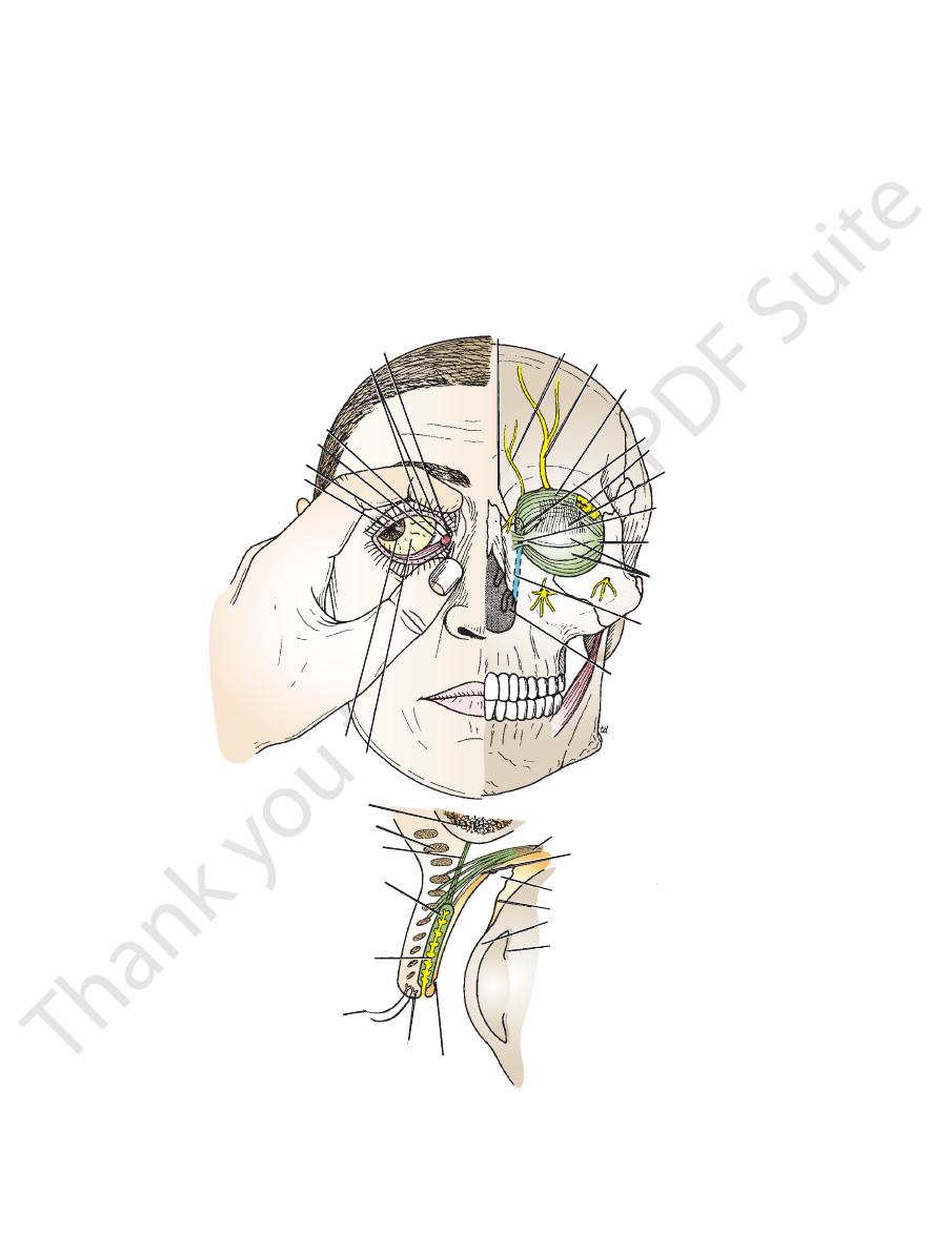
The eyelids protect the eye from injury and excessive
guarded by two thin, movable folds, the eyelids.
and most of the lacrimal apparatus. The orbital opening is
Eyelids
and
-
-
-
-
lacrimal papilla and punctum
orifices of tarsal glands
caruncula lacrimalis
plica semilunaris
iris
pupil
cornea
inferior meatus of nose
lacrimal duct in
nasolacrimal
canal
tarsal plates
lateral palpebral
ligament
medial palpebral
ligament
levator palpebrae
superioris
lacrimal gland
lacrimal sac
orbital septum
supraorbital nerve
supratrochlear nerve
infratrochlear nerve
frontal bone
orbicularis oculi
orbital septum
superior tarsal plate
tarsal gland
eyelash
sebaceous gland
subtarsal
sulcus
iris
cornea
conjunctiva
superior fornix of conjunctiva
smooth muscle
levator palpebrae superioris
A
B
C
conjunctiva covering sclera
FIGURE 11.16
A.
in the levator palpebrae superioris.
Sagittal section through the upper eyelid, and the superior fornix of the conjunctiva. Note the presence of smooth muscle
yellow
duct. Note that a small window has been cut in the orbital septum to show the underlying lacrimal gland and fat (
Left eye, showing the superior and inferior tarsal plates and the lacrimal gland, sac, and
lacrimalis, and puncta lacrimalis.
Right eye, with the eyelids separated to show the openings of the tarsal glands, plica semilunaris, caruncula
B.
).
C.
550
CHAPTER 11
are summarized in Table 11.2.
The origins and insertions of the muscles of the eyelids
attached to the sclera and the lower lid.
lid is pulled downward slightly by the conjunctiva, which is
tinues to cover the upper part of the cornea, and the lower
On looking downward, both lids move, the upper lid con
rioris contracts, and the upper lid moves with the eyeball.
upper lid. On looking upward, the levator palpebrae supe
eye is opened by the levator palpebrae superioris raising the
relaxation of the levator palpebrae superioris muscles. The
closed by the contraction of the orbicularis oculi and the
and the position of the eyeball. The eyelids are
ris muscles
levator palpebrae superio
orbicularis oculi
The position of the eyelids at rest depends on the tone of
Movements of the Eyelids
tarsal plate and the skin (Fig. 11.16).
orbital septum to reach the anterior surface of the superior
pierces the
levator palpebrae superioris muscle
tion of the
(Table 11.16). The aponeurosis of inser
ris oculi muscle
orbicula
septum are covered by the palpebral fibers of the
The superficial surface of the tarsal plates and the orbital
the tarsal plates.
The tarsal glands are embedded in the posterior surface of
to the crest of the lacrimal bone (Fig. 11.16).
bral ligament,
medial palpe
ends of the plates are attached by a band, the
a bony tubercle just within the orbital margin. The medial
to
lateral palpebral ligament,
are attached by a band, the
The lateral ends of the plates
tarsal plates.
rior and inferior
is thickened at the margins of the lids to form the supe
the periosteum at the orbital margins. The orbital septum
(Fig. 11.16). This is attached to
orbital septum
sheet, the
The framework of the eyelids is formed by a fibrous
and is thus clinically important.
small foreign particles introduced into the conjunctival sac
the margin of the lid (Fig. 11.16). The sulcus tends to trap
which runs close to and parallel with
subtarsal sulcus,
Beneath the eyelid is a groove,
palpebral fissure.
open at the
which is
conjunctival sac,
thus forms a potential space, the
ducts of the lacrimal gland (see below). The conjunctiva
upper lateral part of the superior fornix is pierced by the
Its epithelium is continuous with that of the cornea. The
onto the anterior surface of the eyeball (Fig. 11.16).
nices
inferior for
the superior
the eyelids and is reflected at
is a thin mucous membrane that lines
conjunctiva
The
canaliculus carry tears down into the nose (see page 551).
lacrimalis projects into the lacus, and the punctum and
(Figs. 11.16 and 11.17). The papilla
canaliculus lacrimalis
which leads into the
punctum lacrimale,
is a small hole, the
is present. On the summit of the papilla
papilla lacrimalis,
Near the medial angle of the eye a small elevation, the
the caruncle.
lies on the lateral side of
plica semilunaris,
fold, called the
(Figs. 11.16 and 11.17). A reddish semilunar
lacrimalis
caruncula
of which is a small, reddish yellow elevation, the
in the center
lacus lacrimalis,
eyeball by a small space, the
The more rounded medial angle is separated from the
flow of tears and helps make the closed eyelids airtight.
eyelashes (Fig. 11.16). This oily material prevents the over
onto the margin of the lid; their openings lie behind the
modified sebaceous glands that pour their oily secretion
are long,
tarsal glands
rately between adjacent lashes. The
(glands of Moll) are modified sweat glands that open sepa
ciliary glands
open directly into the eyelash follicles. The
taneous junction. The sebaceous glands (glands of Zeis)
They are arranged in double or triple rows at the mucocu
hairs on the free edges of the eyelids (Figs. 11.16 and 11.17).
are short, curved
eyelashes
The
conjunctiva.
called the
and the deep surface is covered by a mucous membrane
The superficial surface of the eyelids is covered by skin,
The Head and Neck
-
-
-
and
-
the
-
-
-
-
the
and the
-
-
-
pupil
sclera
lateral angle of eye
cornea
iris
superficial
conjunctival
plexus of arteries
posterior margin
of eyelid
anterior margin
of eyelid
inferior fornix of conjunctiva
punctum lacrimalis
papilla
lacrimalis
caruncula lacrimalis
caruncula lacrimalis
site for palpation
of medial
palpebral ligament
lacus lacrimalis
lacus lacrimalis
plica semilunaris
superior palpebral sulcus
orbital margin
left eyebrow
A
B
C
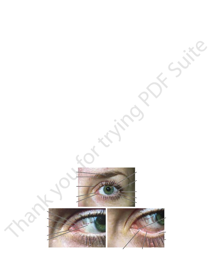
FIGURE 11.17
Left eye of a 29-year-old woman.
The lower eyelid pulled downward and slightly everted to reveal the
view of the medial angle between the eyelids.
An enlarged
The names of structures seen in the examination of the eye.
A.
B.
C.
punctum lacrimale.

Basic Anatomy
551
Muscles of the Eyeball and Eyelids
T A B L E 1 1 . 2
the zygomatic nerve, the zygomaticotemporal nerve, and
nerve, the nerve of the pterygoid canal, the maxillary nerve,
the internal carotid plexus and travels in the deep petrosal
is from
sympathetic postganglionic nerve supply
The
lacrimal gland within the lacrimal nerve.
branch and the zygomaticotemporal nerve. They reach the
join the maxillary nerve. They then pass into its zygomatic
canal. The postganglionic fibers leave the ganglion and
its great petrosal branch and via the nerve of the pterygoid
(sphenopalatine ganglion) via the nervus intermedius and
preganglionic fibers reach the pterygopalatine ganglion
of the facial nerve. The
lacrimal nucleus
derived from the
parasympathetic secretomotor nerve supply
The
the superior fornix of the conjunctiva by 12 ducts.
septum (Fig. 11.16). The gland opens into the lateral part of
anterior and upper part of the orbit posterior to the orbital
palpebrae superioris. It is situated above the eyeball in the
around the lateral edge of the aponeurosis of the levator
which are continuous with each other
palpebral part,
small
orbital part
The lacrimal gland consists of a large
Lacrimal Gland
(see Table 11.4)
Trochlear nerve
Tendinous ring on
Tendinous ring on
Tendinous ring on
Tendinous ring on
Muscle
Origin
Insertion
Nerve Supply
Action
Extrinsic Muscles of Eyeball (Striated Skeletal Muscle)
Superior rectus
posterior wall of
orbital cavity
Superior surface of
eyeball just posterior to
corneoscleral junction
Oculomotor nerve
(3rd cranial nerve)
Raises cornea upward and
medially
Inferior rectus
posterior wall of
orbital cavity
Inferior surface of eyeball just
posterior to corneoscleral
junction
Oculomotor nerve
(3rd cranial nerve)
Depresses cornea
downward and medially
Medial rectus
posterior wall of
orbital cavity
Medial surface of eyeball just
posterior to corneoscleral
junction
Oculomotor nerve
(3rd cranial nerve)
Rotates eyeball so that
cornea looks medially
Lateral rectus
posterior wall of
orbital cavity
Lateral surface of eyeball just
posterior to corneoscleral
junction
Abducent nerve (6th
cranial nerve)
Rotates eyeball so that
cornea looks laterally
Superior oblique
Posterior wall of
orbital cavity
Passes through pulley and
is attached to superior
surface of eyeball beneath
superior rectus
(4th cranial nerve)
Rotates eyeball so that
cornea looks downward
and laterally
Inferior oblique
Floor of orbital cavity
Lateral surface of eyeball
deep to lateral rectus
Oculomotor nerve
(3rd cranial nerve)
Rotates eyeball so that
cornea looks upward and
laterally
Intrinsic Muscles of Eyeball (Smooth Muscle)
Sphincter pupillae
of iris
Parasympathetic via
oculomotor nerve
Constricts pupil
Dilator pupillae
of iris
Sympathetic
Dilates pupil
Ciliary muscle
Parasympathetic via
oculomotor nerve
Controls shape of lens; in
accommodation, makes
lens more globular
Muscles of Eyelids
Orbicularis oculi
Levator palpebrae
superioris
Back of orbital cavity
Anterior surface and upper
margin of superior tarsal
plate
Striated muscle
oculomotor nerve,
smooth muscle
sympathetic
Raises upper lid
Lacrimal Apparatus
and a
is
lacrimal sac
lacrimales pass medially and open into the
The canaliculi
puncta lacrimalis.
through the
lacrimales
From here, the tears enter the
lacus lacrimalis.
The tears circulate across the cornea and accumulate in the
Lacrimal Ducts
finally the lacrimal nerve.
canaliculi
552
CHAPTER 11
the maxilla and the frontal bone.
illa, and the medial margin is formed by the processes of
rior margin is formed by the zygomatic bone and the max
the processes of the frontal and zygomatic bones, the infe
above by the frontal bone, the lateral margin is formed by
is formed
orbital margin
apex posterior (Fig. 11.18). The
The orbit is a pyramidal cavity with its base anterior and its
forced up the duct into the lacrimal sac on blowing the
This prevents air from being
lacrimal fold.
known as the
The opening is guarded by a fold of mucous membrane
bony canal and opens into the inferior meatus of the nose.
The duct descends downward, backward, and laterally in a
emerges from the lower end of the lacrimal sac (Fig. 11.16).
is about 0.5 in. (1.3 cm) long and
nasolacrimal duct
The
nasolacrimal duct.
medial palpebral ligament and is the upper blind end of the
(Fig. 11.16), which lies in the lacrimal groove behind the
The Head and Neck
nose.
The Orbit
Description
-
-
superior oblique
lacrimal gland
levator palpebrae
superioris
superior rectus
lateral rectus
sclera
inferior rectus
inferior oblique
medial rectus
nasociliary nerve
lower division of
oculomotor nerve
infraorbital nerve
inferior oblique
ciliary ganglion
abducent nerve
lateral rectus
trochlear nerve
lacrimal nerve
upper division of
oculomotor nerve
superior rectus
levator palpebrae superioris
supraorbital nerve
trochlear nerve
supratrochlear nerve
optic nerve
trochlea
A
B
C
D
orbital plate of frontal
superior orbital
fissure
greater wing of
sphenoid
zygomatic
optic canal
lesser wing of
sphenoid
maxilla
ethmoid
lacrimal
medial rectus
optic nerve
ophthalmic artery
inferior rectus
lower division of
oculomotor nerve
inferior
orbital
fissure
abducent
nasociliary
upper division of
oculomotor nerve
trochlear nerve
frontal nerve
lacrimal nerve
ophthalmic vein
superior orbital
fissure
superior rectus
supraorbital nerve
supratrochlear nerve
levator palpebrae superioris
superior oblique
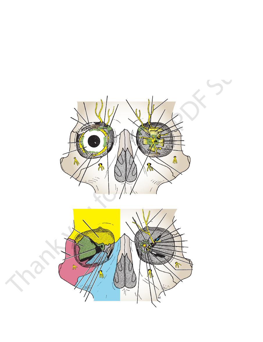
FIGURE 11.18
A.
The optic canal and the superior and inferior orbital fissures on the left side.
forming the walls of the right orbit.
Bones
Muscles and nerves of the left orbit as seen from in front.
Right eyeball exposed from in front. B.
C.
D.