Basic Anatomy
which is about 1 in. (2.5 cm) long. The circular muscle coat
pyloric canal,
is formed by the
pyloric orifice
The
agus (see page 170).
prevents regurgitation of stomach contents into the esoph
be demonstrated here, a physiologic mechanism exists that
stomach (Fig. 5.21). Although no anatomic sphincter can
is where the esophagus enters the
cardiac orifice
The
transverse colon (Fig. 5.11).
extends from the lower part of the greater curvature to the
greater curvature to the spleen, and the greater omentum
omentum (ligament) extends from the upper part of the
the stomach to the pylorus (Fig. 5.21). The gastrosplenic
over the dome of the fundus, and along the left border of
curvature and extends from the left of the cardiac orifice,
is much longer than the lesser
greater curvature
tum. The
5.21). It is suspended from the liver by the lesser omen
ach and extends from the cardiac orifice to the pylorus (Fig.
forms the right border of the stom
lesser curvature
The
(Fig. 5.21).
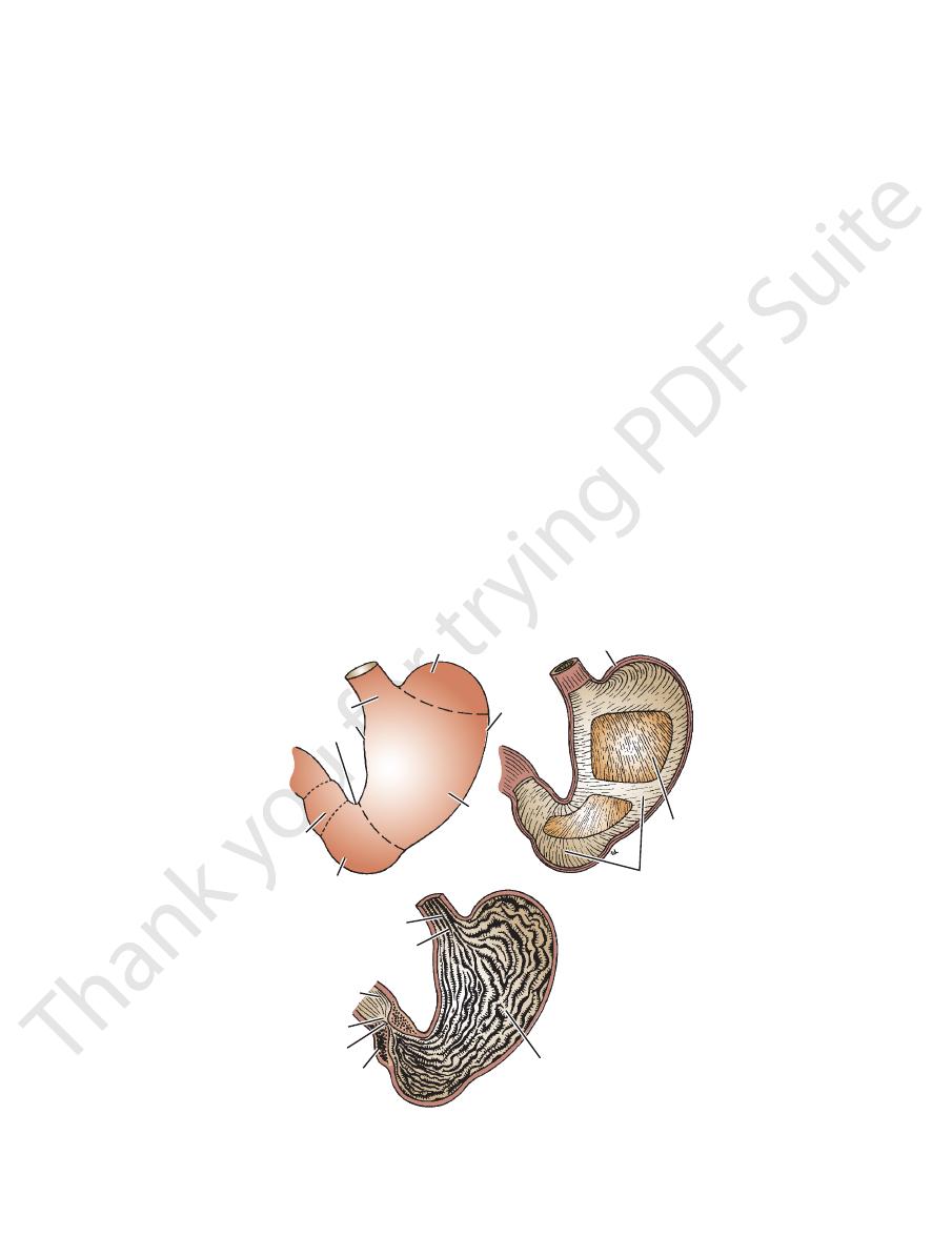
pyloric canal
and the cavity of the pylorus is the
ter,
pyloric sphinc
The thick muscular wall is called the
This is the most tubular part of the stomach.
Pylorus:
ris to the pylorus (Fig. 5.21).
This extends from the incisura angula
Pyloric antrum:
in the lower part of the lesser curvature (Fig. 5.21).
a constant notch
incisura angularis,
to the level of the
This extends from the level of the cardiac orifice
Body:
to the left of the cardiac orifice. It is usually full of gas.
This is dome-shaped and projects upward and
Fundus:
(Fig. 5.21):
The stomach is divided into the following parts
position of the body, and the phase of respiration.
same person and depends on the volume of its contents, the
stomach). Its shape undergoes considerable variation in the
and elongated vertically in the tall, thin person (J-shaped
arranged in the short, obese person (steer-horn stomach)
mobile in between. It tends to be high and transversely
The stomach is relatively fixed at both ends but is very
(Fig. 5.21).
posterior surface
anterior
faces, an
and two sur
lesser curvatures;
greater
curvatures, the
two
pyloric orifices;
cardiac
and has two openings, the
ach lies under cover of the lower ribs. It is roughly J-shaped
into the epigastric and umbilical regions. Much of the stom
men, extending from beneath the left costal margin region
The stomach is situated in the upper part of the abdo
so that efficient digestion and absorption can take place.
trols the rate of delivery of the chyme to the small intestine
and it con
chyme,
gastric secretions to form a semifluid
it has a capacity of about 1500 mL), it mixes the food with
and has three main functions: It stores food (in the adult
The stomach is the dilated portion of the alimentary canal
Location and Description
171
Stomach
-
-
-
and
and
-
and a
■
■
■
■
■
■
-
■
■
-
-
-
-
fundus
cardiac orifice
lesser curvature
incisura angularis
pylorus
antrum
body
greater
curvature
longitudinal muscle coat
oblique muscle
coat
circular muscle coat
esophagus
duodenum
pyloric orifice
pyloric canal
pyloric sphincter
longitudinal folds of mucous coat
cardiac orifice
FIGURE 5.21
Stomach showing the parts, muscular coats, and mucosal lining. Note the increased thickness of the circular
muscle forming the pyloric sphincter.

172
CHAPTER 5
The
passes down to the pylorus (Fig. 5.24).
branch passes up to the liver, and from this a pyloric branch
supply the anterior surface of the stomach. A large hepatic
may be single or multiple, then divides into branches that
on the anterior surface of the esophagus. The trunk, which
rax mainly from the left vagus nerve, enters the abdomen
which is formed in the tho
anterior vagal trunk,
The
and left vagus nerves (Fig. 5.24).
the celiac plexus and parasympathetic fibers from the right
The nerve supply includes sympathetic fibers derived from
Nerve Supply
abdominal wall.
around the root of the celiac artery on the posterior
the stomach eventually passes to the celiac nodes located
ploic nodes, and the short gastric nodes. All lymph from
left and right gastric nodes, the left and right gastroepi
The lymph vessels (Fig. 5.23) follow the arteries into the
Lymph Drainage
joins the superior mesenteric vein.
right gastroepiploic vein
join the splenic vein. The
veins
left gastroepiploic
and the
short gastric veins
vein. The
drain directly into the portal
right gastric veins
and
left
The veins drain into the portal circulation (Fig. 5.22). The
Veins
greater curvature.
left and supplies the stomach along the lower part of the
troduodenal branch of the hepatic artery. It passes to the
arises from the gas
right gastroepiploic artery
The
along the upper part of the greater curvature.
gastrosplenic omentum (ligament) to supply the stomach
artery at the hilum of the spleen and passes forward in the
arises from the splenic
left gastroepiploic artery
The
plenic omentum (ligament) to supply the fundus.
at the hilum of the spleen and pass forward in the gastros
arise from the splenic artery
short gastric arteries
The
stomach.
the lesser curvature. It supplies the lower right part of the
the upper border of the pylorus and runs to the left along
arises from the hepatic artery at
right gastric artery
The
right part of the stomach.
It supplies the lower third of the esophagus and the upper
then descends along the lesser curvature of the stomach.
passes upward and to the left to reach the esophagus and
arises from the celiac artery. It
left gastric artery
The
artery (Fig. 5.20).
The arteries are derived from the branches of the celiac
Arteries
mesocolon, and the transverse colon (Figs. 5.4, 5.6,
kidney, the splenic artery, the pancreas, the transverse
the left suprarenal gland, the upper part of the left
The lesser sac, the diaphragm, the spleen,
Posteriorly:
left lobe of the liver (Figs. 5.2 and 5.6)
margin, the left pleura and lung, the diaphragm, and the
The anterior abdominal wall, the left costal
Anteriorly:
Relations
plenic omentum and the greater omentum.
lesser omentum and the greater curvature as the gastros
rounds the stomach. It leaves the lesser curvature as the
(visceral peritoneum) completely sur
peritoneum
The
nal fibers, circular fibers, and oblique fibers (Fig. 5.21).
contains longitudi
muscular wall of the stomach
The
ten out when the stomach is distended.
mainly longitudinal in direction (Fig. 5.21). The folds flat
that are
rugae,
cular and is thrown into numerous folds, or
of the stomach is thick and vas
mucous membrane
The
sphincter.
nerve plexus in its wall and reflexly cause relaxation of the
the stomach due to filling will stimulate the myenteric
ach and duodenal walls. For example, the stretching of
local nervous and hormonal influences from the stom
from the vagi. In addition, the pylorus is controlled by
fibers from the sympathetic system and inhibitory fibers
tents into the duodenum. The sphincter receives motor
The pyloric sphincter controls the outflow of gastric con
Function of the Pyloric Sphincter
stomach.
be recognized by a slight constriction on the surface of the
pylorus lies on the transpyloric plane, and its position can
(Fig. 5.21). The
pyloric sphincter
tomic and physiologic
of the stomach is much thicker here and forms the ana
The Abdomen: Part II—The Abdominal Cavity
-
-
-
-
-
-
-
-
■
■
■
■
and 5.11)
Blood Supply
-
-
-
-
with peritoneum and has the lesser omentum attached to
in that it is covered on its anterior and posterior surfaces
first inch (2.5 cm) of the duodenum resembles the stomach
curves around the head of the pancreas (Fig. 5.26). The
openings of the bile and pancreatic ducts. The duodenum
long, which joins the stomach to the jejunum. It receives the
The duodenum is a C-shaped tube, about 10 in. (25 cm)
Location and Description
the ileum.
divided into three parts: the duodenum, the jejunum, and
and food absorption takes place in the small intestine. It is
ileocecal junction (Fig. 5.1). The greater part of digestion
canal and extends from the pylorus of the stomach to the
The small intestine is the longest part of the alimentary
the vagi.
from the sympathetic system and inhibitory fibers from
stomach. The pyloric sphincter receives motor fibers
gastric glands and motor to the muscular wall of the
the parasympathetic vagal fibers are secretomotor to the
a proportion of pain-transmitting nerve fibers, whereas
The sympathetic innervation of the stomach carries
(Fig. 5.24).
intestine as far as the splenic flexure and to the pancreas
and superior mesenteric plexuses and is distributed to the
surface of the stomach. A large branch passes to the celiac
then divides into branches that supply mainly the posterior
men on the posterior surface of the esophagus. The trunk
rax mainly from the right vagus nerve, enters the abdo
which is formed in the tho
posterior vagal trunk,
-
-
Small Intestine
Duodenum