Basic Anatomy
181
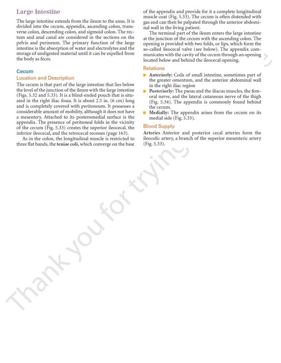
teniae coli
appendices epiploicae
ileocecal valve
frenulum of valve
orifice of appendix
cecum
vascular fold anterior cecal artery
bloodless fold
appendix
appendicular artery
mesoappendix
lymph nodes
ileum
posterior cecal artery
ileal artery
ileocolic artery
colic artery
FIGURE 5.33
. The edge of the
Cecum and appendix. Note that the appendicular artery is a branch of the posterior cecal artery
mesoappendix has been cut to show the peritoneal layers.
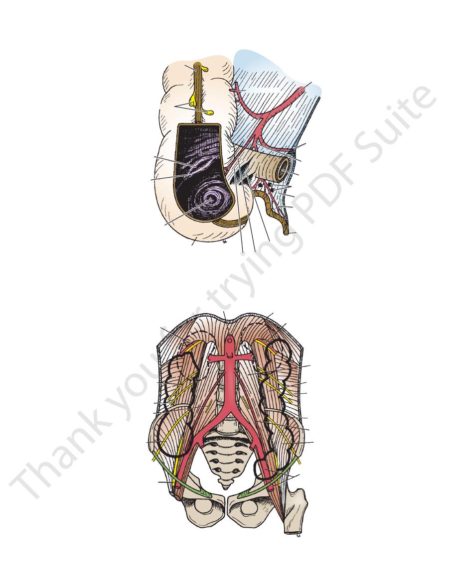
subcostal vessels and
nerve
aorta
diaphragm
rib 12
psoas
quadratus lumborum
transversus muscle
descending colon
testicular artery
iliacus
external iliac artery
femoral nerve
lateral cutaneous
nerve of thigh
genitofemoral nerve
ilioinguinal nerve
iliohypogastric nerve
FIGURE 5.34
Posterior abdominal wall showing posterior relations of the kidneys and the colon.

182
CHAPTER 5
left colic flexure
then ascends to the
by the transverse mesocolon from the pancreas (Fig. 5.6). It
lobe of the liver (Fig. 5.4) and hangs downward, suspended
region. It begins at the right colic flexure below the right
extends across the abdomen, occupying the umbilical
The transverse colon is about 15 in. (38 cm) long and
Location and Description
Transverse Colon
superior mesenteric plexus supply this area of the colon.
Sympathetic and parasympathetic (vagus) nerves from the
superior mesenteric nodes.
course of the colic blood vessels and ultimately reach the
The lymph vessels drain into lymph nodes lying along the
Lymph Drainage
into the superior mesenteric vein.
The veins correspond to the arteries and drain
Veins
rior mesenteric artery (Fig. 5.32) supply this area.
The ileocolic and right colic branches of the supe
Arteries
it (Fig. 5.34).
iliohypogastric and the ilioinguinal nerves cross behind
muscle, and the lower pole of the right kidney. The
lumborum, the origin of the transversus abdominis
The iliacus, the iliac crest, the quadratus
Posteriorly:
omentum, and the anterior abdominal wall (Figs. 5.2
Coils of small intestine, the greater
Anteriorly:
Relations
ascending colon, binding it to the posterior abdominal wall.
colon. The peritoneum covers the front and the sides of the
and becomes continuous with the transverse
colic flexure,
right
of the liver, where it turns to the left, forming the
from the cecum to the inferior surface of the right lobe
in the right lower quadrant (Fig. 5.35). It extends upward
The ascending colon is about 5 in. (13 cm) long and lies
Location and Description
the 10th thoracic segment.
sympathetic nerves and enter the spinal cord at the level of
tion of visceral pain from the appendix accompany the
plexus. Afferent nerve fibers concerned with the conduc
sympathetic (vagus) nerves from the superior mesenteric
The appendix is supplied by the sympathetic and para
enteric nodes.
mesoappendix and then eventually into the superior mes
The lymph vessels drain into one or two nodes lying in the
Lymph Drainage
cecal vein.
The appendicular vein drains into the posterior
Veins
rior cecal artery (Fig. 5.33).
The appendicular artery is a branch of the poste
Arteries
positions are the most common sites.
behind the terminal part of the ileum. The first and second
along the lateral side of the cecum, and (d) in front of or
wall, (b) coiled up behind the cecum, (c) projecting upward
(a) hanging down into the pelvis against the right pelvic
of movement and may be found in the following positions:
The tip of the appendix is subject to a considerable range
Common Positions of the Tip of the Appendix
tinuous longitudinal muscle coat (Figs. 5.32 and 5.33).
base of the appendix, where they converge to form a con
tifying the teniae coli of the cecum and tracing them to the
abdomen, the base of the appendix is easily found by iden
iliac spine to the umbilicus (McBurney’s point). Inside the
of the way up the line joining the right anterior superior
to the anterior abdominal wall its base is situated one third
The appendix lies in the right iliac fossa, and in relation
oappendix contains the appendicular vessels and nerves.
The mes
mesoappendix.
a short mesentery of its own, the
which is attached to the mesentery of the small intestine by
the appendix is free. It has a complete peritoneal covering,
below the ileocecal junction (Fig. 5.33). The remainder of
posteromedial surface of the cecum about 1 in. (2.5 cm)
from 3 to 5 in. (8 to 13 cm). The base is attached to the
ing a large amount of lymphoid tissue. It varies in length
The appendix (Fig. 5.1) is a narrow, muscular tube contain
Location and Description
duced by the stomach, causes relaxation of the muscle tone.
which is pro
gastrin,
the cecum is distended; the hormone
colon. The smooth muscle tone is reflexly increased when
ter and controls the flow of contents from the ileum into the
by physiologists) serves as a sphinc
ileocecal sphincter
The circular muscle of the lower end of the ileum (called
the prevention of reflux of cecal contents into the ileum.
the orifice of the ileum. The valve plays little or no part in
horizontal folds of mucous membrane that project around
A rudimentary structure, the ileocecal valve consists of two
Ileocecal Valve
(vagus) nerves form the superior mesenteric plexus.
Branches from the sympathetic and parasympathetic
and finally reach the superior mesenteric nodes.
The lymph vessels pass through several mesenteric nodes
Lymph Drainage
into the superior mesenteric vein.
The veins correspond to the arteries and drain
Veins
The Abdomen: Part II—The Abdominal Cavity
Nerve Supply
the
-
-
Appendix
-
-
-
-
Blood Supply
-
-
Nerve Supply
-
-
Ascending Colon
■
■
and 5.3).
■
■
Blood Supply
-
Nerve Supply
transverse mesocolon, the position of the transverse colon
the inferior border (Fig. 5.6). Because of the length of the
the posterior layers of the greater omentum are attached to
attached to the superior border of the transverse colon, and
rior border of the pancreas (Fig. 5.6). The mesentery is
verse colon, suspends the transverse colon from the ante
or mesentery of the trans
transverse mesocolon,
The
(Fig. 5.35).
ment
phrenicocolic liga
suspended from the diaphragm by the
left colic flexure is higher than the right colic flexure and is
below the spleen. The
-
-
-
Basic Anatomy
inferior mesenteric artery (Fig. 5.36) supply this area.
The left colic and the sigmoid branches of the
Arteries
nerve (Fig. 5.34) also lie posteriorly.
the lateral cutaneous nerve of the thigh, and the femoral
psoas. The iliohypogastric and the ilioinguinal nerves,
ratus lumborum, the iliac crest, the iliacus, and the left
origin of the transversus abdominis muscle, the quad
The lateral border of the left kidney, the
Posteriorly:
tum, and the anterior abdominal wall (Figs. 5.2 and 5.3)
Coils of small intestine, the greater omen
Anteriorly:
Relations
and the sides and binds it to the posterior abdominal wall.
moid colon, see page 263.) The peritoneum covers the front
it becomes continuous with the sigmoid colon. (For the sig
downward from the left colic flexure, to the pelvic brim, where
in the left upper and lower quadrants (Fig. 5.35). It extends
The descending colon is about 10 in. (25 cm) long and lies
Location and Description
enteric plexus.
thetic pelvic splanchnic nerves through the inferior mes
distal third is innervated by sympathetic and parasympa
vagal nerves through the superior mesenteric plexus; the
The proximal two thirds are innervated by sympathetic and
the colic nodes and then into the inferior mesenteric nodes.
into the superior mesenteric nodes; the distal third drains into
The proximal two thirds drain into the colic nodes and then
Lymph Drainage
into the superior and inferior mesenteric veins.
The veins correspond to the arteries and drain
Veins
artery, a branch of the inferior mesenteric artery (Fig. 5.36).
(Fig. 5.32). The distal third is supplied by the left colic
dle colic artery, a branch of the superior mesenteric artery
The proximal two thirds are supplied by the mid
Arteries
the ileum (Fig. 5.35)
head of the pancreas, and the coils of the jejunum and
The second part of the duodenum, the
Posteriorly:
(Fig. 5.6)
abdominal wall (umbilical and hypogastric regions)
The greater omentum and the anterior
Anteriorly:
Relations
as the pelvis.
is extremely variable and may sometimes reach down as far
183
■
■
■
■
Blood Supply
-
Nerve Supply
-
-
Descending Colon
-
■
■
-
■
■
-
Blood Supply
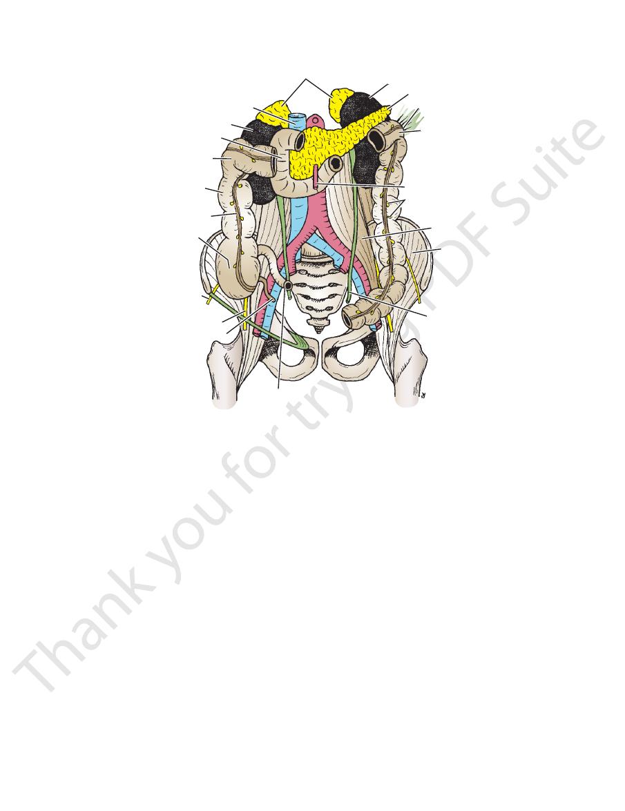
suprarenal glands
left kidney
pancreas
phrenicocolic ligament
left colic flexure
superior mesenteric artery
appendices epiploicae
psoas
iliacus
ureter
ileum
appendix
femoral nerve
lateral cutaneous
nerve of thigh
cecum
teniae coli
ascending colon
right colic flexure
duodenum
right kidney
inferior vena cava
FIGURE 5.35
Abdominal cavity showing the terminal part of the ileum, the cecum, the appendix, the ascending colon, the
right colic flexure, the left colic flexure, and the descending colon. Note the teniae coli and the appendices epiploicae.
184
CHAPTER 5
behind the stomach (Fig. 5.4). On reaching the left kidney,
a wavy course along the upper border of the pancreas and
The large splenic artery runs to the left in
Splenic Artery
artery (Fig. 5.20).
vature of the stomach. It anastomoses with the right gastric
branches, and then turns to the right along the lesser cur
the cardiac end of the stomach, gives off a few esophageal
The small left gastric artery runs to
Left Gastric Artery
hepatic arteries.
It has three terminal branches: the left gastric, splenic, and
celiac plexus and lies behind the lesser sac of peritoneum.
12th thoracic vertebra (Fig. 5.20). It is surrounded by the
commencement of the abdominal aorta at the level of the
The celiac artery or trunk is very short and arises from the
Celiac Artery
down the anal canal.
from the distal one third of the transverse colon to halfway
the artery of the hindgut and supplies the large intestine
of the transverse colon. The inferior mesenteric artery is
second part of the duodenum as far as the distal one third
supplies the gastrointestinal tract from the middle of the
superior mesenteric artery is the artery of the midgut and
far as the middle of the second part of the duodenum. The
tract from the lower one third of the esophagus down as
the artery of the foregut and supplies the gastrointestinal
trated diagrammatically in Figure 5.46. The celiac artery is
development of the different parts of the gut are illus
The arterial supply to the gut and its relationship to the
Arterial Supply
Tract
plexus.
pelvic splanchnic nerves through the inferior mesenteric
The nerve supply is the sympathetic and parasympathetic
teric artery.
mesenteric nodes around the origin of the inferior mesen
Lymph drains into the colic lymph nodes and the inferior
Lymph Drainage
into the inferior mesenteric vein.
The veins correspond to the arteries and drain
Veins
The Abdomen: Part II—The Abdominal Cavity
-
Nerve Supply
Blood Supply of the Gastrointestinal
-
-
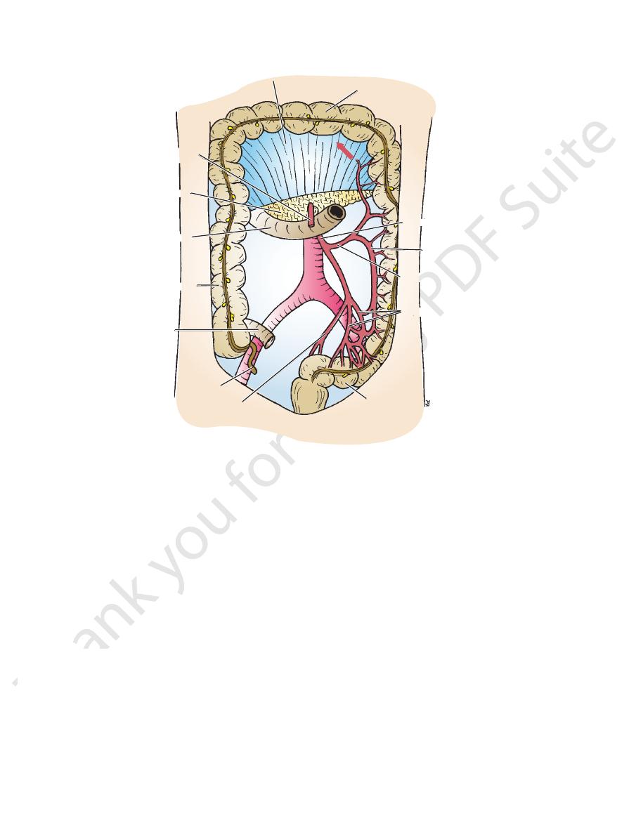
transverse mesocolon
transverse colon
inferior mesenteric artery
marginal artery
left colic artery
sigmoid arteries
sigmoid colon
superior rectal artery
appendix
ileum
ascending colon
duodenum
pancreas
superior mesenteric
artery
FIGURE 5.36
Inferior mesenteric artery and its branches. Note that this artery supplies the large bowel from the distal third of
arrow
artery (
the transverse colon to halfway down the anal canal. It anastomoses with the middle colic branch of the superior mesenteric
).