216
CHAPTER 5
the psoas, following the pelvic brim (Fig. 5.63). It gives off
The external iliac artery runs along the medial border of
riorly by the ureter (Fig. 5.72).
tion, the common iliac artery on each side is crossed ante
into the external and internal iliac arteries. At the bifurca
Each artery ends in front of the sacroiliac joint by dividing
medial border of the psoas muscle (Figs. 5.63 and 5.72).
lumbar vertebra and run downward and laterally along the
branches of the aorta. They arise at the level of the 4th
The right and left common iliac arteries are the terminal
These branches are summarized in Diagram 5.1.
and the median sacral artery (Fig. 5.72)
Three terminal branches: the two common iliac arteries
phrenic artery and four lumbar arteries
Five lateral abdominal wall branches: the inferior
renal artery, and testicular or ovarian artery
Three lateral visceral branches: the suprarenal artery,
The Abdomen: Part II—The Abdominal Cavity
■
■
■
■
■
■
Common Iliac Arteries
-
-
External Iliac Artery
Development of the Suprarenal Glands
pathetic nerve fibers grow into the medulla and influence the
tion and is arranged in cords and clusters. Preganglionic sym
By this means, the medulla comes to occupy a central posi
of the neural crest. These invade the cortex on its medial side.
The cortex develops from the coelomic mesothelium covering
the posterior abdominal wall. At first, a fetal cortex is formed;
later, it becomes covered by a second final cortex. After birth,
the fetal cortex retrogresses, and its involution is largely com-
pleted in the first few weeks of life.
The medulla is formed from the sympathochromaffin cells
-
-
activity of the medullary cells.
Susceptibility to Trauma at Birth
At birth, the suprarenal glands are relatively large because of
the presence of the fetal cortex; later, when this part of the
cortex involutes, the gland becomes reduced in size. During
the process of involution, the cortex is friable and susceptible
to damage and severe hemorrhage.
E M B R Y O L O G I C N O T E S
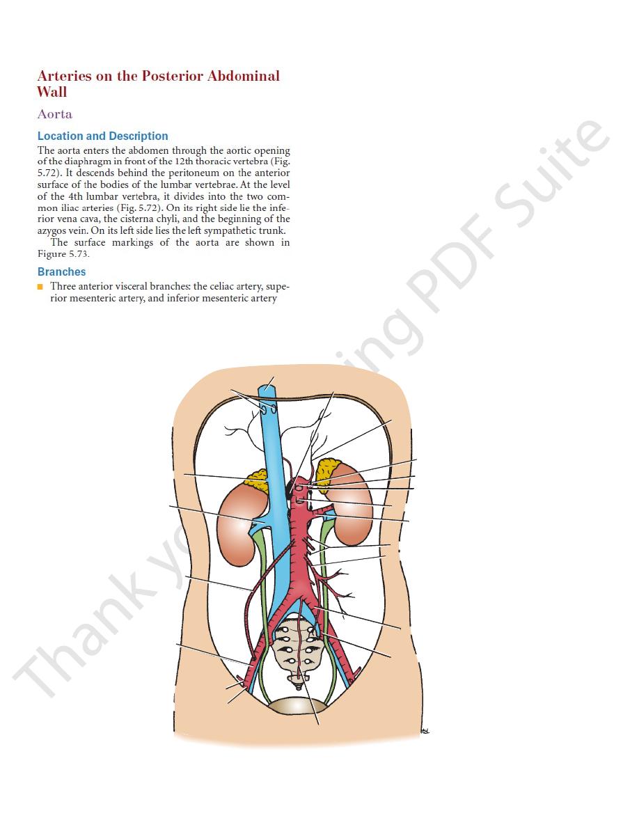
inferior vena cava
hepatic veins
renal vein
testicular artery
external iliac
artery
inferior epigastric artery
median sacral artery
internal iliac artery
common iliac artery
inferior mesenteric artery
lumbar arteries
renal artery
superior mesenteric artery
suprarenal artery
celiac artery
sympathetic trunk
cisterna chyli
inferior phrenic artery
suprarenal vein
deep circumflex iliac artery
y
FIGURE 5.72
Aorta and inferior vena cava.

Basic Anatomy
217
celiac artery
artery
artery
internal iliac
vessels
xiphisternal joint
xiphisternal joint
plane (L1)
plane (L1)
(T9)
(T9)
(L4)
(L4)
inter
intercristal plane
cristal plane
transpyloric
transpyloric
(T12)
mesenteric
inferior mesenteric
(L3)
inferior vena cava
iliac
vessels
anterior superior
iliac spine
common iliac
vessels
aorta
external
superior
(L1)
symphysis pubis
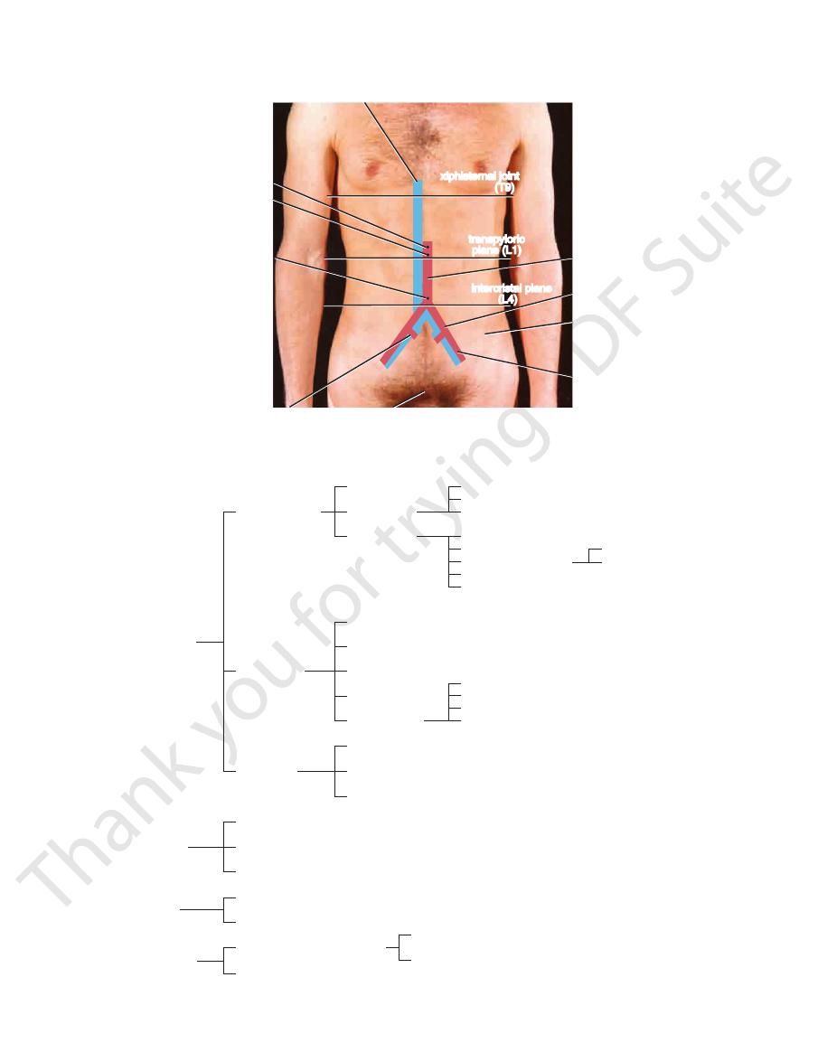
FIGURE 5.73
Surface markings of the aorta and its branches and the inferior vena cava on the anterior abdominal wall.
superior pancreatico-
duodenal artery
1. Three anterior
visceral branches
2. Three lateral
visceral branches
3. Five lateral
abdominal
wall branches
4. Three terminal
branches
a. Celiac artery
b. Superior
mesenteric
artery
c. Inferior
mesenteric
artery
a. Suprarenal artery
b. Renal artery
c. Testicular or ovarian artery
a. Inferior phrenic artery
b. Four lumbar arteries
a. Two common iliac arteries
b. Median sacral artery
left gastric artery
splenic artery
hepatic artery
jejunal and ileal arteries
inferior pancreaticoduodenal artery
middle colic artery
right colic artery
ileocolic artery
left colic artery
sigmoid arteries (two or three)
superior rectal artery
external iliac artery
internal iliac artery
short gastric arteries (six)
splenic arteries (six)
left gastroepiploic artery
cystic artery
right gastric artery
gastroduodenal artery
right hepatic artery
left hepatic artery
anterior cecal artery
posterior cecal artery—appendicular artery
ileal artery
colic artery
right gastroepiploic artery
DIAGRAM 5.1
Branches of Abdominal Aorta

218
CHAPTER 5
abdominal portion of the gastrointestinal tract drains to
If one remembers that the venous blood from the
Diagram 5.2.
The tributaries of the inferior vena cava are summarized in
median sacral vein
Three veins of origin: two common iliac veins and the
phrenic vein and four lumbar veins
Five lateral abdominal wall tributaries: the inferior
drains into the left renal vein)
veins, and right testicular or ovarian vein (the left vein
vein (the left vein drains into the left renal vein), renal
Three lateral visceral tributaries: the right suprarenal
Two anterior visceral tributaries: the hepatic veins
(Fig. 5.72):
The inferior vena cava has the following tributaries
Tributaries
in Figure 5.73.
The surface markings of the inferior vena cava are shown
from the portal vein (Fig. 5.7).
entrance into the lesser sac separates the inferior vena cava
gin and the right ureter lies close to its right border. The
The right sympathetic trunk lies behind its right mar
of the heart.
the 8th thoracic vertebra, and drains into the right atrium
pierces the central tendon of the diaphragm at the level of
vertebra (Fig. 5.72). It ascends on the right side of the aorta,
the right common iliac artery at the level of the 5th lumbar
It is formed by the union of the common iliac veins behind
body below the diaphragm to the right atrium of the heart.
The inferior vena cava conveys most of the blood from the
Location and Description
Inferior Vena Cava
Wall
Veins on the Posterior Abdominal
described on page 256.
front of the sacroiliac joint (Fig. 5.72). Its further course is
The internal iliac artery passes down into the pelvis in
abdominal wall.
and the iliac crest, supplying the muscles of the anterior
5.72). It ascends laterally to the anterior superior iliac spine
iliac artery arises close to the inferior epigastric artery (Fig.
behind the rectus abdominis muscle. The deep circumflex
deep inguinal ring (Fig. 4.4) and enters the rectus sheath
passes upward and medially along the medial margin of the
epigastric artery arises just above the inguinal ligament. It
nal ligament to become the femoral artery. The inferior
The artery enters the thigh by passing under the ingui
(Fig. 5.72).
branches
deep circumflex iliac
inferior epigastric
The Abdomen: Part II—The Abdominal Cavity
the
and
-
Internal Iliac Artery
-
■
■
■
■
■
■
■
■
Aortic Aneurysms
denly narrows may be a lodging site for an embolus discharged
Localized or diffuse dilatations of the abdominal part of the
aorta (aneurysms) usually occur below the origin of the renal
arteries. Most result from atherosclerosis, which causes
weakening of the arterial wall, and occur most commonly
in elderly men. Large aneurysms should be treated by open
surgical repair. Endovascular repair can also be used by the
introduction of a stent graft through one of the iliac arteries
with access through the femoral arteries in the groin.
Embolic Blockage of the Abdominal Aorta
The bifurcation of the abdominal aorta where the lumen sud-
from the heart. Severe ischemia of the lower limbs results.
C L I N I C A L N O T E S
1. Two anterior visceral tributaries—the hepatic veins
2. Three lateral visceral tributaries
4. Three tributaries of origin
a. Right suprarenal vein
(the left drains into the left renal vein)
b. Renal veins
c. Right testicular or ovarian vein
(the left drains into the left renal vein)
a. Inferior phrenic vein
b. Four lumbar veins
a. Two common iliac veins
b. Median sacral vein
external iliac vein
internal iliac vein
3. Five lateral abdominal wall
tributaries
DIAGRAM 5.2
Tributaries of Inferior Vena Cava

Basic Anatomy
lumbar) chain (Fig. 5.76).
a preaortic and a right and left lateral aortic (para-aortic or
The lymph nodes are closely related to the aorta and form
Lymph Nodes
Abdominal Wall
Lymphatics on the Posterior
The portal vein is described on page 194.
Portal Vein
epiploic vein (Fig. 5.22).
the inferior pancreaticoduodenal vein and the right gastro
branches of the superior mesenteric artery and also receives
the portal vein. It receives tributaries that correspond to the
neck of the pancreas, where it joins the splenic vein to form
in front of the third part of the duodenum and behind the
on the right side of the superior mesenteric artery. It passes
within the root of the mesentery of the small intestine and
junction and runs upward on the posterior abdominal wall
culation (Figs. 5.22, 5.26, and 5.48). It begins at the ileocecal
The superior mesenteric vein is a tributary of the portal cir
Superior Mesenteric Vein
enteric vein.
joined by veins from the pancreas and the inferior mes
the neck of the pancreas to form the portal vein. It is
the pancreas. It joins the superior mesenteric vein behind
right within the splenicorenal ligament and runs behind
gastroepiploic veins (Figs. 5.22 and 5.48). It passes to the
eral veins and is then joined by the short gastric and left
It begins at the hilum of the spleen by the union of sev
The splenic vein is a tributary of the portal circulation.
Splenic Vein
that correspond to the branches of the artery.
the splenic vein behind the pancreas. It receives tributaries
mesenteric artery and the duodenojejunal flexure and joins
the posterior abdominal wall on the left side of the inferior
superior rectal vein (Figs. 5.22, 5.26, and 5.48). It passes up
circulation. It begins halfway down the anal canal as the
The inferior mesenteric vein is a tributary of the portal
Inferior Mesenteric Vein
to the branches of the abdominal portion of the aorta.
utaries of the inferior vena cava correspond rather closely
first into the left renal vein, then it is apparent that the trib
that the left suprarenal and testicular or ovarian veins drain
the liver by means of the tributaries of the portal vein, and
219
-
-
-
-
-
Trauma to the Abdominal Aorta
pression and eventual blockage of the inferior vena cava. This
uterus during the later stages of pregnancy. This produces edema
The inferior vena cava is commonly compressed by the enlarged
Because of the multiple anastomoses of the tributaries of the
difficult. Moreover, the thin wall of the vena cava makes it prone
presence of the right costal margin make a surgical approach
denum, and mesentery of the small intestine and the blocking
anatomic inaccessibility of the vessel behind the liver, duo
the fact that the contained blood is under low pressure. The
Injuries to the inferior vena cava are commonly lethal, despite
However, the collateral blood flow does prevent tissue death in
circulation is established, but it is physiologically inadequate.
Because the progress of the disease is slow, some collateral
thromboendarterectomy or a bypass graft should be considered.
arteries. In otherwise healthy individuals, surgical treatment by
impotence, the latter caused by lack of blood in the internal iliac
cal symptoms of pain in the legs on walking (claudication) and
Gradual occlusion of the bifurcation of the abdominal aorta,
caused by the aortic blood pressure. Unless quickly diagnosed
vertebral column, while the delayed rupture of the adventitia is
caused by the sudden compression of the aorta against the
tia. The initial rupture of the intima and media is probably mainly
on automobile crashes. Rupture of the tunica intima and media
Blunt trauma to the aorta is most commonly caused by head-
occurs and is quickly followed by rupture of the turnica adventi-
by MRI, and surgical treatment instituted, death follows.
Obliteration of the Abdominal Aorta and Iliac Arteries
produced by atherosclerosis, results in the characteristic clini-
both lower limbs, although skin ulcers may occur.
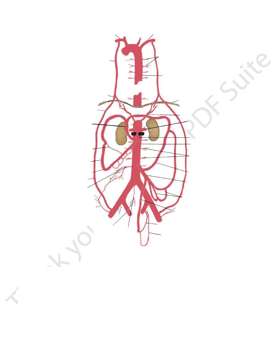
The collateral circulation of the abdominal aorta is shown in
Figure 5.74.
Trauma to the Inferior Vena Cava
-
to extensive tears.
inferior vena cava (Fig. 5.75), it is impossible in an emergency to
ligate the vessel. Most patients have venous congestion of the
lower limbs.
Compression of the Inferior Vena Cava
of the ankles and feet and temporary varicose veins.
Malignant retroperitoneal tumors can cause severe com-
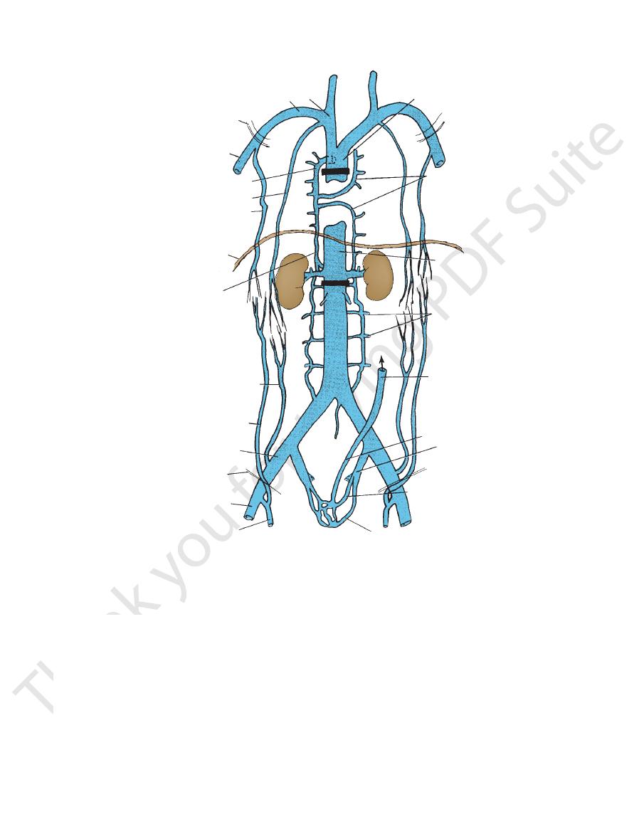
results in the dilatation of the extensive anastomoses of the
tributaries (Fig. 5.75). This alternative pathway for the blood
to return to the right atrium of the heart is commonly referred
to as the caval–caval shunt. The same pathway comes into
effect in patients with a superior mediastinal tumor compressing
the superior vena cava. Clinically, the enlarged subcutaneous
anastomosis between the lateral thoracic vein, a tributary of
the axillary vein; and the superficial epigastric vein, a tributary
of the femoral vein, may be seen on the thoracoabdominal wall
(Fig. 5.75).
C L I N I C A L N O T E S

220
CHAPTER 5
descend from the lower part of the thorax.
and left lumbar trunks, and some small lymph vessels that
The cisterna chyli receives the intestinal trunk, the right
on the right side of the aorta (Fig. 5.76).
diaphragm in front of the first two lumbar vertebrae and
This lies just below the
cisterna chyli.
gated lymph sac, the
commences in the abdomen as an elon
thoracic duct
The
Lymph Vessels
(see Fig. 1.18 and below).
lumbar trunks
left
right
nodes. The efferent lymph vessels form the
vessels of the abdominal walls; and from the common iliac
fundus of the uterus in the female; from the deep lymph
testes in the male and from the ovaries, uterine tubes, and
drain lymph from the kidneys and suprarenals; from the
lateral aortic (para-aortic or lumbar) lymph nodes
The
large intestinal trunk (see Fig. 1.18 and below).
greater part of the liver. The efferent lymph vessels form the
anal canal, and from the spleen, pancreas, gallbladder, and
the lower one third of the esophagus to halfway down the
the lymph from the gastrointestinal tract, extending from
respectively. They drain
inferior mesenteric lymph nodes,
celiac, superior mesenteric,
and are referred to as the
celiac, superior mesenteric, and inferior mesenteric arteries
lie around the origins of the
preaortic lymph nodes
The
The Abdomen: Part II—The Abdominal Cavity
and
and
-
right subclavian artery
left subclavian artery
internal thoracic
artery
posterior intercostal
arteries
thoracic part of aorta
musculophrenic artery
diaphragm
superior epigastric
artery
phrenic artery
left renal artery
middle colic artery
superior mesenteric
artery
marginal artery
abdominal aorta
lumbar arteries
ileocolic artery
left colic artery
inferior mesenteric
artery
sigmoid arteries
superior rectal
artery
middle rectal artery
inferior rectal artery
median
sacral
artery
internal iliac artery
deep circumflex
iliac artery
fourth lumbar
artery
inferior epigastric artery
right colic artery
thoracic
posterior intercos
arteries
thoracic part of
c artery
diaph
c
phre
sig
superio
artery
middle rectal arter
inferior rectal a
median
sacral
artery
l iliac artery
x
FIGURE 5.74
The possible collateral circulations of the abdominal aorta. Note the great dilatation of the mesenteric arteries
black bar
and their branches, which occurs if the aorta is slowly blocked just below the level of the renal arteries (
).

Basic Anatomy
lateral cutaneous nerve of the thigh
labium majus. The
canal to supply the skin of the groin and the scrotum or
wall, and the ilioinguinal nerve passes through the inguinal
supplies the skin of the lower part of the anterior abdominal
abdominal walls (see page 124). The iliohypogastric nerve
(L1) enter the lateral and anterior
ilioinguinal nerves
iliohypogastric
above downward (Fig. 5.34). The
from the lateral border of the psoas, in that order from
cutaneous nerve of the thigh, and femoral nerve emerge
The iliohypogastric nerve, ilioinguinal nerve, lateral
borders of the muscle and from its anterior surface.
branches of the plexus emerge from the lateral and medial
white rami communicantes to the sympathetic trunk. The
from the sympathetic trunk, and the upper two give off
(Fig. 5.77). The anterior rami receive gray rami
communicantes
cle from the anterior rami of the upper four lumbar nerves
ways supplying the lower limb, is formed in the psoas mus
The lumbar plexus, which is one of the main nervous path
Wall
emphasized on page 132.
The importance of the lymph drainage of the testis was
Lymphatic Drainage of the Gonads
221
Nerves on the Posterior Abdominal
Lumbar Plexus
-
-
and
brachiocephalic
vein
subclavian vein
first rib
axillary vein
azygos vein
internal thoracic vein
lateral thoracic vein
diaphragm
ascending lumbar
vein
inferior epigastric vein
superficial epigastric
vein
external iliac vein
inguinal ligament
femoral vein
great saphenous
vein
superior vena cava
hemiazygos veins
inferior vena cava
lumbar veins
inferior mesenteric
vein ascending to
portal vein
superior rectal vein
internal iliac vein
middle rectal vein
inferior rectal vein
FIGURE 5.75
The possible collateral circulations of the superior and inferior venae cavae. Note the alternative pathways that
). Note also the connections that exist between the portal circulation and systemic veins in the anal canal.
black bar
lower
). Similar pathways exist if the inferior vena cava becomes blocked below the renal veins (
upper black bar
azygos vein (
exist for blood to return to the right atrium of the heart if the superior vena cava becomes blocked below the entrance of the
