258
CHAPTER 6
umn to the bony pelvis.
to transmit the weight of the body from the vertebral col
and the joint becomes fibrosed. Their primary function is
these joints. In older people, the synovial cavity disappears
A small but limited amount of movement is possible at
Movements
tip of the fifth lumbar transverse process to the iliac crest.
connects the
iliolumbar ligament
described previously. The
sacrospinous ligaments
sacrotuberous
by the strong
upward (Fig. 6.11). This rotatory movement is prevented
the sacrum downward and rotate the lower end of the bone
The weight of the trunk tends to thrust the upper end of
and lies in front of the joint.
anterior sacroiliac ligament
the two iliac bones. The
suspend the sacrum between
osseous sacroiliac ligaments
inter
posterior
to the stability of the joints. The strong
articular surfaces, the shape of the bones contributes little
the trunk, and, apart from the interlocking of the irregular
the iliac bones (Fig. 6.11). The sacrum carries the weight of
formed between the auricular surfaces of the sacrum and
The sacroiliac joints are strong synovial joints and are
produced by the promontory of the sacrum in the male.
heart shaped in the male because of the indentation
The pelvic inlet is transversely oval in the female but
The false pelvis is shallow in the female and deep in the
prominent bony markings (Figs. 6.1 and 6.4).
in the male are responsible for the thicker bones and more
of the female pelvis for childbearing. The stronger muscles
The more obvious differences result from the adaptation
The sex differences of the bony pelvis are easily recognized.
Extensive flexion and extension are possible at this joint.
Movements
by ligaments.
vertebra. The cornua of the sacrum and coccyx are joined
the bodies of the last sacral vertebra and the first coccygeal
The sacrococcygeal joint is a cartilaginous joint between
Almost no movement is possible at this joint.
Movements
ments that extend from one pubic bone to the other.
a fibrocartilaginous disc. The joint is surrounded by liga
by a layer of hyaline cartilage and are connected together by
pubic bones (Fig. 6.11). The articular surfaces are covered
The symphysis pubis is a cartilaginous joint between the two
The nerve supply is from branches of the sacral spinal nerves.
Nerve Supply
The Pelvis: Part I—The Pelvic Walls
Symphysis Pubis
-
Sacrococcygeal Joint
Sex Differences of the Pelvis
■
■
male.
■
■
Joints of the Pelvis
Sacroiliac Joints
and
-
is thin
and
-
C L I N I C A L N O T E S
muscles (see page 465) hold the hip bones in position while the
flexion. The latter movement causes pain because the hamstring
tion of the vertebral column and is worst at the end of forward
part of the column. In sacroiliac disease, pain is extreme on rota
vertebral column in any direction cause pain in the lumbosacral
surface. In disease of the lumbosacral region, movements of the
terior superior iliac spine is where the joint comes closest to the
The sacroiliac joint is inaccessible to clinical examination.
Obliteration of the cavity in the sacroiliac joint occurs in both
The hormones responsible are estrogen and progesterone
Pelvic Joints
Changes with Pregnancy
During pregnancy, the symphysis pubis and the ligaments of
the sacroiliac and sacrococcygeal joints undergo softening
in response to hormones, thus increasing the mobility and
increasing the potential size of the pelvis during childbirth.
produced by the ovary and the placenta. An additional hor-
mone, called relaxin, produced by these organs can also have
a relaxing effect on the pelvic ligaments.
Changes with Age
sexes after middle age.
Sacroiliac Joint Disease
The sacroiliac joint is innervated by the lower lumbar and
sacral nerves so that disease in the joint can produce low
back pain and pain referred along the sciatic nerve (sciatica).
However, a small area located just medial to and below the pos-
-
sacrum is rotating forward as the vertebral column is flexed.
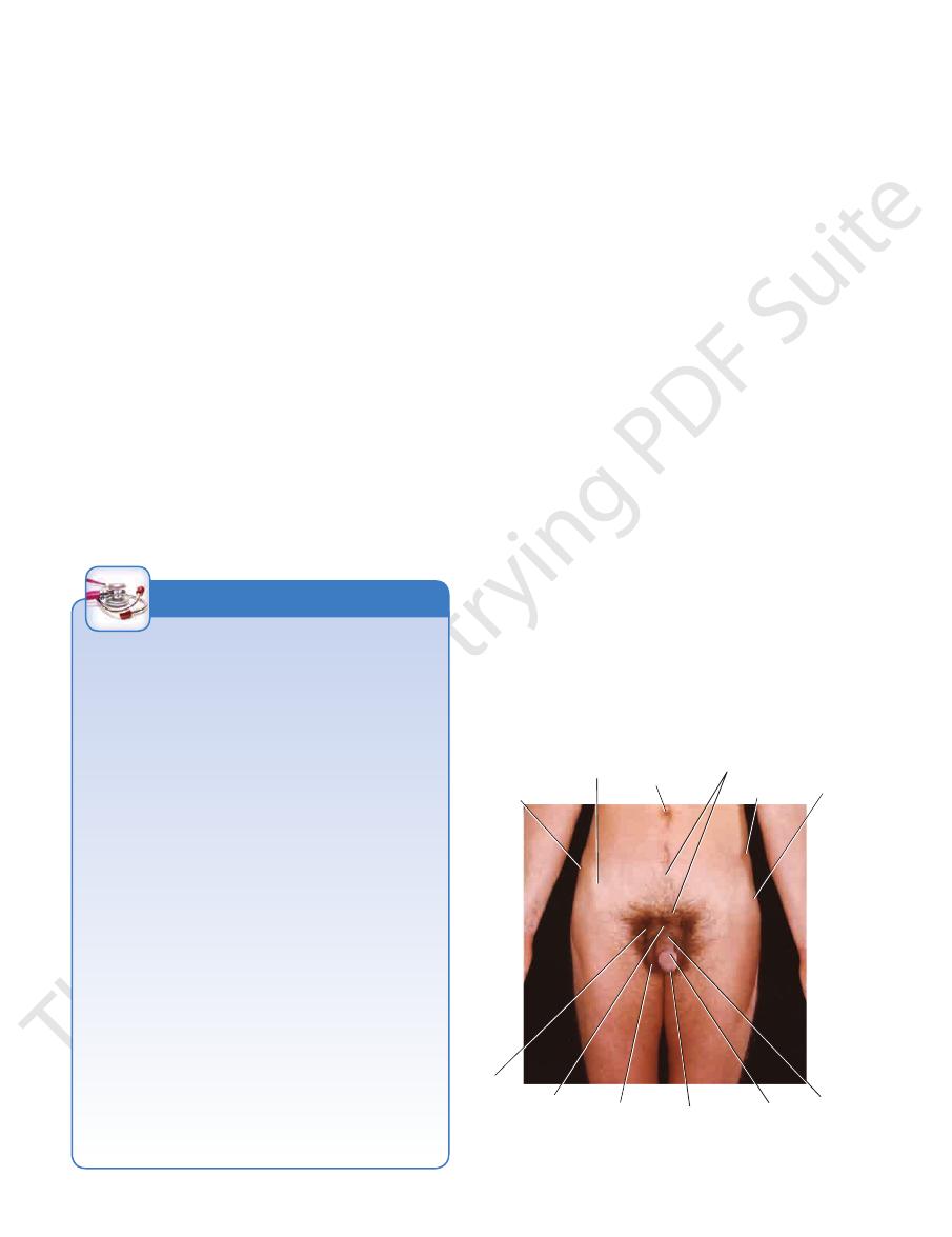
tubercle
of iliac crest
anterior superior
iliac spine
umbilicus
male
distribution
of pubic hair
iliac crest
greater
trochanter
of femur
pubic
tubercle symphysis
pubis
scrotum
external
urethral orifice
glans
penis
body
of penis
FIGURE 6.20
Anterior view of the pelvis of a 27-year-old man.
Surface Anatomy
259
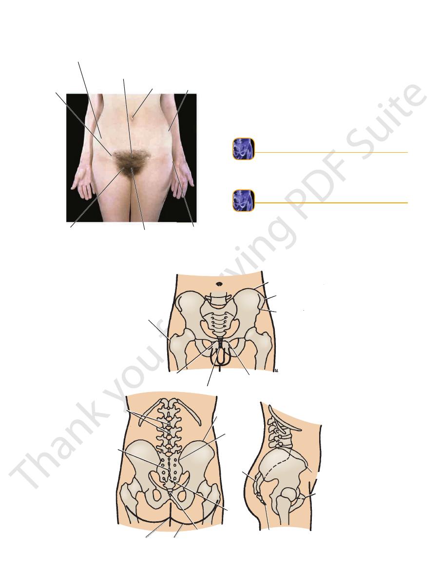
mons pubis showing
female distribution
of pubic hair
anterior superior
iliac spine
site of
inguinal
ligament
pubic tubercle
umbilicus
iliac crest
greater
trochanter
of femur
symphysis pubis
FIGURE 6.21
Anterior view of the pelvis of a 29-year-old woman.
wider in the female than in the male.
The subpubic angle, or pubic arch, is more rounded and
The sacrum is shorter, wider, and flatter in the female
the male they are turned in.
In the female the ischial tuberosities are everted and in
The pelvic outlet is larger in the female than in the male.
is much shorter.
male, and the distance between the inlet and the outlet
The pelvic cavity is roomier in the female than in the
■
■
■
■
■
■
than in the male.
■
■
natomy
aphic
adiog
R
R
a
Radiographic anatomy of the pelvis is fully described on
page 297.
natomy
face
s
uR
a
Surface Landmarks
length (Figs. 6.20, 6.21, and 6.22).
The iliac crest can be felt through the skin along its entire
Iliac Crest
greater trochanter of femur
iliac crest
tubercle of iliac crest
anterior superior iliac spine
pubic tubercle
symphysis pubis
pubic crest
lumbar spines
sacral
spines
natal cleft
fold of buttock
coccyx
sacral
hiatus
sacrum
coccyx
pubic tubercle
anterior superior
iliac spine
posterior superior
iliac spine
iliac crest
FIGURE 6.22
Relationship between different parts of the pelvis and the body surface.
260
CHAPTER 6
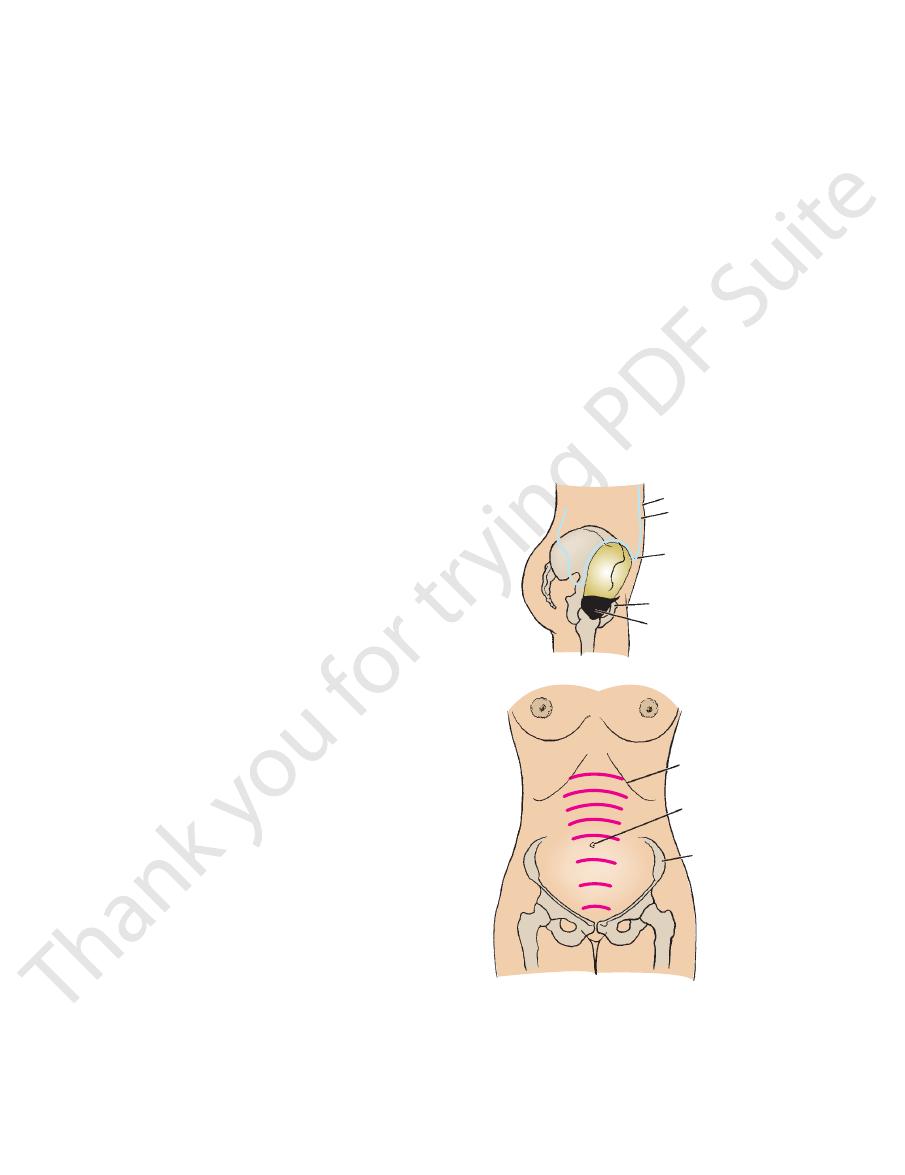
of the uterus can be palpated through the lower part of the
Toward the end of the 2nd month of pregnancy, the fundus
of the symphysis pubis.
of the bladder lies just below the level of the upper border
the pelvic cavity is not great enough to contain it. The neck
abdominal organ even when empty because the capacity of
In children, until the age of 6 years, the bladder is an
the abdominal wall (see page 272).
wall so that the front of the bladder is in direct contact with
bladder becomes peeled off from the anterior abdominal
pubis (Fig. 6.23). The peritoneum covering the distended
through the anterior abdominal wall above the symphysis
out of the pelvis into the abdomen, where it can be palpated
terior to the symphysis pubis. As the bladder fills, it rises up
In adults, the empty bladder is a pelvic organ and lies pos
Viscera
The Pelvis: Part I—The Pelvic Walls
Urinary Bladder
-
Uterus
umbilicus
peritoneum
superior wall of distended
bladder
body of pubis
urinary bladder
A
B
costal margin
umbilicus
iliac crest
months
9
10
8
7
6
5
4
3
FIGURE 6.23
coccyx can be palpated with the gloved finger in the anal
(2.5 cm) behind the anus. The anterior surface of the
be palpated in the cleft between the buttocks about 1 in.
The inferior surface and tip of the coccyx (Fig. 6.22) can
between the buttocks.
above the tip of the coccyx and beneath the skin of the cleft
terminates (Fig. 6.22). The hiatus lies about 2 in. (5 cm)
the lower end of the sacrum, where the extradural space
The sacral hiatus is situated on the posterior aspect of
most part of the cleft between the buttocks.
crest. The crest can be felt beneath the skin in the upper
with each other in the midline to form the median sacral
The spinous processes of the sacrum (Fig. 6.22) are fused
region.
as a solid structure through the fat that is present in this
between the bodies of the pubic bones and can be palpated
The symphysis pubis (Figs. 6.1 and 6.22) lies in the midline
of the pubic bone, medial to the pubic tubercle (Figs. 6.1
The pubic crest is the ridge of bone on the superior surface
majus.
can be palpated through the lateral margin of the labium
with the examining finger. In the female, the pubic tubercle
easily in the male by invaginating the scrotum from below
end of the inguinal ligament. The tubercle can be palpated
pubis (Figs. 6.20, 6.21, and 6.22). Attached to it is the medial
The pubic tubercle can be felt on the upper border of the
Pubic Tubercle
sacroiliac joint.
space; it also coincides with the level of the middle of the
which coincides with the lower limit of the subarachnoid
small skin dimple and on a level with the 2nd sacral spine,
end of the iliac crest (Fig. 6.22). It lies at the bottom of a
The posterior superior iliac spine is situated at the posterior
fold of the groin (Figs. 6.20, 6.21, and 6.22).
end of the iliac crest and lies at the upper lateral end of the
The anterior superior iliac spine is situated at the anterior
comes to lie in direct contact with the abdominal wall.
the anterior abdominal wall so that the front of the bladder
covering the distended bladder becomes peeled off from
various months of pregnancy. Note that the peritoneum
Height of the fundus of the uterus at
Surface anatomy of the empty bladder and
A.
the full bladder B.
Anterior Superior Iliac Spine
Posterior Superior Iliac Spine
Pubic Crest
and 6.22).
Symphysis Pubis
Spinous Processes of Sacrum
-
Sacral Hiatus
Coccyx
canal.

Surface Anatomy
pelvic viscera; they are described in detail on pages 311
inations are extremely valuable methods of palpating the
Bimanual rectoabdominal and vaginal–abdominal exam
Means of Palpating the Pelvic Viscera
Rectal and Vaginal Examinations as a
the fundus of the uterus also descends.
part of the fetus, usually the head, descends into the pelvis,
month of pregnancy (Fig. 6.23). Later, when the presenting
cus and reaches the region of the xiphoid process by the 9th
of the uterus, the fundus rises above the level of the umbili
anterior abdominal wall. With the progressive enlargement
261
-
-
and 326.
www.thePoint.lww.com/Snell9e.
Clinical Cases
and
Review Questions
are available online at