
3
Lecture
Spinal Cord (Internal Structure)
TRACTS OF SPINAL CORD
The white matter of the spinal cord consist of a mixture of Nerve fibers and are
arranged in tracts and for purpose of description, spinal tracts are divided
into:
-
- Ascending tracts
-
- Descending tracts
-
- Intersegmental tracts
-
Considerable overlap between tracts are present

Transverse section of the spinal cord at the midcervical level
showing the general arrangement of the ascending tracts on
the right and the descending tracts on the left

- On entering the spinal cord, the sensory nerve fibers of different sizes and
functions are sorted out and segregated into nerve bundles or tracts in the
white matter
-
Some of the nerve fibers serve to link different segments of the spinal cord,
while others ascend from the spinal cord to higher centers and thus connect
the spinal cord with the brain
Ascending Tracts
The information (of the ascending pathway) may be divided into two main
groups:
(1)
exteroceptive information, which originates from outside the body, such as
pain, temperature, and touch.
(2)
proprioceptive information, which originates from inside the body, for
example, from muscles and joints.
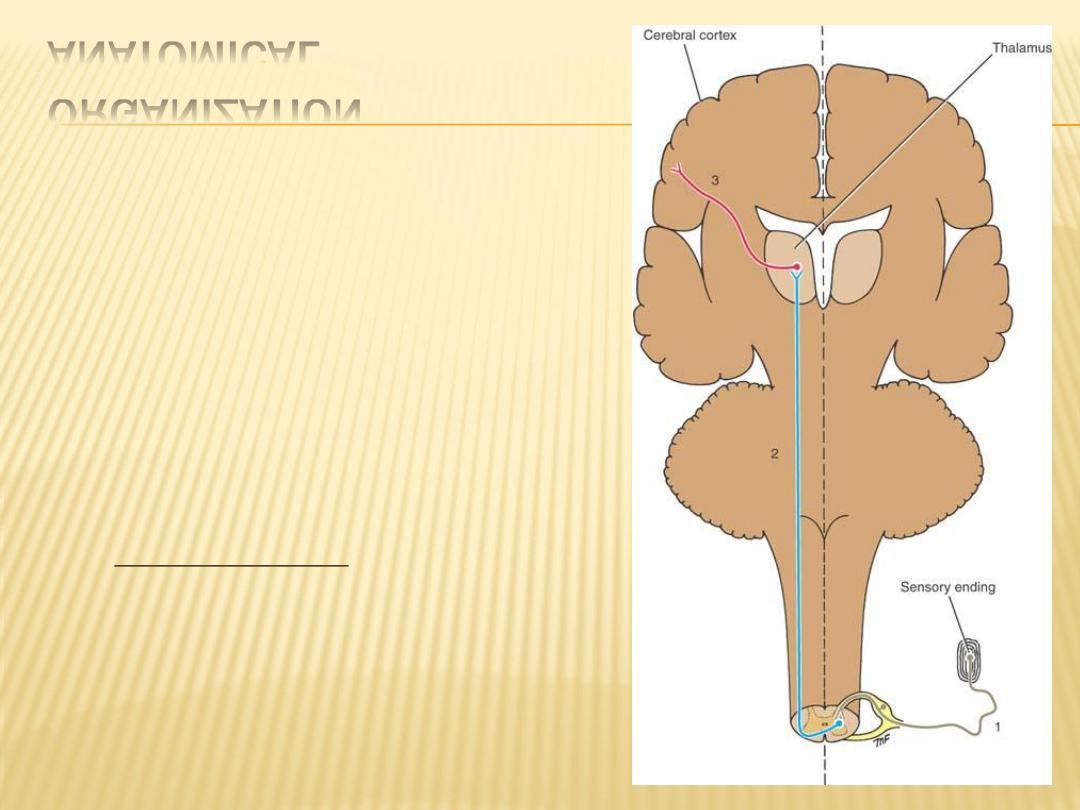
ANATOMICAL
ORGANIZATION
first-order neuron
second-order neuron
third-order neuron
-
- This three-neuron chain is the most
common arrangement, but some afferent
pathways use more or fewer neurons
-
-
- Many of the neurons in the ascending
pathways branch and give a major input
, which, in turn,
reticular formation
into the
activates the cerebral cortex, maintaining
wakefulness. Other branches pass to motor
neurons and participate in reflex muscular
activity
Pain and Temperature Pathways
-
- Lateral spinothalamic tract.
-
(contribute to posterolateral tract of Lissauer)
-
Axons of 2
nd
order n. cross obliquely to the opposite side within one spinal
segment, 2
nd
order neuron located at substania gelatinosa, at higher level at
medulla oblongata it accompanied with ant. spinothalamic and spinotectal
tracts to form the Spinal lemniscus that ascending through the posterior
part of Pons then end of this neuron at thalamus, 3
rd
order neuron then
pass through post. limb of internal capsule and the corona radiata to reach
the somesthetic area of precentral gyrus of cerebral cortex
-
Types of pain, - fast pain and slow pain
-
Light (Crude) Touch and Pressure Pathways
-
Anterior Spinothalamic Tract
-
The axons enter the spinal cord from the posterior root ganglion
-
divide into ascending and descending branches
-
travel for a distance of one or two segments of the spinal cord,
-
The axons of the second-order neuron cross very obliquely to the opposite
side in the anterior gray and white commissures within several spinal
segments and ascend in the opposite anterolateral white column as the
anterior spinothalamic tract
-
contributing to the posterolateral tract of Lissauer
-
form part of spinal lemniscus
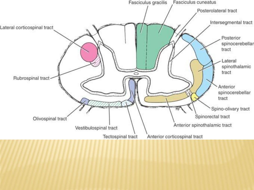
Transverse section of the spinal cord at the midcervical level
showing the general arrangement of the ascending tracts on
the right and the descending tracts on the left
Discriminative Touch, Vibratory Sense, and Conscious Muscle Joint Sense
Posterior White Column: Fasciculus Gracilis and Fasciculus Cuneatus
They are separated by a septum
Ascending ipsilateraly and 2
nd
order n. terminate and synapse at gracilis and
cunatus nuclei
Decussation occur as sensory decussation to form medial lemniscus at level of
medulla oblongata pass to thalamus and 3
rd
order n. to somesthetic area of
precentral gyrus of cerebral cortex
Muscle Joint Sense Pathways to the Cerebellum
Posterior Spinocerebellar Tract (2
nd
order n – nucleus dorsalis (Clarke's column)
Anterior Spinocerebellar Tract (nucleus dorsalis)
Cuneocerebellar Tract originate in the nucleus cuneatus and enter the cerebellum
through the inferior cerebellar peduncle of the same side
The fibers are known as the posterior external arcuate fibers, and their function is
to convey information of muscle joint sense to the cerebellum
Other Ascending Pathways
Spinotectal Tract
(afferent information for spinovisual reflexes and brings about
movements of the eyes and head toward the source of the stimulation)
Spinoreticular Tract
(The spinoreticular tract provides an afferent pathway for
the
reticular formation
, which plays an important role in influencing levels of
consciousness)
Spino-olivary Tract
(conveys information to the cerebellum from cutaneous and
proprioceptive )
Visceral Sensory Tracts
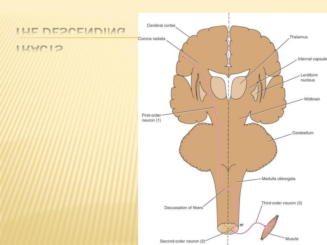
THE DESCENDING
TRACTS
ANATOMICAL
ORGANIZATION
first-order neuron
second-order neuron
third-order neuron
Corticospinal tracts
- Fibers of the corticospinal tract arise as axons of pyramidal cells situated in
the fifth layer of the cerebral cortex
- Pass within Corona radiata
-
- then pass within internal capsule (the fibers are organized so that those
closest to the genu are concerned with cervical portions of the body, while
those situated more posteriorly are concerned with the lower extremity)
-
- basis pedunculi of the midbrain
-
- in pons, the tract is broken into many bundles by the transverse
pontocerebellar fibers

-
- In the medulla oblongata, the bundles become grouped together along the
anterior border to form a swelling known as the pyramid (pyramidal tract)
-
At the junction of the medulla oblongata and the spinal cord, most of the fibers
cross the midline at the decussation of the pyramids and enter the lateral
white column
-
- lateral corticospinal tract
-
- anterior corticospinal tract
Reticulospinal Tracts
From a collection of nerves at the midbrain, pons, and medulla oblongata called the
reticular formation
-
- pontine reticulospinal tract
-
- medullary reticulospinal tract
-
Surve as a pathway by which the hypothalamus can control the sympathetic
outflow and the sacral parasympathetic outflow

Tectospinal Tract
Fibers of this tract arise from nerve cells in the superior colliculus of the midbrain
These fibers are believed to be concerned with reflex postural movements in
response to visual stimuli
Rubrospinal Tract
the red nucleus is situated in the tegmentum of the midbrain at the level of the
superior colliculus
tract facilitates the activity of the flexor muscles and inhibits the activity of the
extensor or antigravity muscles
Vestibulospinal Tract
The vestibular nuclei are situated in the pons and medulla oblongata beneath the
floor of the fourth ventricle
this tract, facilitate the activity of the extensor muscles and inhibit the activity of
the flexor muscles in association with the maintenance of balance
Olivospinal Tract

Descending Autonomic Fibers
The higher centers of the central nervous system associated with the control of
autonomic activity are situated in the cerebral cortex, hypothalamus,
amygdaloid complex, and reticular formation
Intersegmental Tracts
Short ascending and descending tracts that originate and end within the spinal
cord exist in the anterior, lateral, and posterior white columns. The function
of these pathways is to interconnect the neurons of different segmental
levels, and the pathways are particularly important in intersegmental spinal
reflexes
REFLEX ARC
A reflex may be defined as an involuntary response to a stimulus. It depends on
the integrity of the reflex arc. In its simplest form, a reflex arc consists of the
following anatomical structures:
(1) a receptor organ,
(2) an afferent neuron,
(3) an effector neuron,
(4) an effector organ
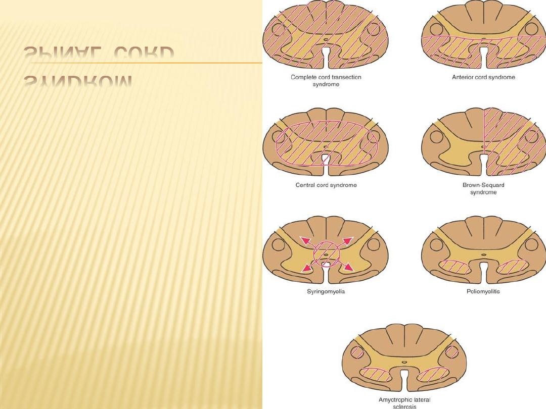
SPINAL CORD
SYNDROM
-
1- Complete cord transection
-
2- ant. cord syndrom
-
3- central cord syndrom
-
4- Brown squard syndrom