
4
Lecture
The BRAINSTEM
Medulla Oblongata
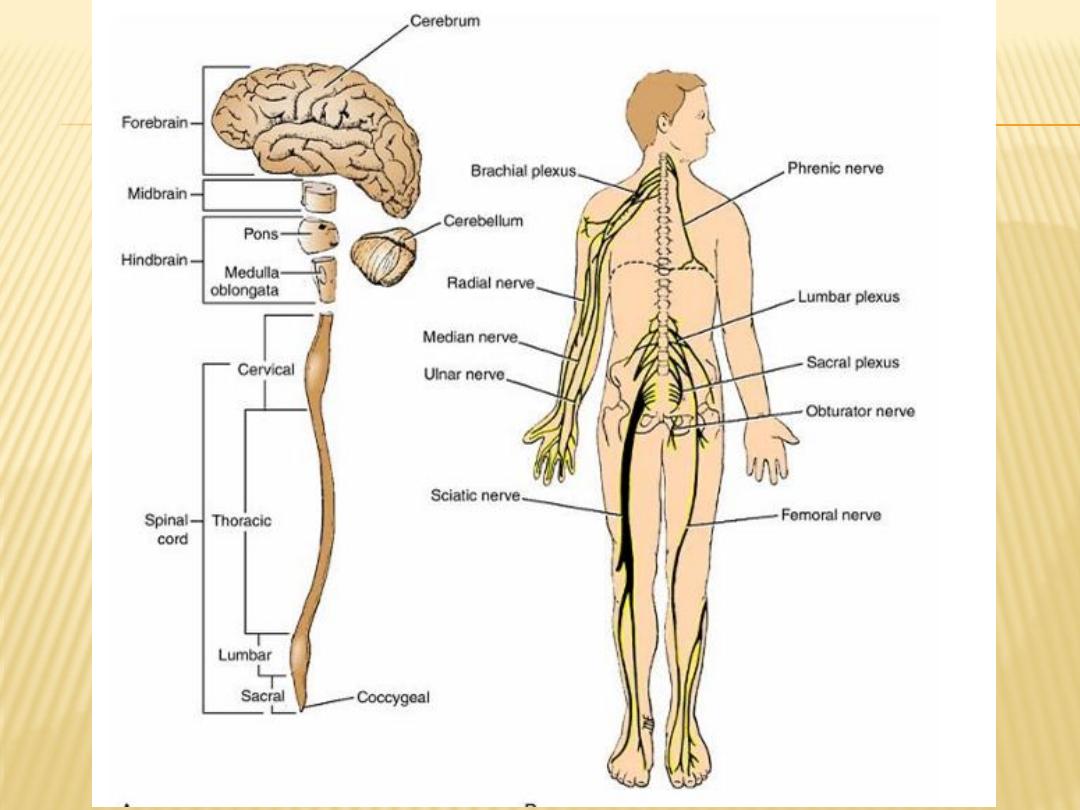


Introduction to brainstem
-
1- Medulla oblongata
-
2- Pons
-
3- Midbrain
-
occupies the posterior cranial fossa of the skull.
-
connects the narrow spinal cord with the expanded forebrain.
-
Function of Brainstem
-
(1) it serves as a conduit for the ascending tracts and descending tracts
connecting the spinal cord to the different parts of the higher centers in the
forebrain
(2) it contains important reflex centers associated with the control of
respiration and the cardiovascular system and with the control of
consciousness
(3) it contains the important nuclei of cranial nerves III through XII.
-
-
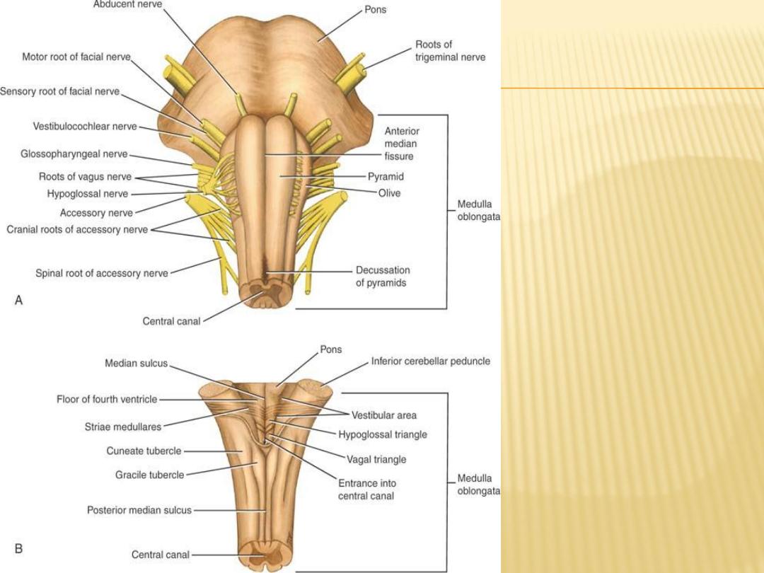
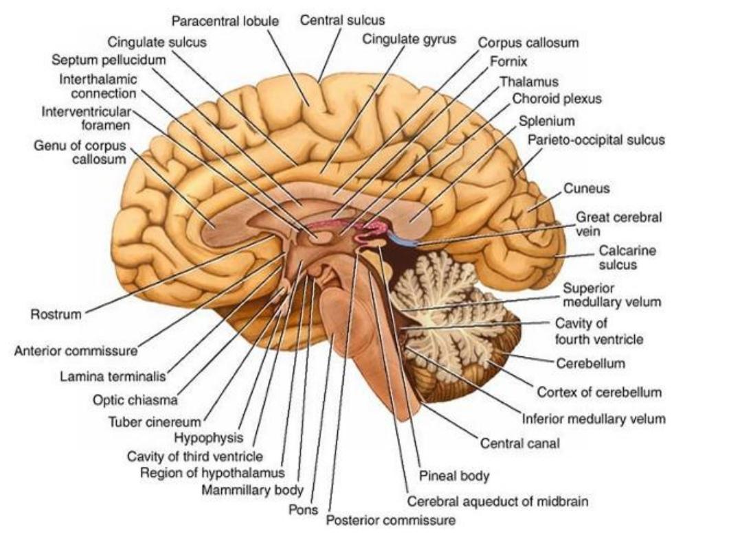
Medulla
oblongata
A- ant view
B- post view

Medulla Oblongata
Gross appearnse:
- Connect the pons sup to spinal cord inf
- About 2.5 cm in length
- The junction of the medulla and spinal cord is at the origin of the anterior and
posterior roots of the first cervical spinal nerve at level of foramen magnum
-
- It is conical in shape
-
- central canal
-
- cavity of fourth ventricle
-
Anteriorly:
-
- ant median fissure
-
- pyramid
-
- decussation of the pyramids
-
- Posterolateral to the pyramids are the olives
-
Posteriorly:
-
- sup is the floor 4
th
ventricle
-
- inf the median sulcus
-
- gracile tubercle and lat to it the cuneate tubercle

The internal structure of the medulla oblongata is considered at four levels:
1)
level of decussation of pyramids
2)
level of decussation of lemnisci
3)
level of the olives
4)
level just inferior to the pons.

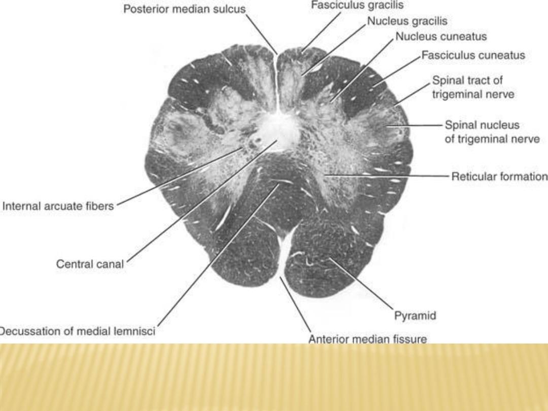
level of decussation of pyramids
A transverse section through the inferior half of the medulla oblongata
passes through the decussation of the pyramids, the great motor
decussation.
It contain the following nuclei:
Nucleus gracilis, nucleus cuneatus, spinal nucleus of cranial nerve V,
accessory nucleus
level of decussation of lemnisci
A transverse section through the inferior half of the medulla oblongata, a
short distance above the level of the decussation of the pyramids, passes
through the decussation of lemnisci, the great sensory decussation The
decussation of the lemnisci takes place anterior to the central gray matter
and posterior to the pyramids.
It contain the following nuclei:
Nucleus gracilis, nucleus cuneatus, spinal nucleus of cranial nerve V,
accessory nucleus, hypoglossal nucleus

level of the olives
A transverse section through the olives passes across the inferior part of the
fourth ventricle
The amount of gray matter has increased at this level owing to the presence of
the many nuclei
It contain the following nuclei:
Inferior olivary nucleus, spinal nucleus of cranial nerve V, vestibular nucleus,
glossopharyngeal nucleus, vagal nucleus, hypoglossal nucleus, nucleus
ambiguus, nucleus of tractus solitarius
level just inferior to the pons.
There are no major changes, in comparison to the previous level, in the
distribution of the gray and white matter
It contain the following nuclei:
Lateral vestibular nucleus, cochlear nuclei
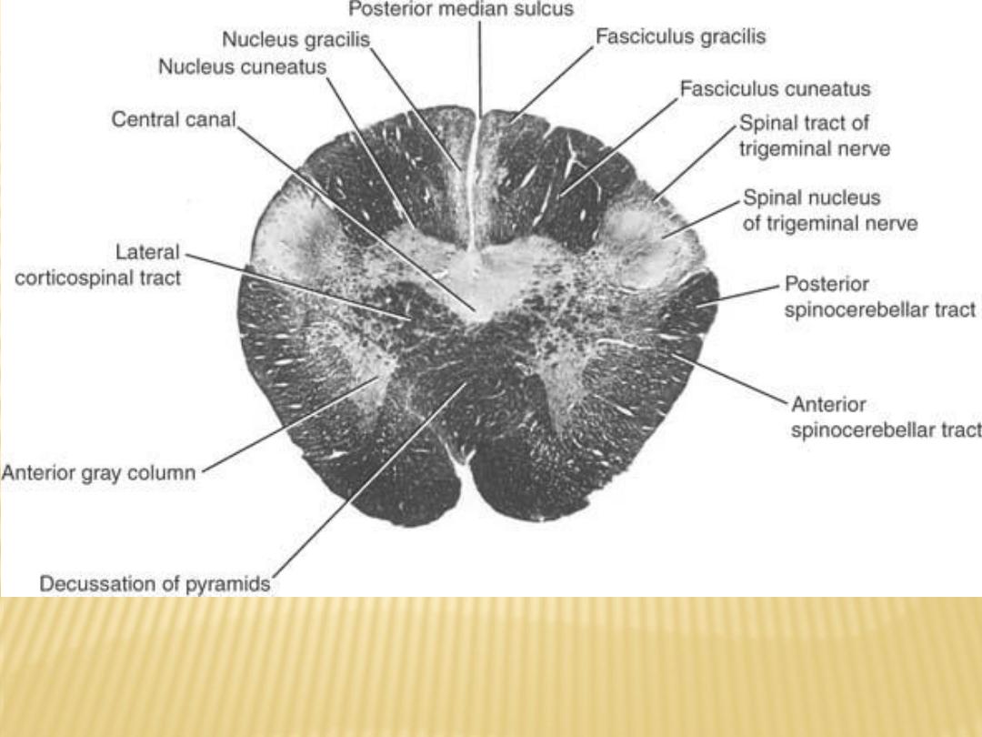
Transverse section of the medulla oblongata
at the level of decussation of the pyramids
Transverse section of the medulla oblongata at
the level of decussation of the medial lemnisci
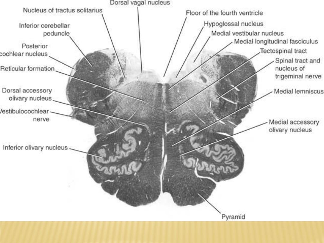
Transverse section of the medulla oblongata at the level of the middle of the olivary
nuclei

Blood supply of Medulla oblongata:
1) ventrally: branches from vertibral and basilar arteries, Also branches from
ant spinal artery
artery
2) dorsolaterally: by post inf cerebellar artery
-
Venous drainage:
-
1)ventrally: basilar venous plexus
and inf petrosal sinus
2) Dorsally and dorsolaterally to occipital sinus
3) Medullary veins communicate with sinuses and spinal veins
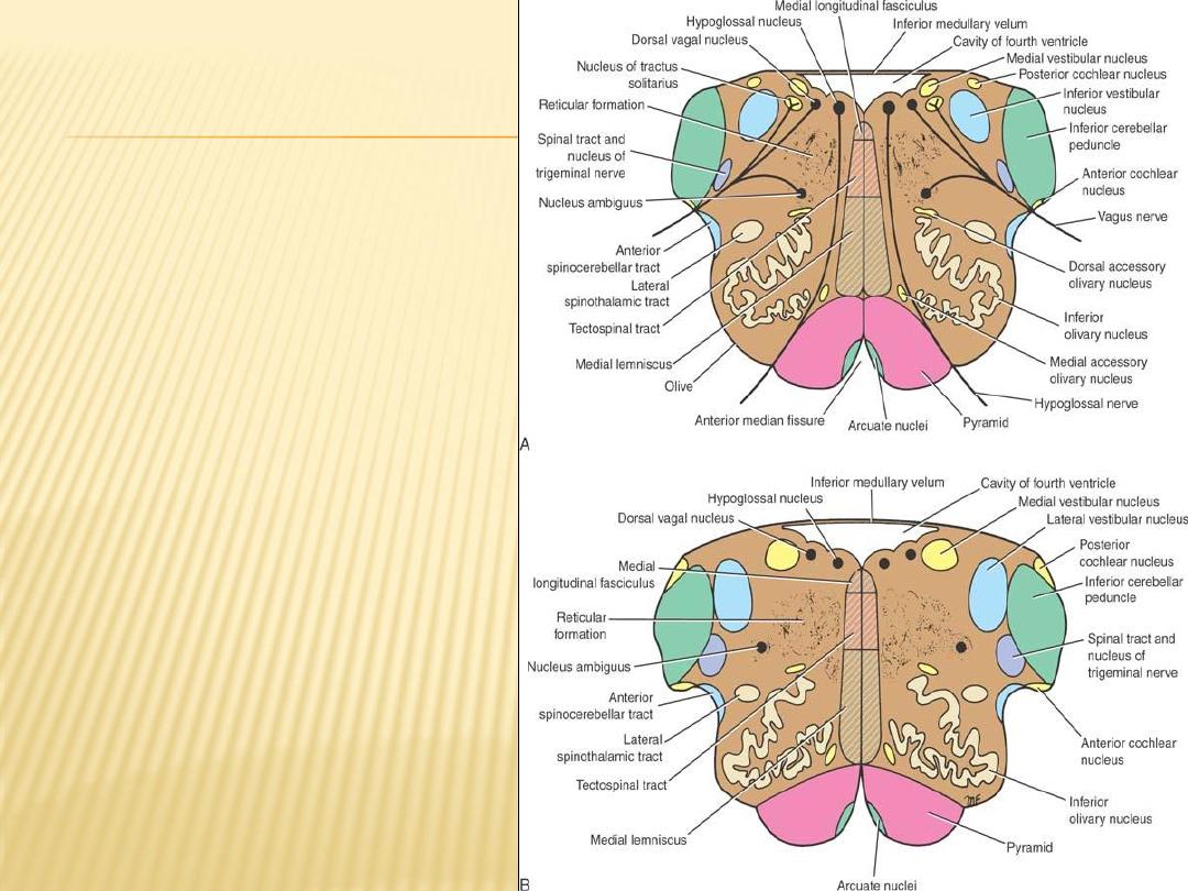
Olivary Nuclear Complex
- The largest nucleus of this
complex is the inferior olivary
nucleus
- Smaller dorsal and medial
accessory olivary nuclei also
are present
- The function of the olivary
nuclei is associated with
voluntary muscle movement.

Vestibulocochlear Nuclei
Vestibular comples:
(1) medial vestibular nucleus
(2) inferior vestibular nucleus
(3) lateral vestibular nucleus
(4) superior vestibular nucleus
Cochlear nuclei:
(1) anterior cochlear nucleus
(2) posterior cochlear nucleus
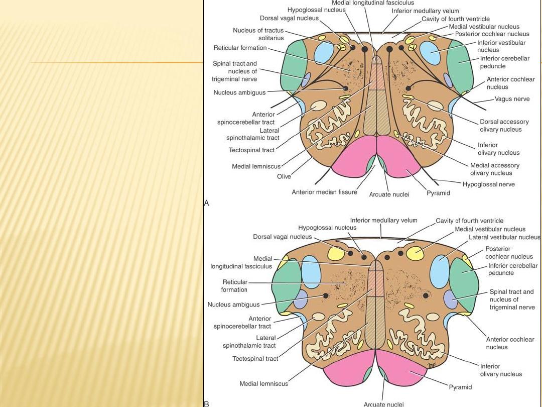
The Nucleus Ambiguus
The nucleus ambiguus
consists of large motor
neurons and is situated
deep within the reticular
formation
The emerging nerve fibers join
the glossopharyngeal,
vagus, and cranial part of
the accessory nerve and
are distributed to voluntary
skeletal muscle

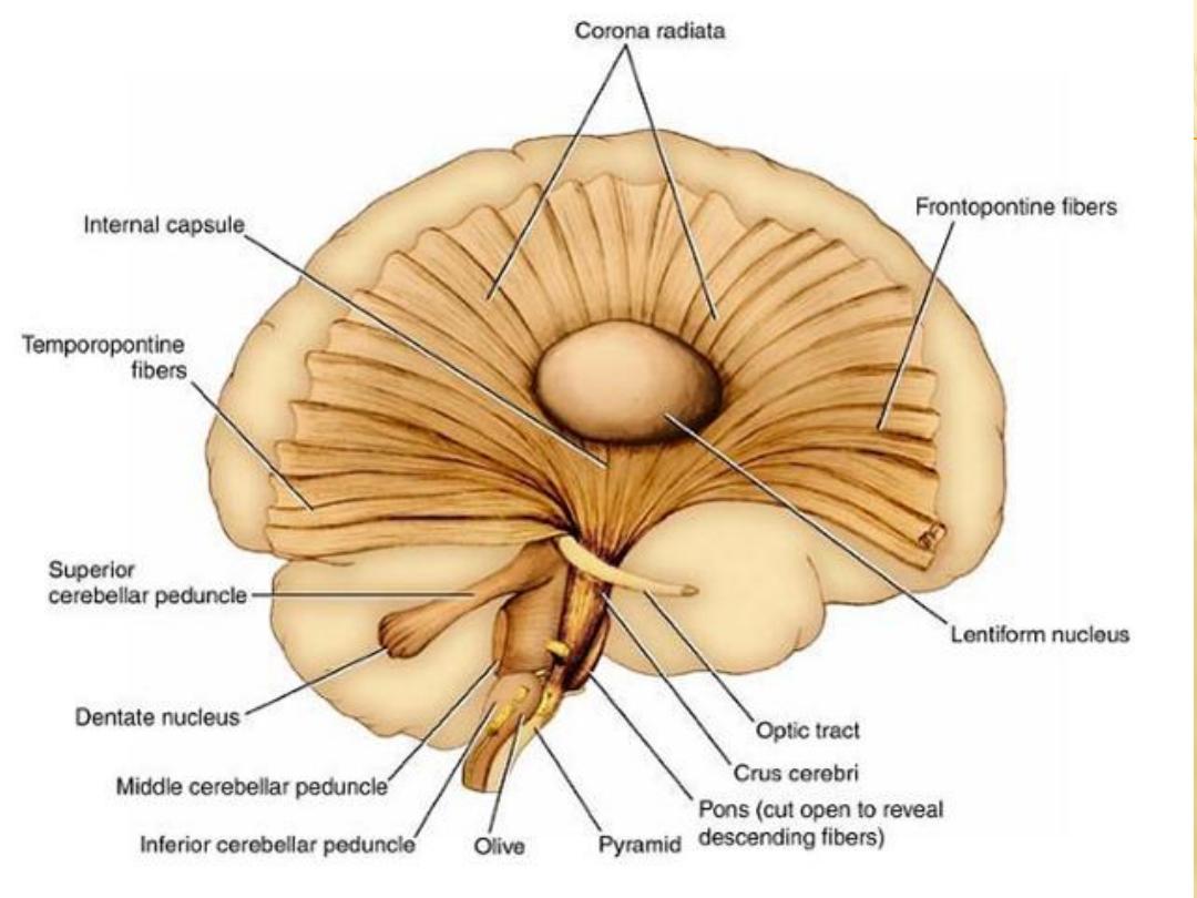
The medial lemniscus
forms a flattened tract on each side of the midline posterior
to the pyramid, These fibers emerge from the decussation of the lemnisci and
convey sensory information to the thalamus.
The medial longitudinal fasciculus
forms a small tract of nerve fibers situated on
each side of the midline posterior to the medial lemniscus and anterior to the
hypoglossal nucleus It consists of ascending and descending fibers
The inferior cerebellar peduncle
is situated in the posterolateral corner of the
section on the lateral side of the fourth ventricle
The spinal tract of the trigeminal nerve
and its nucleus are situated on the
anteromedial aspect of the inferior cerebellar peduncle
The reticular formation
, consisting of a diffuse mixture of nerve fibers and small
groups of nerve cells, is deeply placed posterior to the olivary nucleus The
reticular formation represents, at this level, only a small part of this system
the reticular formation is present at all levels of Medulla oblongata, it also
present in the pons and midbrain

5
Lecture
The BRAINSTEM
Pons and Midbrain
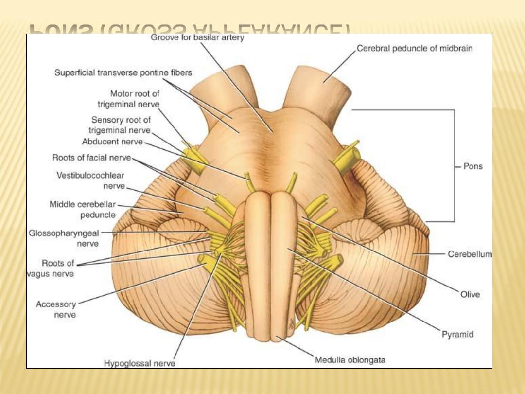
PONS (GROSS APPEARANCE)
Anterior surface of the brainstem showing the pons

Pons connect the medulla oblongata to midbrain and lie ant to cerebellum
It is about 1 inch (2.5 cm) long and owes its name to the appearance presented on
the anterior surface, which is that of a bridge connecting the right and left
cerebellar hemispheres
Ant it is convex with multiple transverse fibers that converge on each side to form
the middle cerebellar peduncle
a shallow groove in the midline, the basilar groove, which lodges the basilar artery
anterolateral surface of the pons, the trigeminal nerve emerges on each side. Each
nerve consists of a smaller, medial part, known as the motor root, and a larger,
lateral part, known as the sensory root
the groove between the pons and the medulla oblongata, there emerge, from
medial to lateral, the abducent, facial, and vestibulocochlear nerves
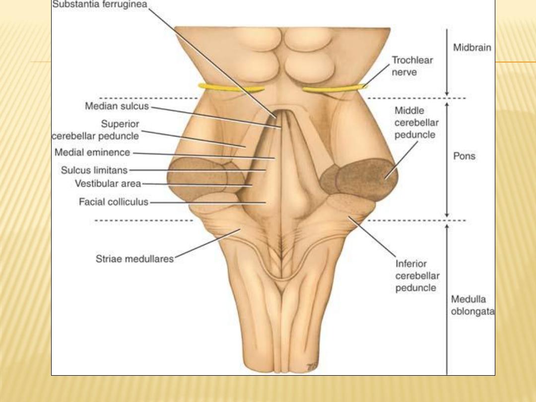
Pos. surface of the brainstem showing the pons. The cerebellum has been removed

Post it is triangular in shape forming the upper half of the floor of the fourth
ventricle, it is hidden by the cerebellum
posterior surface is limited laterally by the
superior
cerebellar peduncles
and is
divided into symmetrical halves by a median sulcus
Lateral to this sulcus is an elongated elevation, the
medial eminence
, which is
bounded laterally by the
sulcus limitans
Facial colliculus:
is produced by the root of the facial nerve winding around the
nucleus of the abducent nerve
The floor of the superior part of the sulcus limitans is bluish-gray in color and is
called the
substantia ferruginea
area vestibuli :
Lateral to the sulcus limitans, produced by the underlying vestibular
nuclei

Internal structure of the pons
pons is commonly divided into a posterior part, the tegmentum, and an anterior
basal part by the transversely running fibers of the trapezoid body
The structure of the pons may be studied at two levels: (1) transverse section
through the caudal part, passing through the facial colliculus, and (2) transverse
section through the cranial part, passing through the trigeminal nuclei
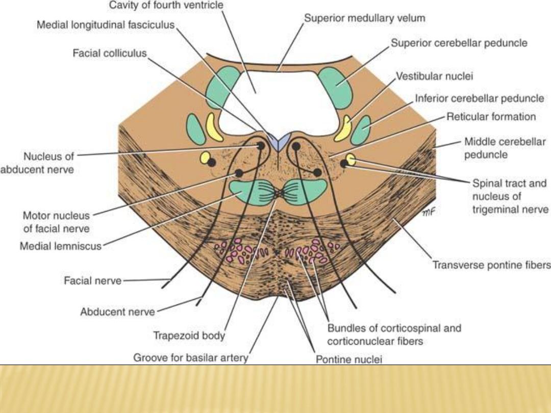
Transverse section through the caudal part of the pons at the level of the facial
colliculus
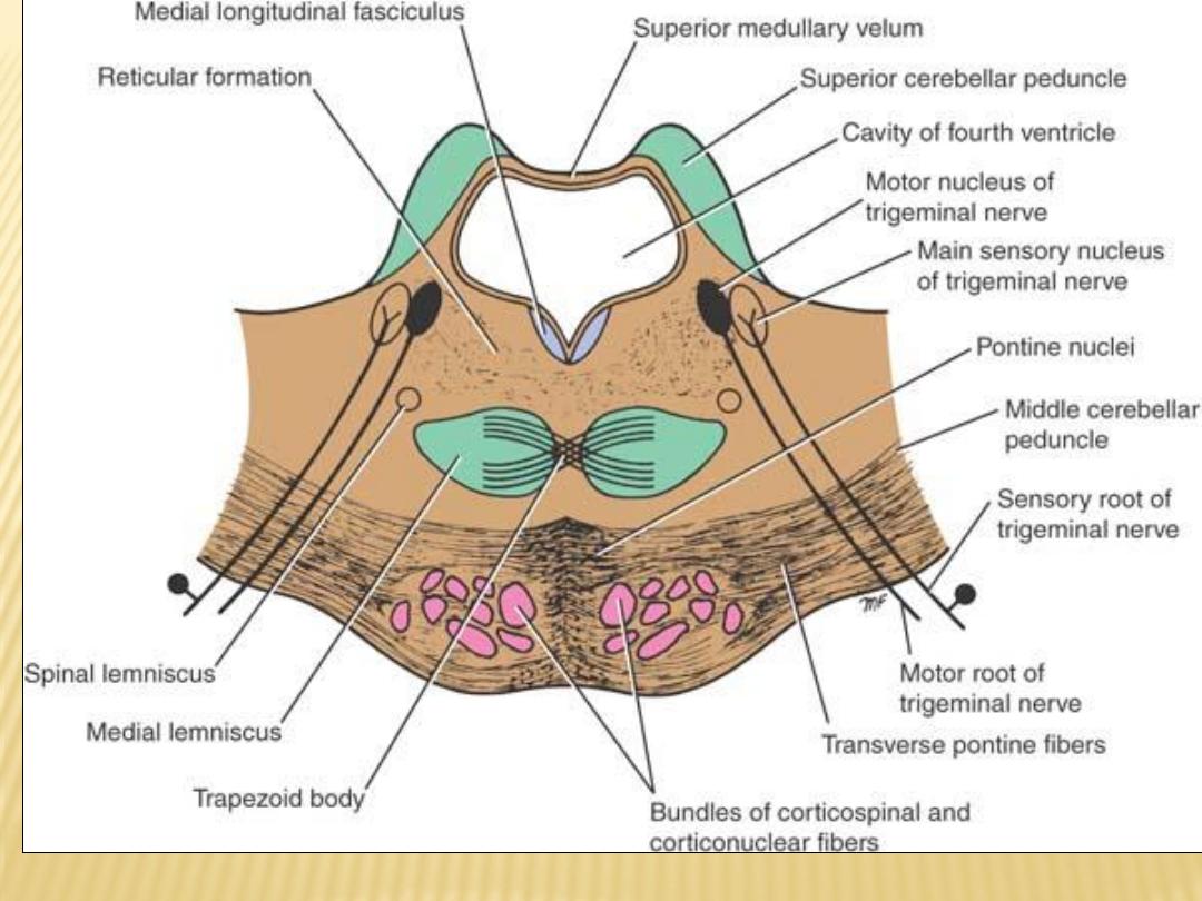
Transverse section through the pons at the level of the trigeminal nuclei
Comparison of the Different Levels of the Pons
Showing the Major Structures at Each Level
Level
Cavity
Nuclei
Motor Tracts
Sensory Tracts
Facial
colliculus
Fourth ventricle
Facial nucleus, abducent
nucleus, medial
vestibular nucleus, spinal
nucleus of cranial nerve
V, pontine nuclei,
trapezoid nuclei
Corticospinal and
corticonuclear tracts,
transverse pontine fibers,
medial longitudinal
fasciculus
Spinal tract of cranial
nerve V; lateral, spinal,
and medial lemnisci
Trigemin
al nuclei
Fourth ventricle
Main sensory and motor
nucleus of cranial nerve
V, pontine nuclei,
trapezoid nuclei
Corticospinal and
corticonuclear tracts,
transverse pontine fibers,
medial longitudinal
fasciculus
Lateral, spinal, and
medial lemnisci
a
Note that the reticular formation is present at all levels.
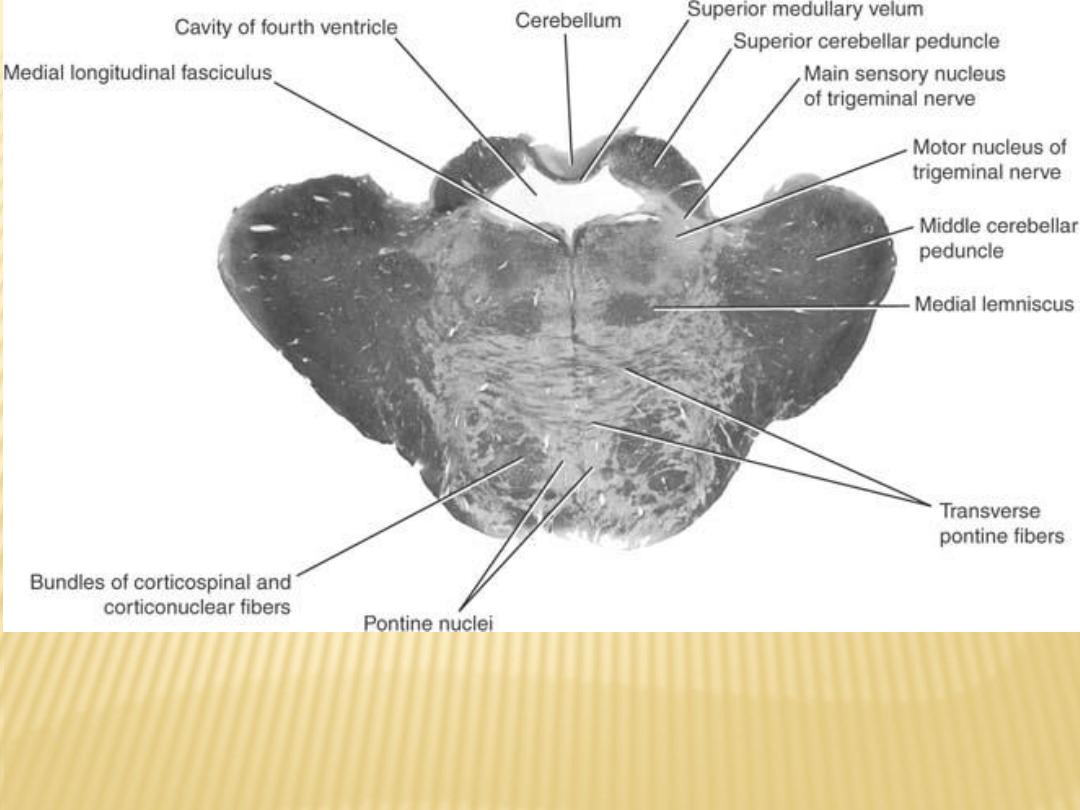
Photomicrograph of a transverse section of the pons at
the level of the trigeminal nuclei
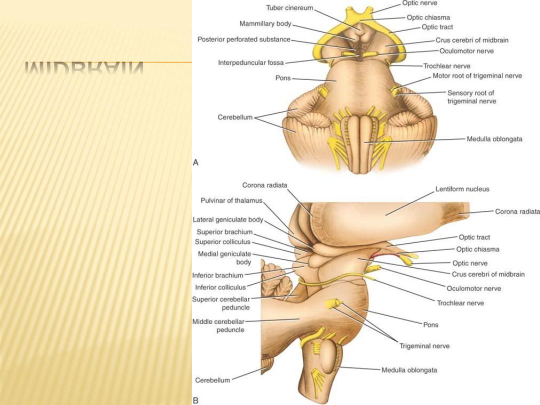
MIDBRAIN

The midbrain measures about 0.8 inch (2 cm) in length and connects the pons
and cerebellum with the forebrain
The midbrain is traversed by a narrow channel, the cerebral aqueduct, which is
filled with cerebrospinal fluid which connects the third and fourth ventricles
The midbrain comprises two lateral halves, called the cerebral peduncles;
The midbrain is devided by the cerebral aquiduct into ventral and dorsal part
Ventrally:
divided into an anterior part, the crus cerebri, and a posterior part, the
tegmentum, by a pigmented band of gray matter, the substantia nigra.,.
Dorsally:
The tectum consist of four small surface swellings, two superior and two
Inferior colliculi
For description, two levels are studied for the internal structure of midbrain
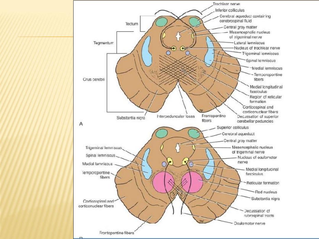
- Transverse Section of the Midbrain at the Level of the Inferior Colliculi
- Transverse Section of the Midbrain at the Level of the superior Colliculi
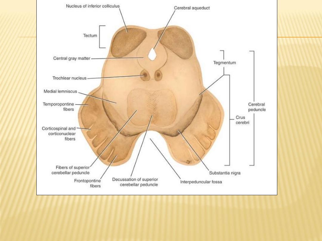
Transverse section of the midbrain through the inferior colliculi shows the division
of the midbrain into the tectum and the cerebral peduncles. Note that the
cerebral peduncles are subdivided by the substantia nigra into the tegmentum
and the crus cerebri

The
Interpeduncular fossa
, is a deep depression in the midline on the anterior
aspect of the midbrain it is bounded on either side by the
crus cerebri
.
Many small blood vessels perforate the floor of the interpeduncular fossa, and this
region is termed the
posterior perforated substance
The oculomotor nerve emerges from a groove on the medial side of the crus cerebri
and passes forward in the lateral wall of the cavernous sinus.
Comparison of Two Levels of the Midbrain
Showing the Major Structures at Each Level
Level
Cavity
Nuclei
Motor Tract
Sensory Tracts
Inferior
colliculi
Cerebral aqueduct
Inferior colliculus,
substantia nigra, trochlear
nucleus, mesencephalic
nuclei of cranial nerve V
Corticospinal and
corticonuclear tracts,
temporopontine,
frontopontine, medial
longitudinal fasciculus
Lateral, trigeminal,
spinal, and medial
lemnisci; decussation
of superior cerebellar
peduncles
Superior
colliculi
Cerebral aqueduct
Superior colliculus,
substantia nigra,
oculomotor nucleus,
Edinger-Westphal nucleus,
red nucleus, mesencephalic
nucleus of cranial nerve V
Corticospinal and
corticonuclear tracts,
temporopontine,
frontopontine, medial
longitudinal fasciculus,
decussation of
rubrospinal tract
Trigeminal, spinal, and
medial lemnisci
a
Note that the reticular formation is present at all levels.
Transverse
sections of the
midbrain. A: At
the level of the
inferior
colliculus. B:
At the level of
the superior
colliculus.
Note that
trochlear
nerves
completely
decussate
within the
superior
medullary
velum
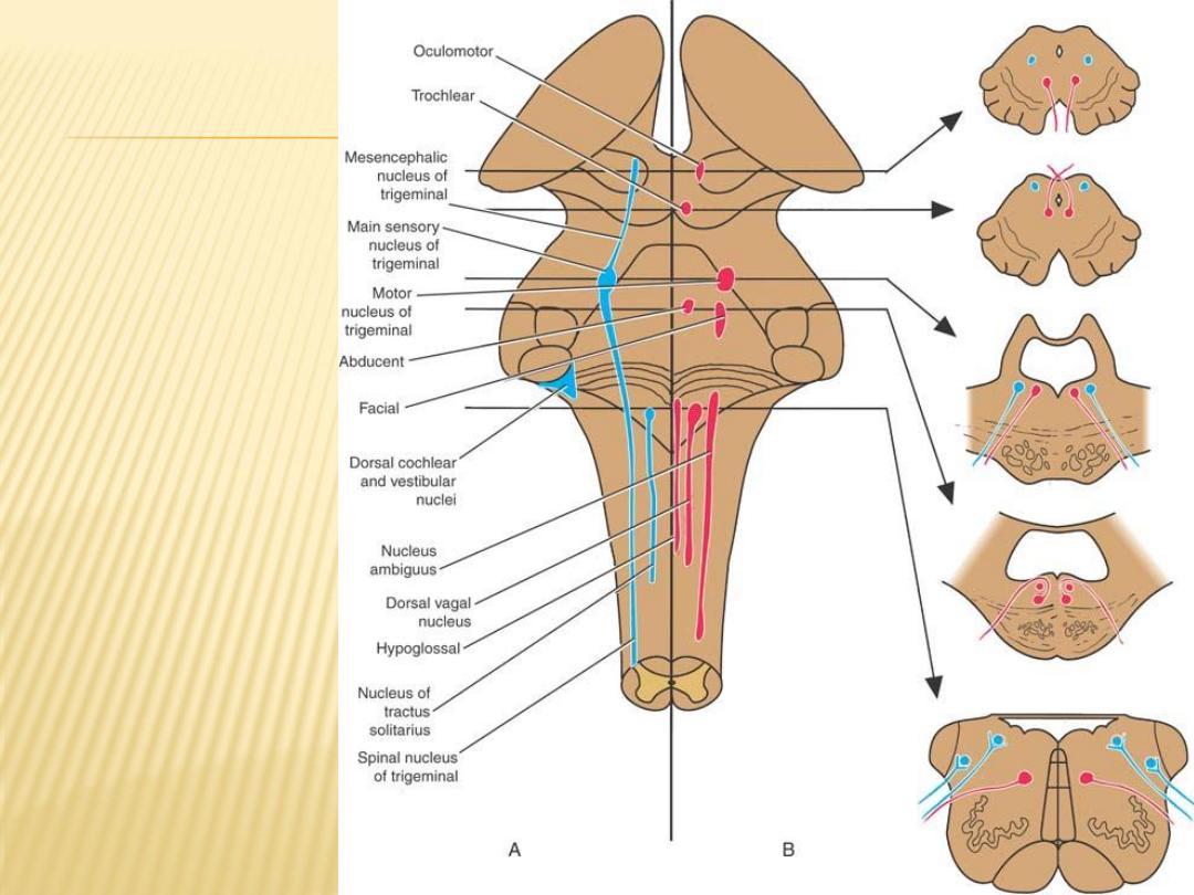
Position of some of
the cranial nerve
nuclei in the
brainstem. A:
Surface projection
on the posterior
aspect of the
brainstem. B:
Cross sections.
The motor nuclei
are in red and the
sensory nuclei in
blue

MCQ Question (sample)
base of skull
1)
The interior of the base of the skull is divided into two cranial fossae:
anterior, and posterior
2)
anterior cranial fossa lodges the frontal lobes of the cerebral hemispheres
3)
optic nerve pass through optic canal
4)
occipital bones make a major part of middle cranial fossa