
7
Lecture
Cranial Nerves
(1
st
–6
th
)

Cranial Nerves
There are 12 pairs of cranial nerves, which leave the brain and pass through
foramina and fissures in the skull. All the nerves are distributed in the head
and neck, except cranial nerve X, which also supplies structures in the
thorax and abdomen. The cranial nerves are named as follows:
1.
Olfactory
2.
Optic
3.
Oculomotor
4.
Trochlear
5.
Trigeminal
6.
Abducent
7.
Facial
8.
Vestibulocochlear
9.
Glossopharyngeal
10.
Vagus
11.
Accessory
12.
Hypoglossal

Organization of the Cranial Nerves
The olfactory, optic, and vestibulocochlear nerves are entirely sensory.
The oculomotor, trochlear, abducent, accessory, and hypoglossal nerves are
entirely motor.
The trigeminal, facial, glossopharyngeal, and vagus nerves are both sensory
and motor nerves.

Olfactory Nerves (Cranial Nerve I)
The olfactory nerves arise from the olfactory receptor nerve cells in the
olfactory mucous membrane located in the upper part of the nasal cavity
above the level of the superior concha
Each receptor cell consists of a small bipolar nerve cell.
olfactory hairs
: a number of short cilia arise from the coarse peripheral process
of the bipolar cell
olfactory nerve fibers
: Bundles of these nerve fibers pass through the openings
to enter the olfactory bulb. The
bone
ethmoid
cribriform plate of the
of the
olfactory nerve fibers are unmyelinated and are covered with Schwann cells
Olfactory Bulb
This ovoid structure possesses several types of nerve cells, the largest of which
,
tufted cells and granular cells
, Smaller nerve cells, called
the mitral cell
is
The olfactory bulb, in addition, receives axons from the contralateral
olfactory bulb through the olfactory tract.
Olfactory Tract
narrow band of white matter runs from the posterior end of the olfactory bulb
beneath the inferior surface of the frontal lobe of the brain
It consists of the central axons of the mitral and tufted cells of the bulb and
some centrifugal fibers from the opposite olfactory bulb
As the olfactory tract reaches the anterior perforated substance, it divides into
carries the axons to the
stria
. The lateral
striae
medial and lateral olfactory
and
periamygdaloid
olfactory area of the cerebral cortex, namely, the
The medial
(primary olfactory cortex)
areas often known as the
prepiriform
olfactory stria carries the fibers that cross the median plane in the anterior
commissure to pass to the olfactory bulb of the opposite side
The entorhinal area (area 28) of the parahippocampal gyrus, which receives
numerous connections from the primary olfactory cortex, is called the
(secondary olfactory cortex)
The primary olfactory cortex sends nerve fibers to many other centers within
the brain to establish connections for emotional and autonomic responses
to olfactory sensations.
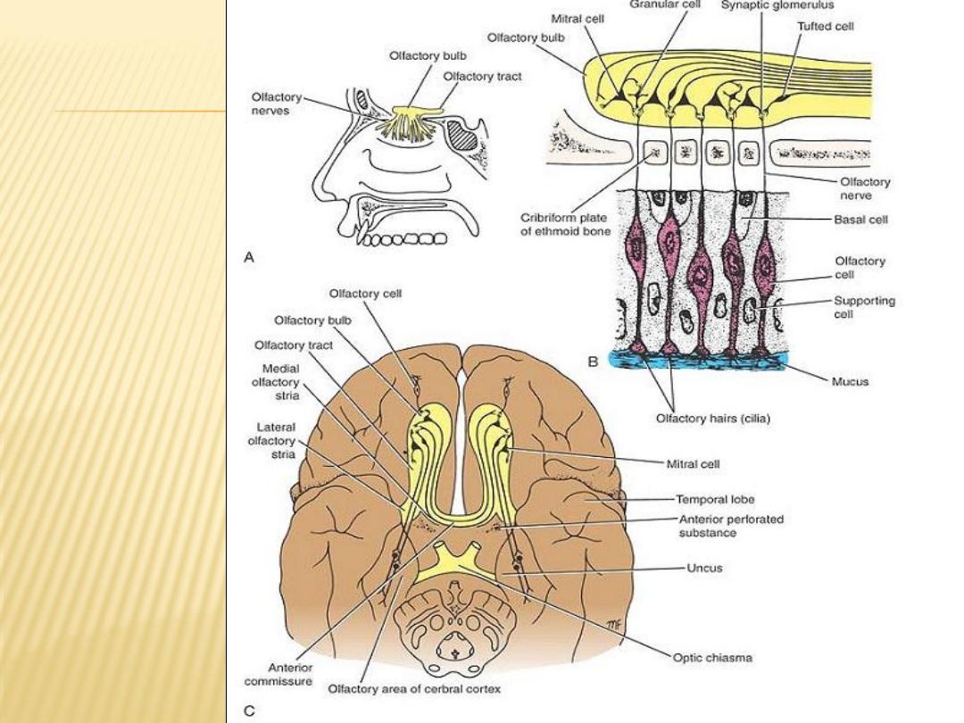
A: Distribution of
olfactory
nerves on the
lateral wall of
the nose.
B: Connections
between the
olfactory cells
and the neurons
of the olfactory
bulb.
C: Connections
between the
olfactory cell
and the rest of
the olfactory
system.

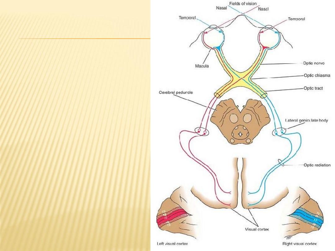
Optic Nerve (Cranial Nerve II)
Origin of the Optic Nerve
The fibers of the optic nerve are the axons of the cells in the ganglionic layer of
the retina. They converge on the optic disc and exit from the eye, about 3 or
optic nerve
mm to the nasal side of its center, as the
4
The fibers of the optic nerve are myelinated, but the sheaths are formed from
oligodendrocytes rather than Schwann cells, since the optic nerve is
comparable to a tract within the central nervous system.
The optic nerve leaves the orbital cavity through the optic canal and unites with
the optic nerve of the opposite side to form the
optic chiasma

Optic Chiasma
In the chiasma, the fibers from the nasal (medial) half of each retina, including the
nasal half of the macula, cross the midline and enter the optic tract of the
opposite side, while the fibers from the temporal (lateral) half of each retina,
including the temporal half of the macula, pass posteriorly in the optic tract of
the same side
Optic Tract
The optic tract emerges from the optic chiasma and passes posterolaterally around
the cerebral peduncle. Most of the fibers now terminate by synapsing with nerve
, which is a small projection from the posterior
lateral geniculate body
cells in the
nucleus and the
pretectal
part of the thalamus. A few of the fibers pass to the
and are concerned with light reflexes
of the midbrain
colliculus
superior
Lateral Geniculate Body
The lateral geniculate body is a small, oval swelling projecting from the pulvinar of
the thalamus. It consists of six layers of cells, on which synapse the axons of the
optic tract. The axons of the nerve cells within the geniculate body leave it to
form the
optic radiation
Optic Radiation:
The fibers of the optic radiation are the axons of the nerve cells of the
lateral geniculate body. The tract passes posteriorly through the retrolenticular part
of the internal capsule and terminates in the visual cortex (area 17),
Optic Pathway
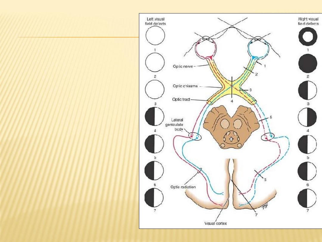
Visual field defects associated with
lesions of the optic pathways.
1. Right-sided circumferential
blindness due to retrobulbar
neuritis.
2. Total blindness of the right eye due
to division of the right optic nerve.
3. Right nasal hemianopia due to a
partial lesion of the right side of
the optic chiasma.
4. Bitemporal hemianopia due to a
complete lesion of the optic
chiasma.
5. Left temporal hemianopia and right
nasal hemianopia due to a lesion
of the right optic tract.
6. Left temporal and right nasal
hemianopia due to a lesion of the
right optic radiation.
7. Left temporal and right nasal
hemianopia due to a lesion of the
right visual cortex.

Neurons of the Visual Pathway
Four neurons conduct visual impulses to the visual cortex:
(1)
rods and cones, which are specialized receptor neurons in the retina;
(2)
bipolar neurons, which connect the rods and cones to the ganglion cells;
(3)
ganglion cells, whose axons pass to the lateral geniculate body; and
(4)
neurons of the lateral geniculate body, whose axons pass to the cerebral
cortex

Visual Reflexes
Direct and Consensual Light Reflexes
If a light is shone into one eye, the pupils of both eyes normally constrict.
The constriction of the pupil on which the light is shone is called the direct
light reflex; the constriction of the opposite pupil, even though no light fell on
that eye, is called the consensual light reflex
Accommodation Reflex
When the eyes are directed from a distant to a near object, contraction of
the medial recti brings about convergence of the ocular axes; the lens
thickens to increase its refractive power by contraction of the ciliary muscle;
and the pupils
Corneal Reflex
Light touching of the cornea or conjunctiva results in blinking of the eyelids
Visual Body Reflexes
The automatic scanning movements of the eyes and head that are made
when reading, the automatic movement of the eyes, head, and neck toward
the source of the visual stimulus, and the protective closing of the eyes and
even the raising of the arm for protection
Pupillary Skin Reflex
The pupil will dilate if the skin is painfully stimulated by pinching

Oculomotor Nerve (Cranial Nerve III)
The oculomotor nerve has two motor nuclei:
(1)
the main motor nucleus
(2)
the accessory parasympathetic nucleus.
Course of the Oculomotor Nerve
The oculomotor nerve emerges on the anterior surface of the midbrain. It
passes forward between the posterior cerebral and the superior cerebellar
arteries. It then continues into the middle cranial fossa in the lateral wall of
the cavernous sinus. Here, it divides into a superior and an inferior ramus,
.
superior orbital fissure
which enter the orbital cavity through the
the
nerve supplies the following extrinsic muscles of the eye:
oculomotor
The
, superior rectus, medial rectus, inferior rectus,
superioris
palpebrae
levator
ciliary
. It also supplies, through its branch to the
and inferior oblique
ganglion and the short ciliary nerves, parasympathetic nerve fibers to the
following intrinsic muscles: the constrictor pupillae of the iris and ciliary
muscles.
Therefore, the oculomotor nerve is entirely motor and is responsible for lifting
the upper eyelid; turning the eye upward, downward, and medially;
constricting the pupil; and accommodating the eye.
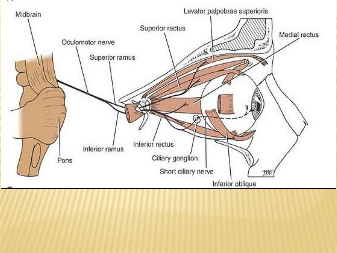
The distribution of the oculomotor nerve
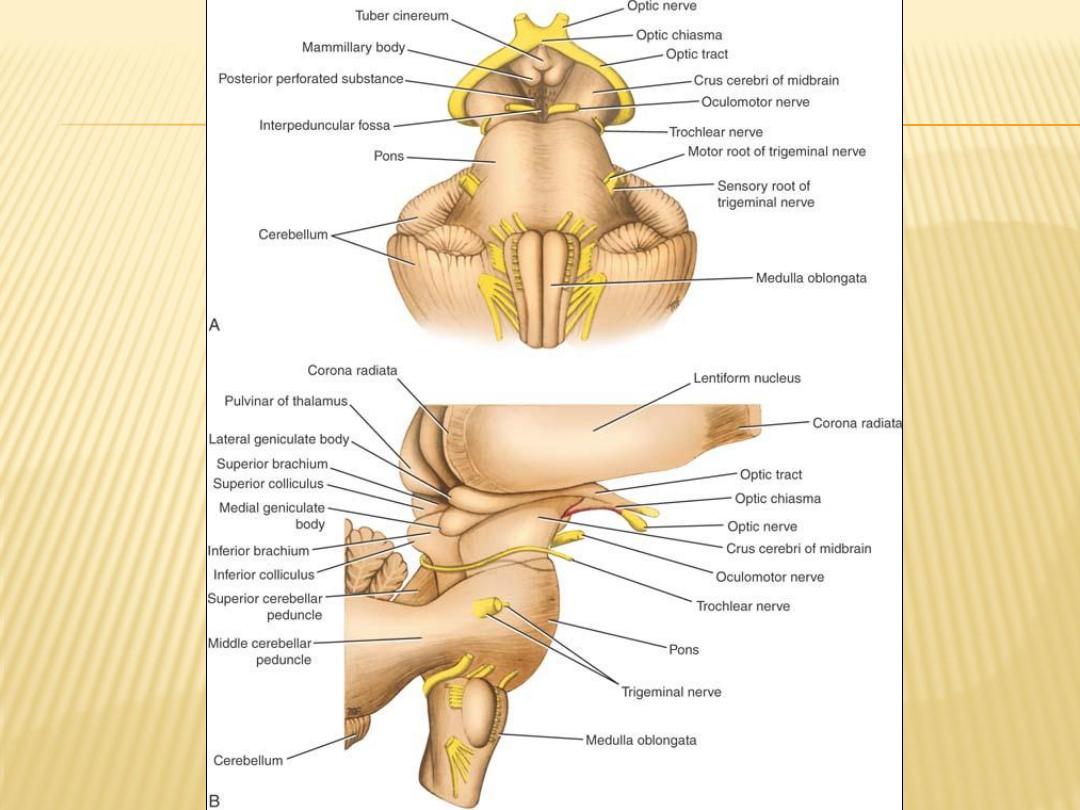

Trochlear Nerve (Cranial Nerve IV)
The trochlear nucleus is situated in the anterior part of the gray matter that
surrounds the cerebral aqueduct of the midbrain
The nerve fibers, after leaving the nucleus, pass posteriorly around the central
gray matter to reach the posterior surface of the midbrain
The trochlear nerve, the most slender of the cranial nerves and the only one to
leave the posterior surface of the brainstem emerges from the midbrain and
immediately decussates with the nerve of the opposite side. The trochlear
nerve passes forward through the middle cranial fossa in the lateral wall of
superior orbital
the cavernous sinus and enters the orbit through the
muscle of the eyeball. The
superior oblique
. The nerve supplies the
fissure
trochlear nerve is entirely motor and assists in turning the eye downward
and laterally.
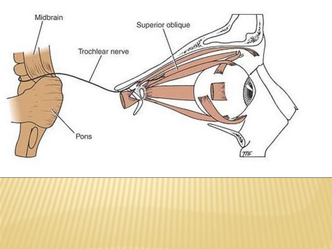
Distribution of the trochlear nerve

Trigeminal Nerve (Cranial Nerve V)
is the largest cranial nerve and contains both sensory and motor fibers. It is the
sensory nerve to the greater part of the head and the motor nerve to several
muscles, including the muscles of mastication
Trigeminal Nerve Nuclei
(1)
the main sensory nucleus,
(2)
the spinal nucleus,
(3)
the mesencephalic nucleus
(4)
the motor nucleus

Course of the Trigeminal Nerve
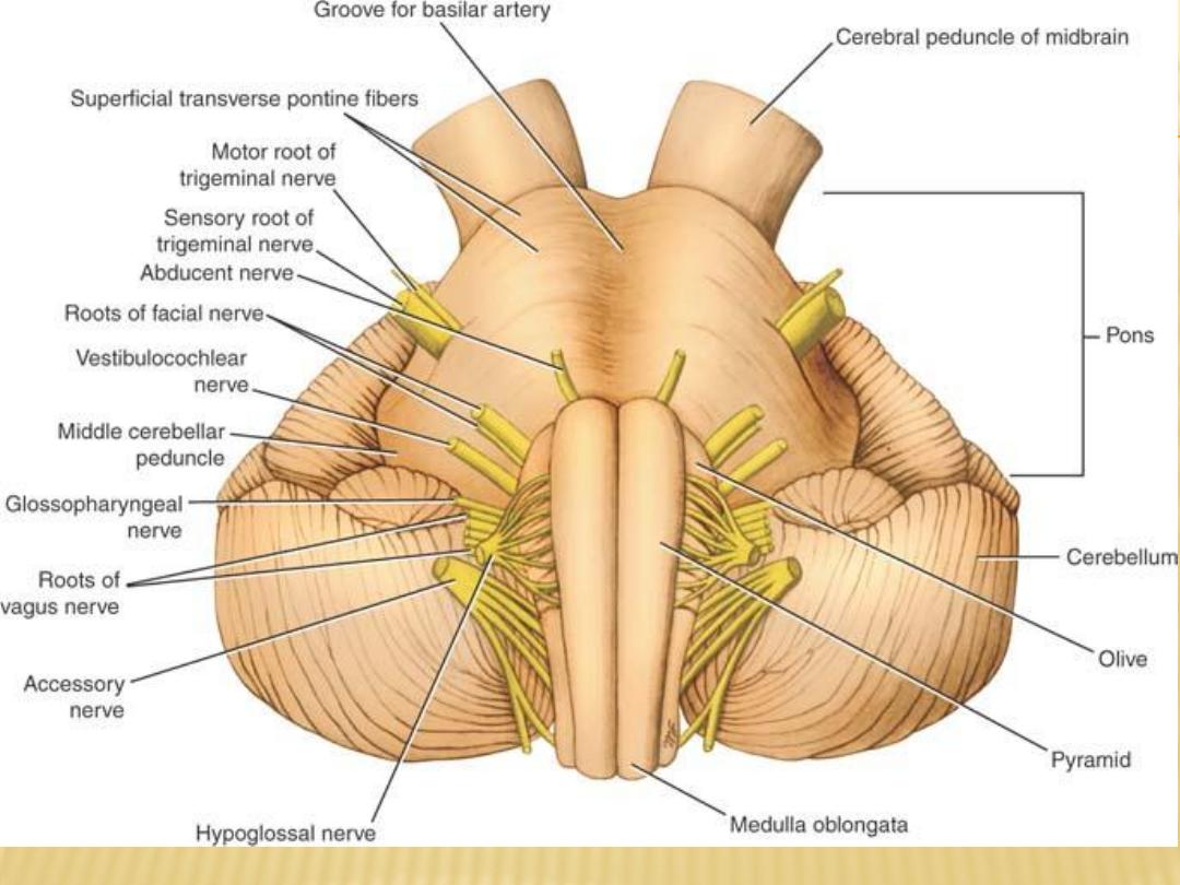
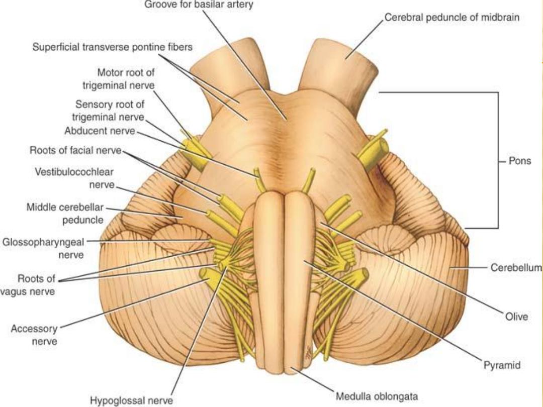
The trigeminal nerve leaves the anterior aspect of the pons as a small motor
root and a large sensory root
The nerve passes forward out of the posterior cranial fossa and rests on the
upper surface of the apex of the petrous part of the temporal bone in the
middle cranial fossa. The large sensory root now expands to form the
crescent-shaped trigeminal ganglion, which lies within a pouch of dura
mater called the
trigeminal or Meckel cave
Branches arise from the anterior border or the ganglion:
The ophthalmic nerve (V1)
contains only sensory fibers and leaves the skull
to enter the orbital cavity.
superior orbital fissure
through the
The maxillary nerve (V2)
also contains only sensory fibers and leaves the skull
.
rotundum
foramen
through the
The mandibular nerve (V3)
contains both sensory and motor fibers and leaves
ovale
foramen
the skull through the
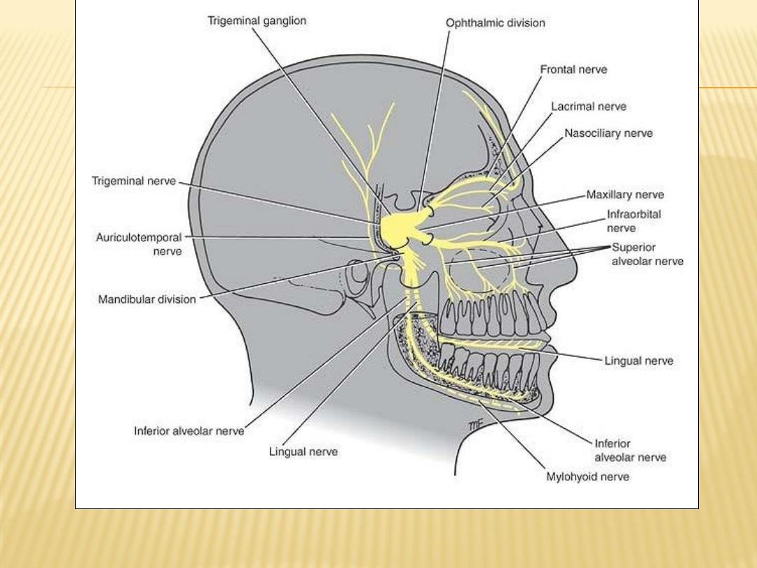
Distribution of the trigeminal nerve.
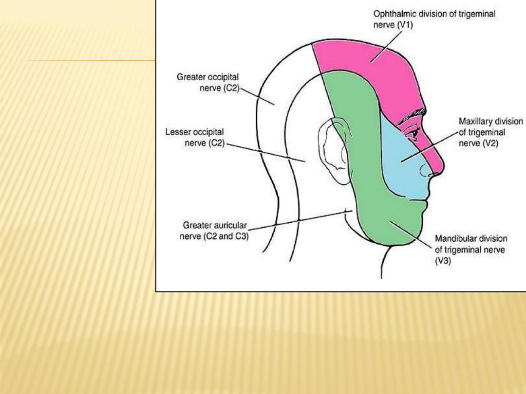
Sensory nerve supply to the skin of the head and
neck. Note that the skin over the angle of the jaw is
supplied by the great auricular nerve (C2 and C3)
and not by branches of the trigeminal nerve.
The sensory fibers to
the skin of the face
from each division
supply a distinct
zone, there being
little or no overlap
of the dermatomes
(compare with the
overlap of the
dermatomes of the
spinal nerves).
As noted previously,
the motor fibers in
the mandibular
division are mainly
distributed to
muscles of
mastication

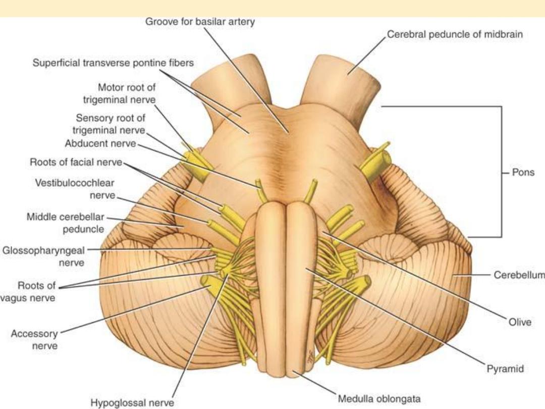
Abducent Nerve (Cranial Nerve VI)
The abducent nerve is a small motor nerve that supplies the lateral rectus
muscle of the eyeball
The small motor nucleus is situated beneath the floor of the upper part of
the fourth ventricle, close to the midline and beneath the colliculus facialis
Course:
The fibers of the abducent nerve pass anteriorly through the pons and
emerge in the groove between the lower border of the pons and the medulla
oblongata It passes forward through the cavernous sinus, lying below and
lateral to the internal carotid artery. The nerve then enters the orbit through
superior orbital fissure.
the
The abducent nerve is entirely a motor nerve and supplies the lateral rectus
muscle and, therefore, is responsible for turning the eye laterally.
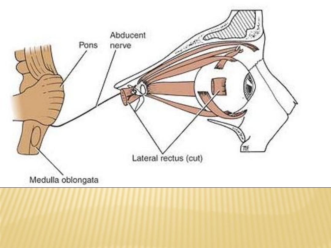
Distribution of the abducent nerve

8
Lecture
Cranial Nerves
(7
th
–12
th
)

Facial Nerve (Cranial Nerve VII)
The facial nerve has three nuclei:
(1)
the main motor nucleus
(2)
the parasympathetic nuclei
(3)
the sensory nucleus
1- Main Motor Nucleus
lies deep in the reticular formation of the lower part of the pons
The part of the nucleus that supplies the muscles of the upper part of the face
receives corticonuclear fibers from both cerebral hemispheres
The part of the nucleus that supplies the muscles of the lower part of the face
receives only corticonuclear fibers from the opposite cerebral hemisphere
2- Parasympathetic Nuclei
lie posterolateral to the main motor nucleus
They are the superior salivatory and lacrimal nuclei
3- Sensory Nucleus
The sensory nucleus is the upper part of the nucleus of the tractus solitarius
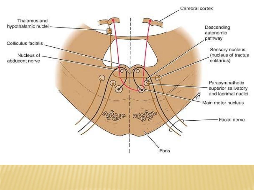
Facial nerve nuclei and their central connections

Course of the Facial Nerve
abducent
travel posteriorly around the medial side of the
fibers of the motor root
nucleus. pass around the nucleus beneath the colliculus facialis in the floor of
the fourth ventricle then emerge from the brainstem
) is formed of the central processes of the
s
intermediu
nervus
The sensory root (
unipolar cells of the geniculate ganglion. It also contains the efferent
preganglionic parasympathetic fibers from the parasympathetic nuclei
-
Two roots (sensory and motor) form the facial nerve that emerge from the
anterior surface of the brain between the pons and the medulla oblongata
-
enter the internal acoustic meatus in the petrous part of the temporal bone
laterally with the vestibular nerve
-
nerve enters the facial canal and runs laterally through the inner ear
-
On medial wall of the tympanic cavity, the nerve expands to form the sensory
geniculate ganglion
-
the facial nerve turns downward on the medial side of the aditus of the mastoid
antrum, descends behind the pyramid, and emerges from the stylomastoid
foramen
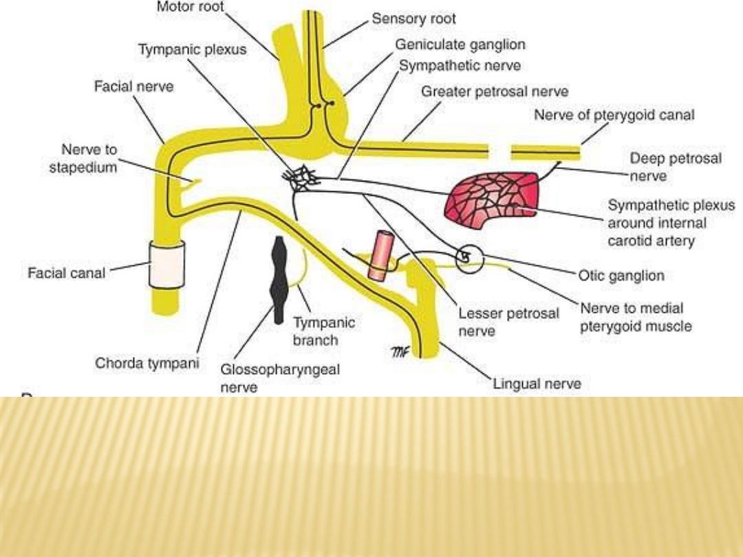
Branches of the facial nerve within the petrous part of the temporal bone;
the taste fibers are shown in black. The glossopharyngeal nerve is also
shown
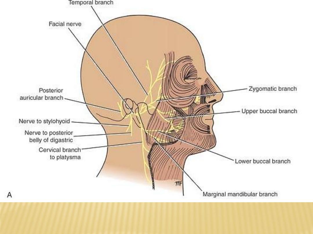
Distribution of the facial nerve

Distribution of the Facial Nerve
supplies the muscles of facial expression, the auricular
The motor nucleus
muscles, the stapedius, the posterior belly of the digastric, and the
stylohyoid muscles
nucleus supplies the submandibular and sublingual
salivatory
The superior
salivary glands and the nasal and palatine glands.
nucleus supplies the lacrimal gland.
The lacrimal
thirds of the
-
receives taste fibers from the anterior two
The sensory nucleus
tongue, the floor of the mouth, and the palate.

Vestibulocochlear Nerve (Cranial Nerve VIII)
-
the vestibular nerve
- the cochlear nerve
the vestibular nerve
The vestibular nerve conducts nerve impulses from the utricle and saccule that
provide information concerning the position of the head; the nerve also
conducts impulses from the semicircular canals that provide information
concerning movements of the head.
They enter the anterior surface of the brainstem in a groove between the lower
border of the pons and the upper part of the medulla oblongata
The Vestibular Nuclear Complex
(1)
the lateral vestibular nucleus,
(2)
the superior vestibular nucleus
(3)
the medial vestibular nucleus, and
(4)
the inferior vestibular nucleus
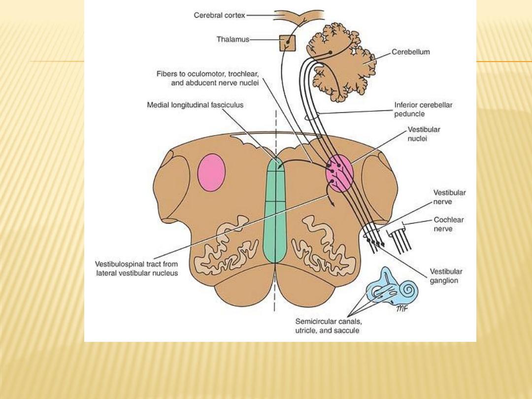
Vestibular nerve nuclei and their central connections

Cochlear Nerve
The cochlear nerve conducts nerve impulses concerned with sound from the organ
of Corti in the cochlea. The fibers of the cochlear nerve are the central
processes of nerve cells located in the spiral ganglion of the cochlea
They enter the anterior surface of the brainstem at the lower border of the pons on
the lateral side of the emerging facial nerve and are separated from it by the
vestibular nerve
Cochlear Nuclei
Anterior and posterior cochlear nuclei
Course of the Vestibulocochlear Nerve
The vestibular and cochlear parts of the nerve leave the anterior surface of the
brain between the lower border of the pons and the medulla oblongata
They run laterally in the posterior cranial fossa and enter the internal acoustic
meatus with the facial nerve. The fibers are then distributed to the different
parts of the internal ear
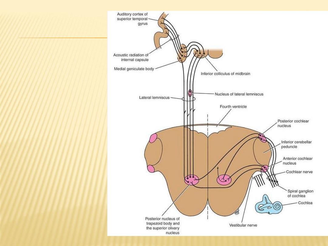
Cochlear nerve
nuclei and
their central
connections
Distribution of the vestibulocochlear nerve

Glossopharyngeal Nerve (Cranial Nerve IX)
The glossopharyngeal nerve has three nuclei:
(1)
the main motor nucleus: supply the stylopharyngeus muscle
(2)
the parasympathetic nucleus
(3)
the sensory nucleus: is part of the nucleus of the tractus solitarius
Course
It leaves the anterolateral surface of the upper part of the medulla oblongata
as a series of rootlets in a groove between the olive and the inferior
cerebellar peduncle
jugular foramen
leaves the skull through the
The nerve then descends through the upper part of the neck in company with
the internal jugular vein and the internal carotid artery to reach the posterior
. The nerve then
muscle, which it supplies
stylopharyngeus
border of the
passes forward between the superior and middle constrictor muscles of the
sensory branches to the mucous membrane of the pharynx
pharynx to give
and the posterior third of the tongue

Nerve (Cranial Nerve X)
Vagus
The vagus nerve has three nuclei:
1)
the main motor nucleus: is formed by the nucleus ambiguus The efferent fibers supply
the constrictor muscles of the pharynx and the intrinsic muscles of the larynx
2)
the parasympathetic nucleus:
are distributed to the involuntary muscle of the bronchi, heart, esophagus,
efferent fibers
stomach, small intestine, and large intestine as far as the distal one-third of the
transverse colon
nerve (carotid
glossopharyngeal
from the hypothalamus and from the
afferent fibers
sinus reflex).
3)
the sensory nucleus
Nerve
Vagus
of the
Course
Leave medulla oblongata as a series of rootlets in a groove between the olive and the inferior
cerebellar peduncle
jugular foramen
leaves the skull through the
two sensory ganglia, a rounded superior ganglion, and a cylindrical inferior ganglion
the cranial root of the accessory nerve joins the vagus nerve and is distributed mainly in its
pharyngeal and recurrent laryngeal branches
with the internal
the carotid sheath
nerve descends vertically in the neck within
vagus
The
jugular vein and the internal and common carotid arteries
The right vagus nerve The left vagus nerve

Distribution
of the vagus
nerve

Accessory Nerve (Cranial Nerve XI)
The accessory nerve is a motor nerve that is formed by the union of a cranial
and a spinal root.
The cranial root (part) is formed from the axons of nerve cells of the nucleus
ambiguus
The spinal root (part) is formed from axons of nerve cells in the spinal nucleus
Hypoglossal Nerve (Cranial Nerve XII)
The hypoglossal nerve is a motor nerve that supplies all the intrinsic muscles of
the tongue as well as the styloglossus, the hyoglossus, and the genioglossus
muscles.
The hypoglossal nerve fibers emerge on the anterior surface of the medulla
oblongata between the pyramid and the olive
hypoglossal canal
leaves the skull through the
In the upper part of its course, the hypoglossal nerve is joined by C1 fibers to
from the cervical plexus
the hypoglossal nerve controls the movements and shape of the tongue
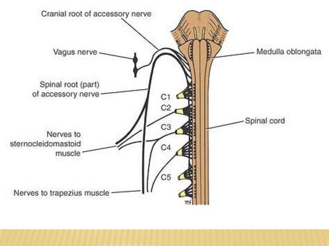
Distribution of the accessory nerve
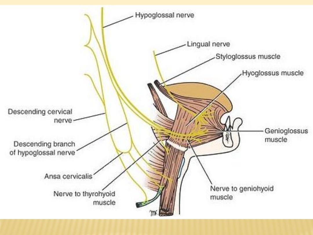
Distribution of hypoglassal nerve