
9
Lecture
Reticulat formation
&
Autonomic Nervous
System
RETICULAR FORMATION
The reticular formation, as its name would suggest, resembles a net (reticular) that
is made up of nerve cells and nerve fibers. The net extends up through the axis
of the central nervous system from the spinal cord to the cerebrum. It is
strategically placed among the important nerve tracts and nuclei. It receives
input from most of the sensory systems and has efferent fibers that descend
and influence nerve cells at all levels of the central nervous system.
The exceptionally long dendrites of the neurons of the reticular formation permit
input from widely placed ascending and descending pathways.
Through its many connections, it can influence skeletal muscle activity, somatic and
visceral sensations, the autonomic and endocrine systems, and even the level of
consciousness
Gross appearance
consists of a deeply placed continuous network of nerve cells and fibers that extend
from the spinal cord through the medulla, the pons, the midbrain, the
subthalamus, the hypothalamus, and the thalamus. The diffuse network may be
divided into three longitudinal columns:
1- median column, consisting of intermediate-size neurons
2- medial column, containing large neurons
3- lateral column, mainly small neurons
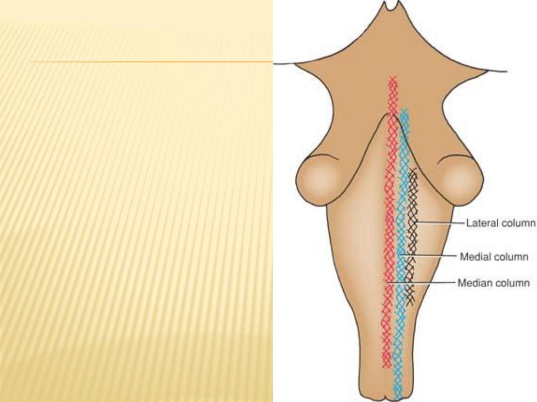
Diagram showing the
approximate positions of
the median, medial, and
lateral columns of the
reticular formation in the
brainstem.

Afferent Projections
afferent pathways project onto the reticular formation from most parts of the
central nervous system
spinothalamic
tracts, the
spinoreticular
there are the
,
From the spinal cord
tracts, and the medial lemniscus
, there are ascending afferent tracts, which
From the cranial nerve nuclei
include the vestibular, acoustic, and visual pathways
pathway
cerebelloreticular
, there is the
From the cerebellum
Efferent Projections
Multiple efferent pathways extend down to the brainstem and spinal cord
tracts to neurons in the motor
reticulospinal
and
reticulobulbar
through the
nuclei of the cranial nerves and the anterior horn cells of the spinal cord
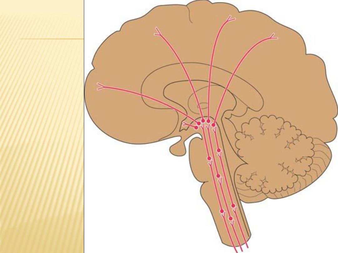
Diagram
showing
the
afferent
fibers of
the
reticular
formation

Functions of the Reticular Formation
1-
Control of skeletal muscle, modulating muscle tone and reflex activity. It can
also bring about reciprocal inhibition; for example, when the flexor muscles
contract, the antagonistic extensors relax
2-
Control of somatic and visceral sensations
3-
Control of the autonomic nervous system
4-
Control of the endocrine nervous system. Either directly or indirectly through
the hypothalamic nuclei
5-
Influence on the biologic clocks. By means of its multiple afferent and
efferent pathways to the hypothalamus
6-
The reticular activating system. Arousal and the level of consciousness are
controlled by the reticular formation
Autonomic Nervous System
The autonomic nervous system is distributed throughout the central and
peripheral nervous systems.
It is divided into two parts, the sympathetic and the parasympathetic
consist of both afferent and efferent fibers.
This division between sympathetic and parasympathetic is made on the basis of
anatomical differences, differences in the neurotransmitters, and differences in the
physiologic effects
Both the sympathetic and parasympathetic divisions produce opposite effects in
most organs and are thus considered as physiologic antagonists
Sympathetic Part
is the larger of the two parts of the autonomic system and is widely
distributed throughout the body
innervating the heart and lungs, the muscle in the walls of many blood
vessels, the hair follicles and the sweat glands, and many abdominopelvic
viscera
dilate the pupils; inhibit smooth muscle of the bronchi, intestine, and
bladder wall; and close the sphincters. The hair is made to stand on end,
and sweating occurs

Efferent Nerve Fibers (Sympathetic Outflow)
The lateral gray columns (horns) of the spinal cord from the first thoracic segment to
the second lumbar segment (sometimes third lumbar segment) possess the cell
bodies of the sympathetic connector neurons
Then axons of these cells leave the cord in the anterior nerve roots and pass via the
white rami communicantes to the paravertebral ganglia of the sympathetic trunk
they are distributed as follows:
1-
They synapse with an excitor neuron in the ganglion, They are distributed in branches
of the spinal nerves to smooth muscle in the blood vessel walls, sweat glands, and
arrector muscles of the hairs of the skin
2-
They travel cephalad in the sympathetic trunk to synapse in ganglia in the cervical
region
postganglionic nerve fibers communicantes to join the cervical spinal nerves
Again, the postganglionic nerve fibers communicantes to join the lumbar, sacral, and
coccygeal spinal nerves
3-
They may pass through the ganglia of the sympathetic trunk without synapsing
they leave the sympathetic trunk as the greater splanchnic, lesser splanchnic, and
lowest or least splanchnic nerves
A few preganglionic fibers, traveling in the greater splanchnic nerve, end directly on the
cells of the suprarenal medulla. These medullary cells, which may be regarded as
modified sympathetic excitor neurons, are responsible for the secretion of
epinephrine and norepinephrine
Afferent Nerve Fibers
Afferent myelinated nerve fibers travel from the viscera through the sympathetic ganglia
without synapsing.
They pass to the spinal nerve and reach posterior root ganglion of the corresponding
spinal nerve
The central axons then enter the spinal cord
They either:
form the afferent component of a local reflex arc
or ascend to higher centers, such as the hypothalamus
Sympathetic Trunks
are two ganglionated nerve trunks that extend the whole length of the vertebral column
In the neck, each trunk has 3 ganglia; in the thorax, 11 or 12; in the lumbar region, 4 or
5; and in the pelvis, 4 or 5
Relations of sympathetic trunk
In the neck, the trunks lie anterior to the transverse processes of the cervical
vertebrae;
in the thorax, they are anterior to the heads of the ribs or lie on the sides of the
vertebral bodies;
in the abdomen, they are anterolateral to the sides of the bodies of the lumbar
vertebrae;
in the pelvis, they are anterior to the sacrum.
Below, the two trunks end by joining together to form a single ganglion, the ganglion
impar

Parasympathetic Part of the Autonomic System
The activities of the parasympathetic part of the autonomic system are directed
toward conserving and restoring energy. The heart rate is slowed, pupils are
constricted, peristalsis and glandular activity is increased, sphincters are
opened, and the bladder wall is contracted
Efferent Nerve Fibers (Craniosacral Outflow)
The connector nerve cells of the parasympathetic part of the autonomic nervous
system are located in the brainstem and the sacral segments of the spinal cord
•
oculomotor (parasympathetic or Edinger-Westphal nucleus),
•
facial (superior salivatory nucleus and lacrimatory nucleus),
•
glossopharyngeal (inferior salivatory nucleus),
•
vagus nerves (dorsal nucleus of the vagus).
The axons of these connector nerve cells are myelinated and emerge from the brain
within the cranial nerves
•
The sacral connector nerve cells are found in the gray matter of the second,
third, and fourth sacral segments of the spinal cord.
The cranial parasympathetic ganglia are the ciliary, pterygopalatine,
submandibular, and otic
In certain locations, the ganglion cells are placed in nerve plexuses, such as the
cardiac plexus, pulmonary plexus, myenteric plexus (Auerbach plexus), and
mucosal plexus (Meissner plexus);
Afferent Nerve Fibers
travel from the viscera to their cell bodies, located either in the sensory ganglia of
the cranial nerves or in the posterior root ganglia of the sacrospinal nerves
they may take part in the formation of local reflex arcs or pass to higher centers of
the autonomic nervous system, such as the hypothalamus
The Large Autonomic Plexuses
Large collections of sympathetic and parasympathetic efferent nerve fibers and
their associated ganglia, together with visceral afferent fibers, form autonomic
nerve plexuses in the thorax, abdomen, and pelvis. Branches from these
plexuses innervate the viscera
In the thorax: cardiac, pulmonary, and esophageal plexuses.
In the abdomen:the plexuses are associated with the aorta and its branches, celiac,
superior mesenteric, inferior mesenteric, and aortic plexuses.
In the pelvis: the superior and inferior hypogastric plexuses
Autonomic Ganglia
The autonomic ganglion is the site where preganglionic nerve fibers synapse on
postganglionic neurons
Sympathetic ganglia form part of the sympathetic trunk or are prevertebral in
position
Parasympathetic ganglia are situated close to or within the walls of the viscera.
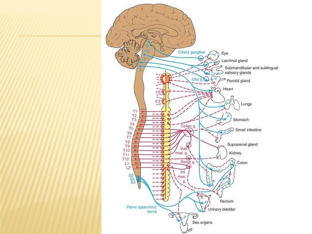
Efferent part of the
autonomic nervous
system.
Preganglionic
parasympathetic
fibers are shown in
solid blue, and
postganglionic
parasympathetic
fibers are shown in
interrupted blue.
Preganglionic
sympathetic fibers
are shown in solid
red, and
postganglionic
sympathetic fibers
are shown in
interrupted red