
10
Lecture
Cerebrum

Cerebrum
Subdivisions of the Cerebrum
The cerebrum is the largest part of the brain, situated in the
anterior and middle cranial fossae of the skull and occupying
the whole concavity of the vault of the skull. It may be divided
into two parts:
the diencephalon
, which forms the central core,
and
the telencephalon
, which forms the cerebral hemispheres.
The diencephalon
consists of the third ventricle and the structures that form its boundaries It
extends posteriorly to the point where the third ventricle becomes
continuous with the
cerebral aqueduct
and anteriorly as far as
the
interventricular foramina
Thus, the diencephalon is a midline structure with
symmetrical right and left halves
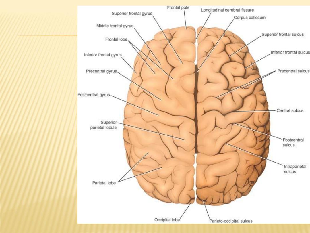
Superior view
of the
cerebral
hemispheres

Gross Features of diencephalon
inferior surface
The only area exposed to the surface in the intact brain.
formed by
hypothalamic
and other structures, which include, from anterior to
posterior, the
optic chiasma
, with the
optic tract
on either side; the
infundibulum
, with
the tuber cinereum
; and the
mammillary bodies

superior surface of the diencephalon
is concealed by the
fornix
Fornix
is thick bundle of fibers that originates in the hippocampus of the
temporal lobe and arches posteriorly over the
thalamus
to join the
mammillary body
Sup wall formed by
roof of the third ventricle
lateral surface of the diencephalon
bounded by the internal capsule of white matter
Medial surface
Since the diencephalon is divided into symmetrical halves by the slitlike third
ventricle, it also has a medial surface. The medial surface of the
diencephalon (i.e., the lateral wall of the third ventricle) is formed in its
superior part by the medial surface of the thalamus and in its inferior part
by the hypothalamus, These two areas are separated from one another by a
shallow sulcus, the hypothalamic sulcus

The diencephalon can be divided into four major parts:
(1)
the thalamus,
(2)
the subthalamus,
(3)
the epithalamus,
(4)
the hypothalamus
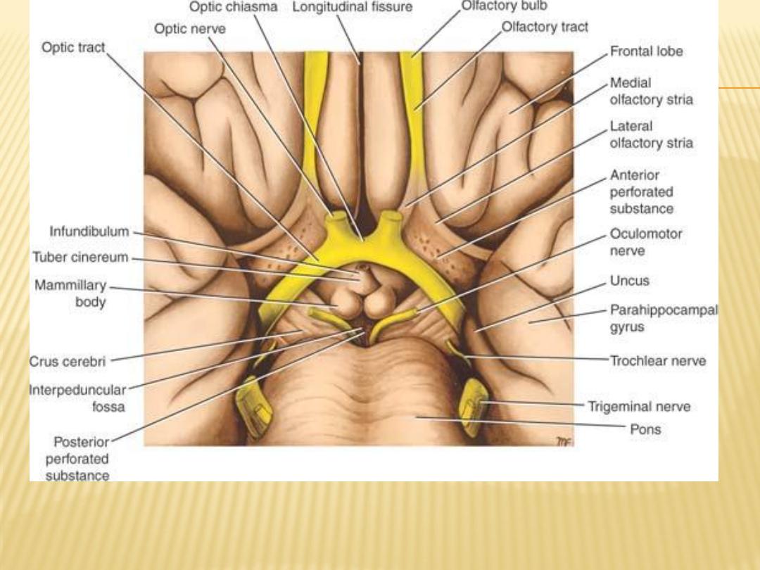
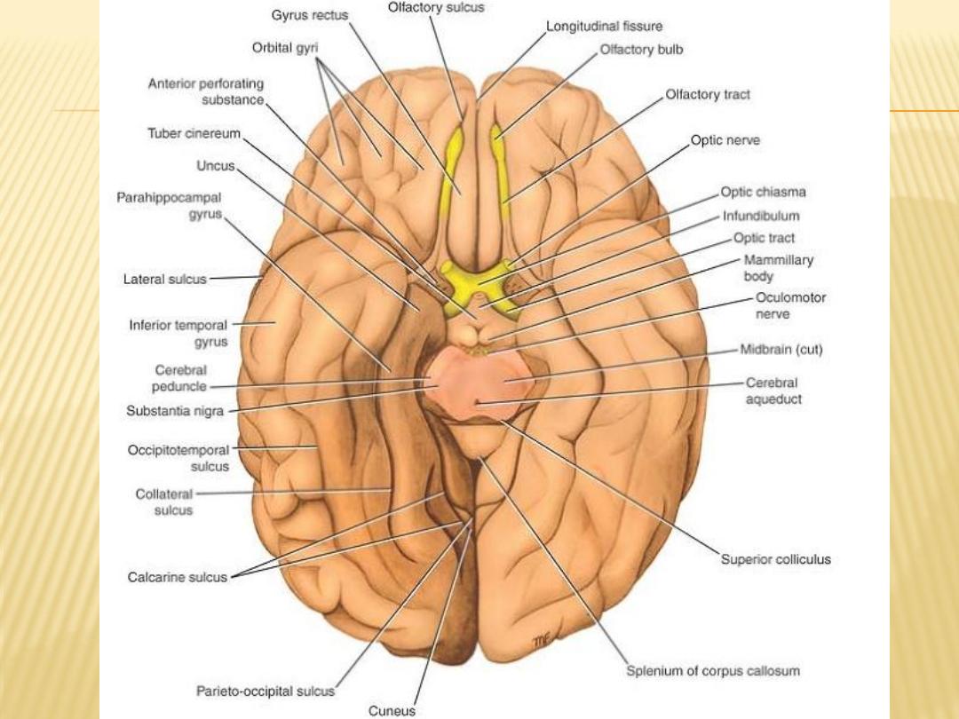
Inferior surface of the brain showing parts of the diencephalon
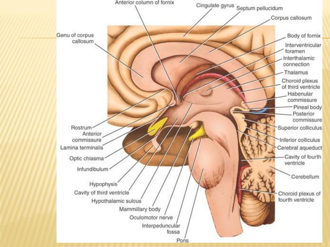
Sagittal section of
the brain showing
the medial
surface of the
diencephalon

Thalamus
-
a large ovoid mass of gray matter that forms the major part of the
diencephalon
It is a region of great functional importance and serves as a cell station to
all the main sensory systems (except the olfactory pathway)
-
The activities of the thalamus are closely related to that of the cerebral
cortex and damage to the thalamus causes great loss of cerebral function
-
pulvinar:
is the expanded posterior end of the thalamus
-
lateral geniculate body:
a small elevation on the under aspect of the lateral
portion of the pulvinar
The thalamus contain many important nuclei that will be discussed in future

As a relation,
superior surface
is covered medially by the tela choroidea and the fornix, and laterally, it is
covered by ependyma and forms part of the floor of the lateral ventricle
The inferior surface
is continuous with the tegmentum of the midbrain
medial surface of the thalamus
forms the superior part of the lateral wall of the third ventricle and is usually
connected to the opposite thalamus by a band of gray matter, the
interthalamic connection
lateral surface of the thalamus
is separated from the lentiform nucleus by the very important band of white
matter called the internal capsule

Function of thalamus
1- very important cell station that receives the main sensory tracts (except the
olfactory pathway)
2- plays a key role in the integration of visceral and somatic functions
Other functions will be discussed later
Subthalamus
inferior to the thalamus
between the thalamus and the tegmentum of the midbrain
craniomedially, it is related to the hypothalamus
Among the collections of nerve cells found in the subthalamus are
- cranial ends of the red nuclei
- substantia nigra
- subthalamic nucleus involved in the control of muscle activity
- The subthalamus also contains many important tracts that pass up from the
tegmentum to the thalamic nuclei; the cranial ends of the medial, spinal,
and trigeminal lemnisci are examples
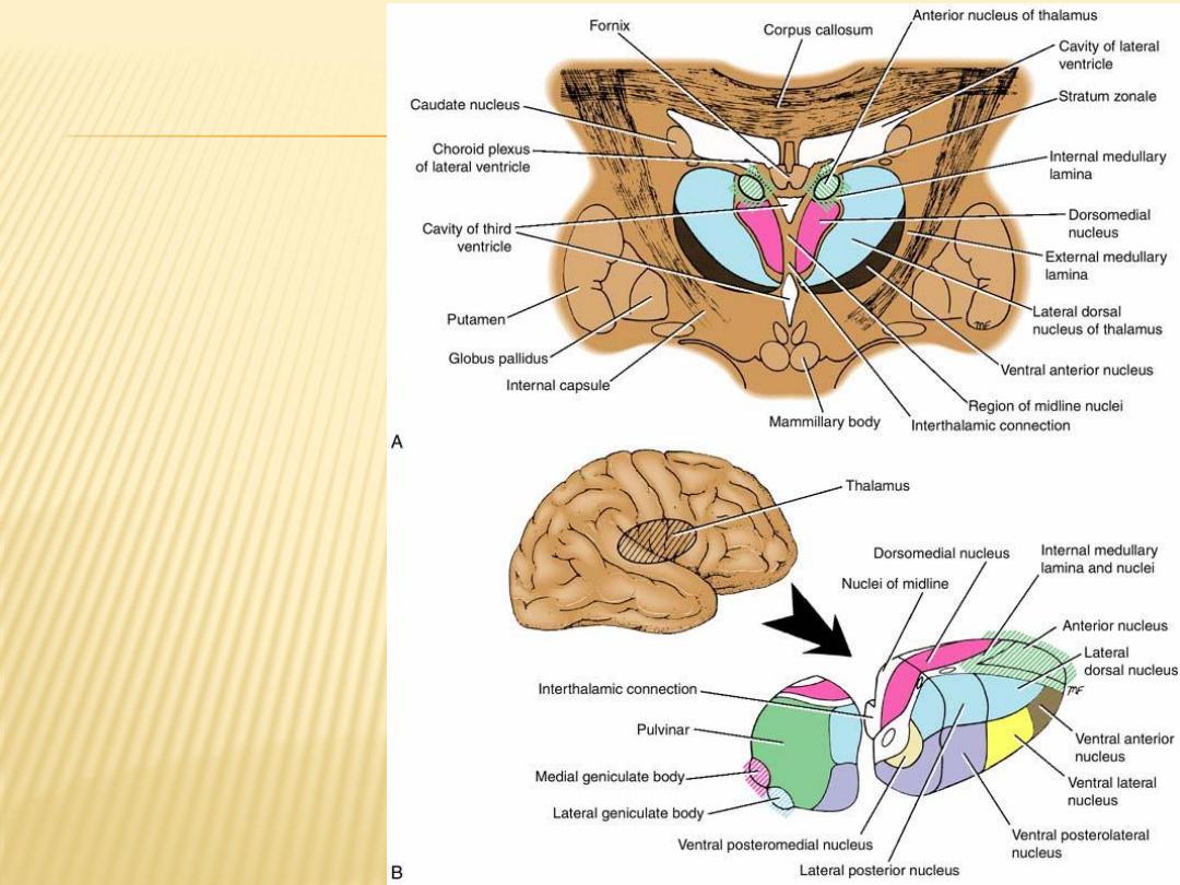
Nuclei of the
thalamus

Epithalamus
-
consists of the habenular nuclei and their connections and the pineal gland
Habenular Nucleus
small group of neurons situated just medial to the posterior surface of the
thalamus
-
Pineal Gland (Body)
a small, conical structure that is attached by the pineal stalk to the
diencephalon
Relations:
- dorsally by splenium of corpus callosum
- ventrally by midbrain
- sup, post part of 3
rd
ventricle
- inf, cerebellar vermis
- consist of pinealocytes and the glial cells
- Calcifications may be found normally at age of 16
- cysts may be found (pineal cyst)
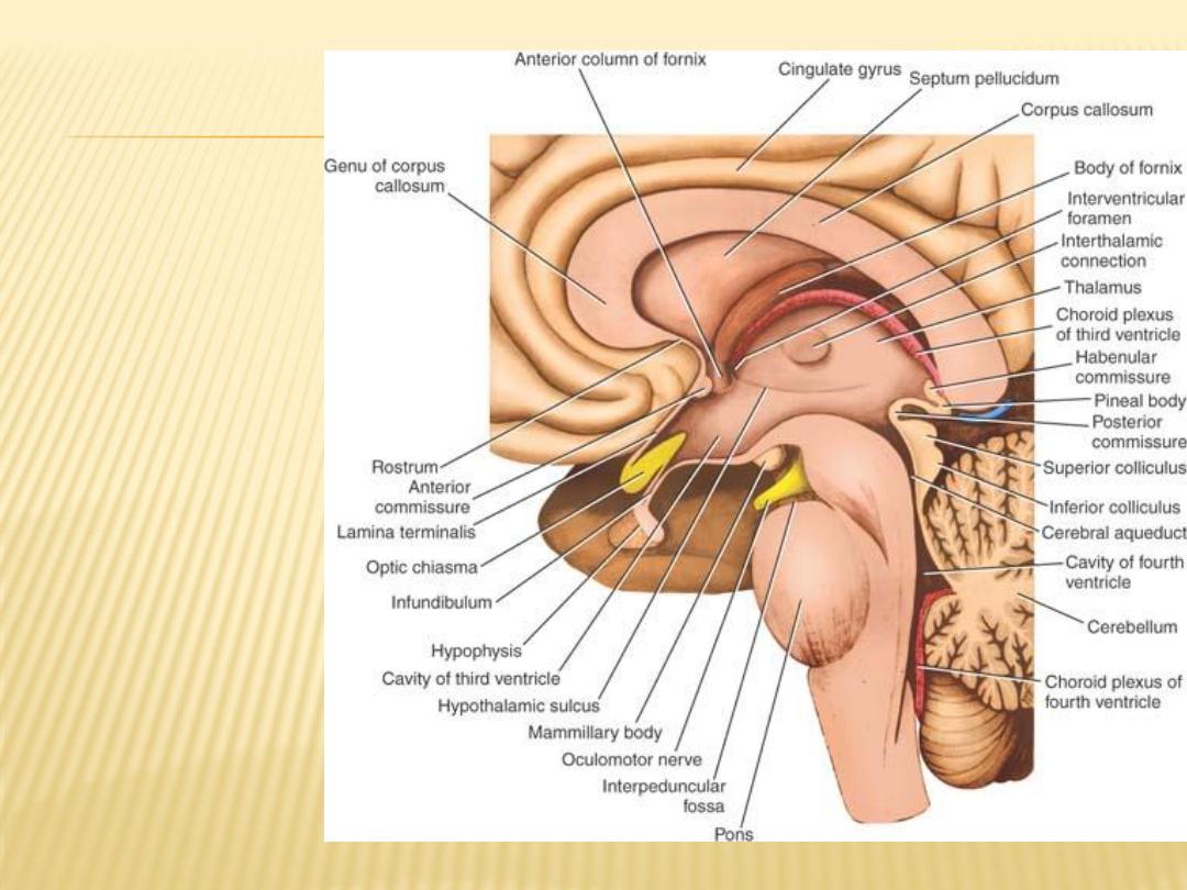
Sagittal section
of the brain
showing the
medial
surface of the
diencephalon
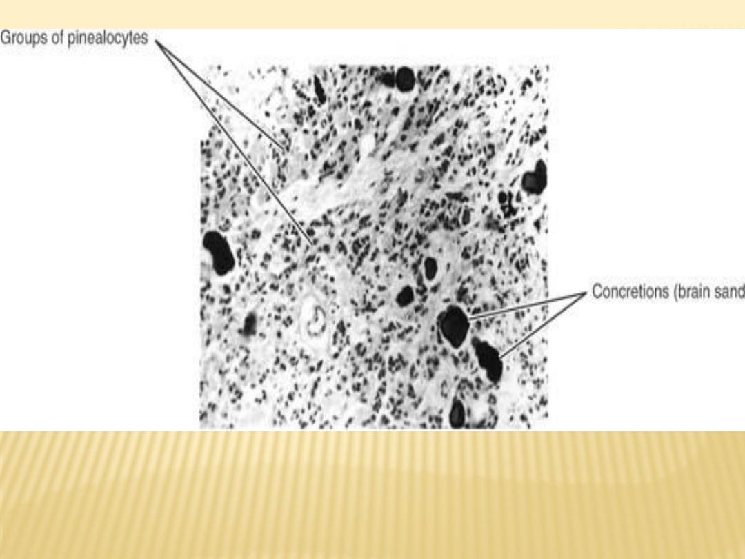
Photomicrograph of a section of the pineal gland stained with hematoxylin
and eosin

Functions of the Pineal Gland
- an important endocrine gland capable of influencing the activities of the
pituitary gland. the islets of Langerhans of the pancreas, the parathyroids,
the adrenal cortex and the adrenal medulla, and the gonads.
- Melatonin and the enzymes needed for its production are present in high
concentrations within the pineal gland
- pineal gland plays an important role in the regulation of reproductive function

Hypothalamus
is that part of the diencephalon that extends from the region of the optic
chiasma to the caudal border of the mammillary bodies
It lies below the hypothalamic sulcus on the lateral wall of the third ventricle
Physiologically, there is hardly any activity in the body that is not influenced by
the hypothalamus
The hypothalamus controls and integrates the functions of the autonomic
nervous system and the endocrine systems and plays a vital role in
maintaining body homeostasis. It is involved in such activities as regulation
of body temperature, body fluids, drives to eat and drink, sexual behavior,
and emotion

Relations of the Hypothalamus
area
preoptic
to the hypothalamus:
Anterior
of the midbrain
tegmentum
, the hypothalamus merges into the
Caudally
is the thalamus
Superiorly
region
subthalamic
lies the
Inferolaterally
When observed from below, the hypothalamus is seen to be related to the
following structures, from anterior to posterior: (1) the optic chiasma, (2) the
infundibulum and the tuber cinereum, and (3) the mammillary bodies
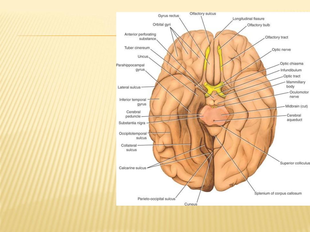
Optic Chiasma
The optic chiasma is a
flattened bundle of nerve
fibers situated at the
junction of the anterior
wall and floor of the third
ventricle
Tuber Cinereum
a convex mass of gray matter,
as seen from the inferior
surface
It is continuous inferiorly with
the infundibulum
Mammillary Bodies
two small hemispherical
bodies situated side by
side posterior to the tuber
cinereum
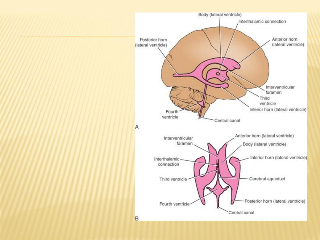
Ventricular cavities of the
brain.
A: Lateral view.
B: Superior view
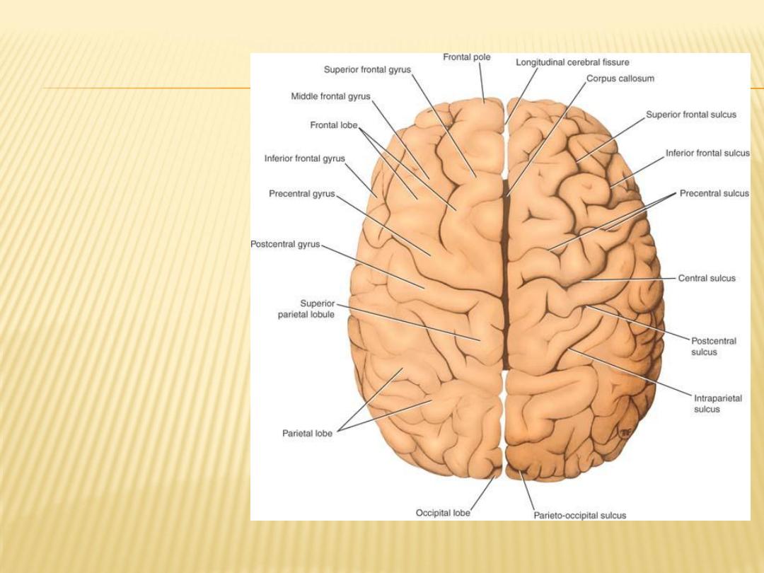
Superior view of the
cerebral
hemispheres.
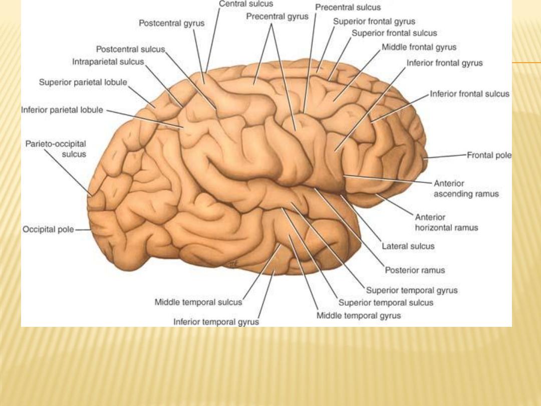
Lateral view of the right cerebral hemisphere
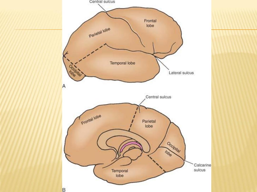
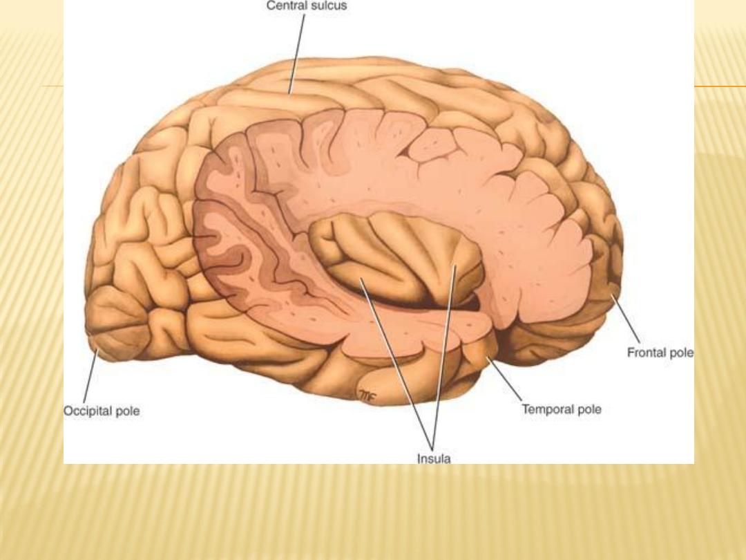
Lateral view of the right cerebral hemisphere dissected to reveal the right
insula

11
Lecture
Cerebrum, continuation

General Appearance of the Cerebral Hemispheres
the largest part of the brain; they are separated by a deep midline sagittal
fissure, the longitudinal cerebral fissure
The fissure contains the sickle-shaped fold of dura mater, the falx cerebri, and
the anterior cerebral arteries.
great commissure, the corpus callosum, connects the hemispheres across the
midline
A second horizontal fold of dura mater separates the cerebral hemispheres
from the cerebellum and is called the tentorium cerebelli
To increase the surface area of the cerebral cortex maximally, the surface of
each cerebral hemisphere is thrown into folds or gyri, which are separated
from each other by sulci or fissures
each hemisphere are devided into lobes, which are named according to the
cranial bones under which they lie.
The central, parieto-occipital, lateral and calcarine sulci are boundaries used
for the division of the cerebral hemisphere into frontal, parietal, temporal,
and occipital lobes

Main Sulci
The central sulcus
(fissure of Rolanad) it begin aproximately midway between frontal and occopital pole
and terminates just above the lateral sulcus, separate between frontal and parietal,
motor (area 4) and sensory (area 1-3), precentral and postcentral gyrus
Clinically it is related to anterior (frontal) branch of the middle meningeal artery which is
important as a common site of injury during head trauma resulting in extradural
hematoma.
,
The lateral sulcus
a deep cleft found mainly on the inferior and lateral surfaces of the cerebral
hemisphere, divides into the anterior horezontal ramus (anterior) and the anterior
ascending ramus (ascending) and continues as the posterior ramus
Anterir to the anterior horezontal ramus (anterior) is the pars orbitalis, between the
anterior horezontal ramus (anterior) and the anterior ascending ramus (ascending)
is the pars triangularis (motor speech area – brochas area), posterior to the anterior
ascending ramus (ascending) is pars opercularis
Clinically it is related to middle cerebral artery
An area of cortex called the insula lies at the bottom of the deep lateral sulcus and
cannot be seen from the surface unless the lips of the sulcus are separated

occipital sulcus
-
parieto
The
begins on the superior medial margin of the hemisphere about 2 inches (5 cm)
anterior to the occipital pole, It passes downward and anteriorly on the
medial surface to meet the calcarine sulcus forming a Y shaped appearance
sulcus
calcarine
The
is found on the medial surface of the hemisphere
It commences under the posterior end of the corpus callosum and arches
upward and backward to reach the occipital pole
The calcarine sulcus is joined at an acute angle by the parieto-occipital sulcus
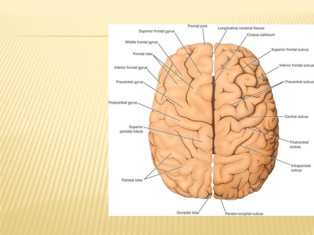
Superior view of
the cerebral
hemispheres
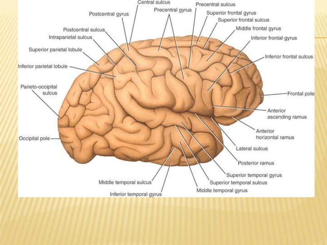
Lateral view of the right cerebral hemisphere
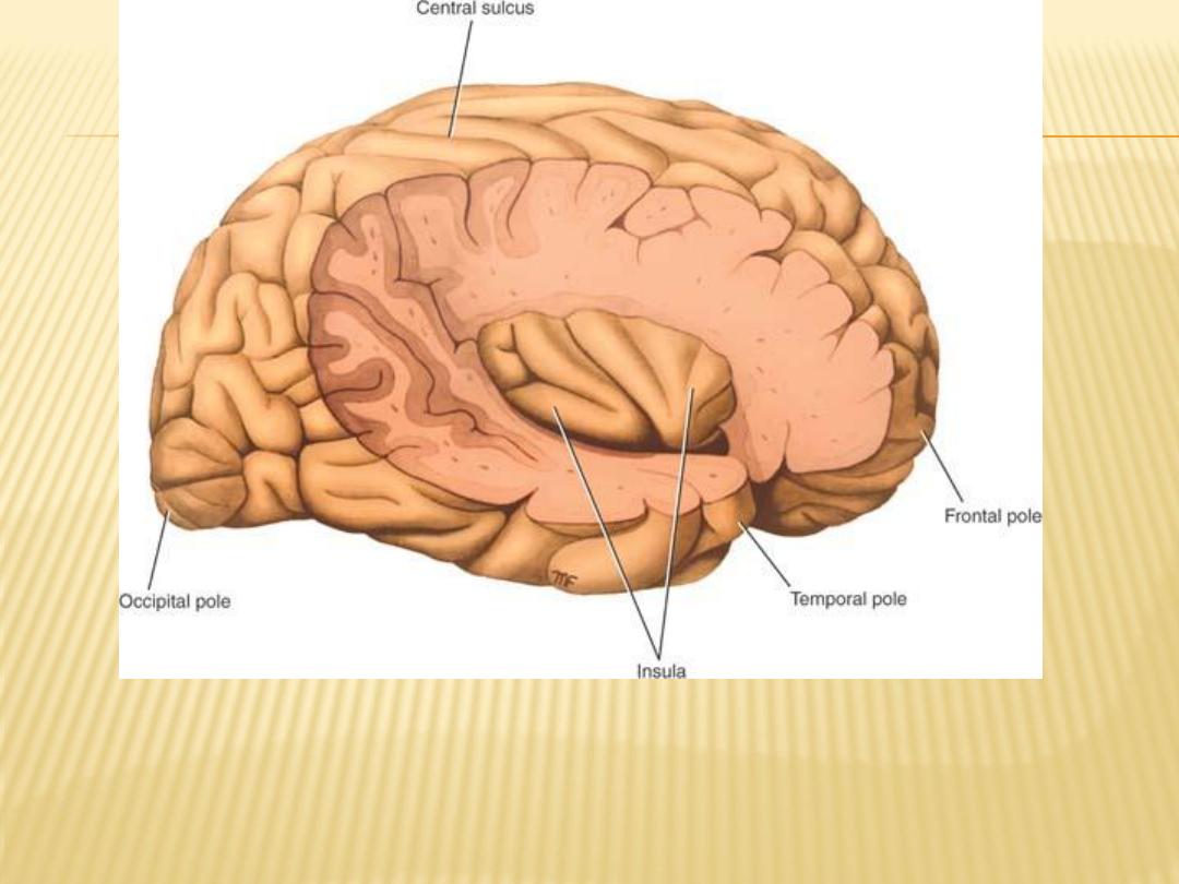
Lateral view of the right cerebral hemisphere
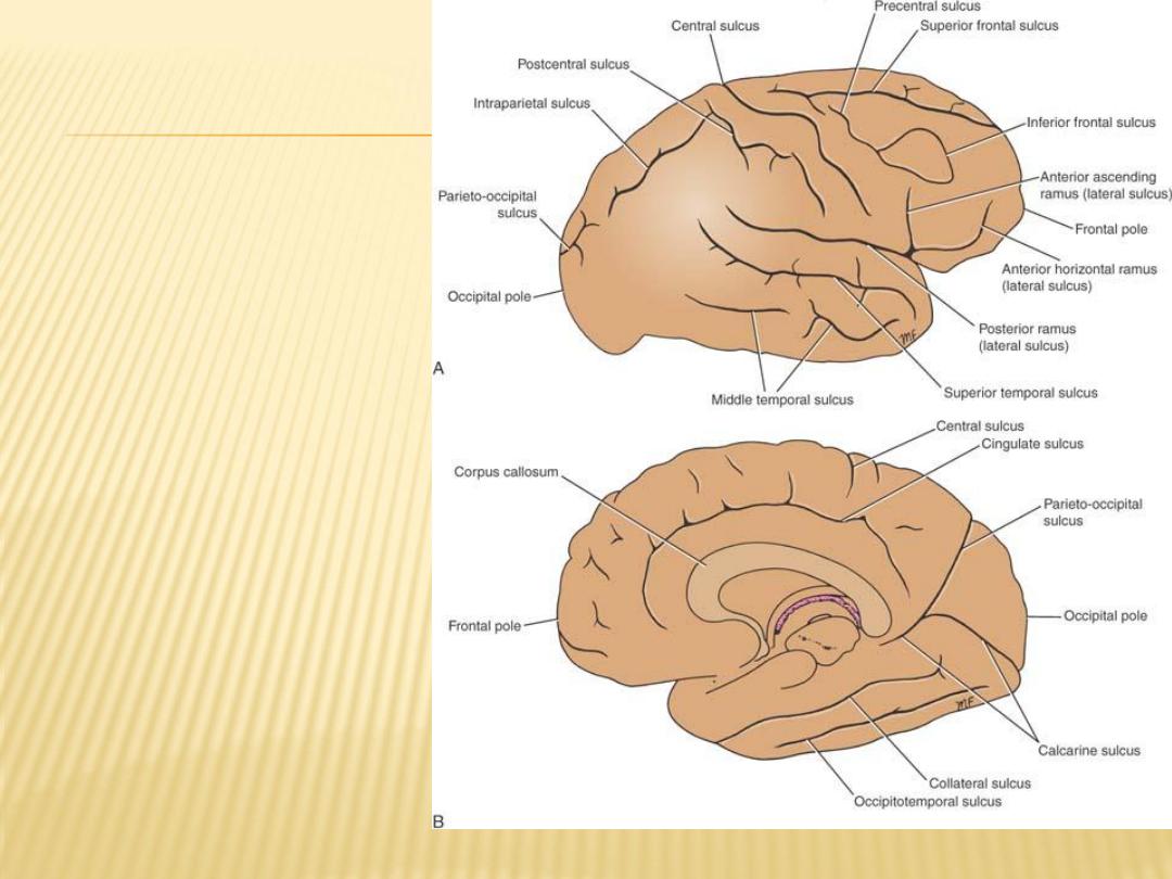
Lateral view of the right
cerebral hemisphere
showing the main sulci. B:
Medial view of the right
cerebral hemisphere
showing the main sulci.

Lobes of the Cerebral Hemisphere
The frontal lobe
occupies the area anterior to the central sulcus and superior to the lateral sulcus
(precentral gyrus , superior frontal gyrus , middle frontal gyrus and inferior frontal gyrus )
The parietal lobe
occupies the area posterior to the central sulcus and superior to the lateral sulcus; it
extends posteriorly as far as the parieto-occipital sulcus
(postcentral gyrus, superior parietal lobule (gyrus), and the inferior parietal lobule
(gyrus).)
The superior and inferior parietal gyri are separated by the intra-parietal sulcus
The temporal lobe
occupies the area inferior to the lateral sulcus, superior and middle (inferior) temporal
sulcus devide the lobe into superior, middle, and inferior temporal gyri
The occipital lobe the occipitotemporal sulcus forms the medial and lateral
occipitotemporal gyri (on the inferior surface of the cerebrum)
The occipital lobe contain the lunate sulcus (behind which is the visual area –area 17)
on superolateral sulcus

Medial and Inferior Surfaces of the Hemisphere
many important areas that should be recognized
The corpus callosum, which is the largest commissure of the brain, forms a
striking feature on this surface
cingulate gyrus begins beneath the anterior end of the corpus callosum and
continues above the corpus callosum until it reaches its posterior end, The
gyrus is separated from the corpus callosum by the callosal sulcus. The
cingulate gyrus is separated from the superior frontal gyrus by the cingulate
sulcus
Inferior surface of the frontal lobe (orbital surface) devided by orbital sulci (H-
shaped sulci) into anterior orbital, posterior orbital, lateral orbital and
medial orbital gyei
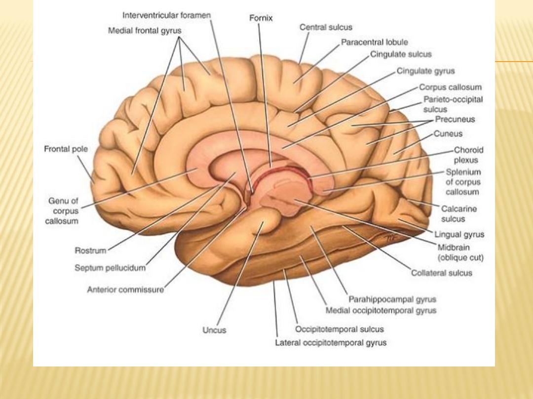
Medial view of the cerebral hemisphere

The precuneus:
is an area of cortex bounded anteriorly by the upturned posterior end of the
cingulate sulcus and posteriorly by the parieto-occipital sulcus
The cuneus:
is a triangular area of cortex bounded above by the parieto-occipital sulcus,
inferiorly by the calcarine sulcus, and posteriorly by the superior medial
margin.
The collateral sulcus
is situated on the inferior surface of the hemisphere. It runs anteriorly below
the calcarine sulcus.
lingual gyrus
Between the collateral sulcus and the calcarine sulcus.
Anterior to the lingual gyrus is the parahippocampal gyrus; the latter terminates
in front as the hooklike uncus
