13
Lecture
Ventricular System

Ventricular system
Origin and circulation of the
cerebrospinal fluid (pink).

The ventricles consist of four fluid-filled cavities located within the brain;
1.
the two lateral ventricles
2.
the third ventricle
3.
the fourth ventricle
-The two lateral ventricles communicate through the interventricular foramina
(phoramen of Monro) with the third ventricle
-
The third ventricle is connected to the fourth ventricle by the narrow
cerebral aqueduct (aqueduct of Sylvius)
The fourth ventricle, is continuous with the narrow central canal of the spinal
cord and, through the three foramina in its roof, with the subarachnoid
lateral
and the
Magendie
the median aperture or foramen of
space (
)
Luschka
openings of the fourth ventricle, or the foramina of
is a small dilatation at the inferior end of central canal
terminal ventricle
The ventricles are lined throughout with ependyma and are filled with
cerebrospinal fluid
Lateral Ventricles
C-shaped cavity with a body, which occupies the parietal lobe and three horn
(anterior, posterior, and inferior horns) extend into the frontal, occipital, and
temporal lobes, respectively

the ventricular
cavities of the
brain.
A: Lateral view
B: Anterior view
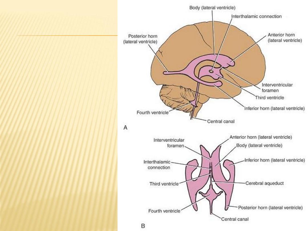
Ventricular cavities
of the brain.
A: Lateral view
B: Superior view

Choroid Plexus of the Lateral Ventricle
is a vascular fringe composed of pia mater covered with the ependymal lining
of the ventricular cavity
It is the irregular lateral edge of the tela choroidea, which is a two-layered fold
of pia mater situated between the fornix superiorly and the upper surface of
the thalamus
The function of the choroid plexus is to produce cerebrospinal fluid
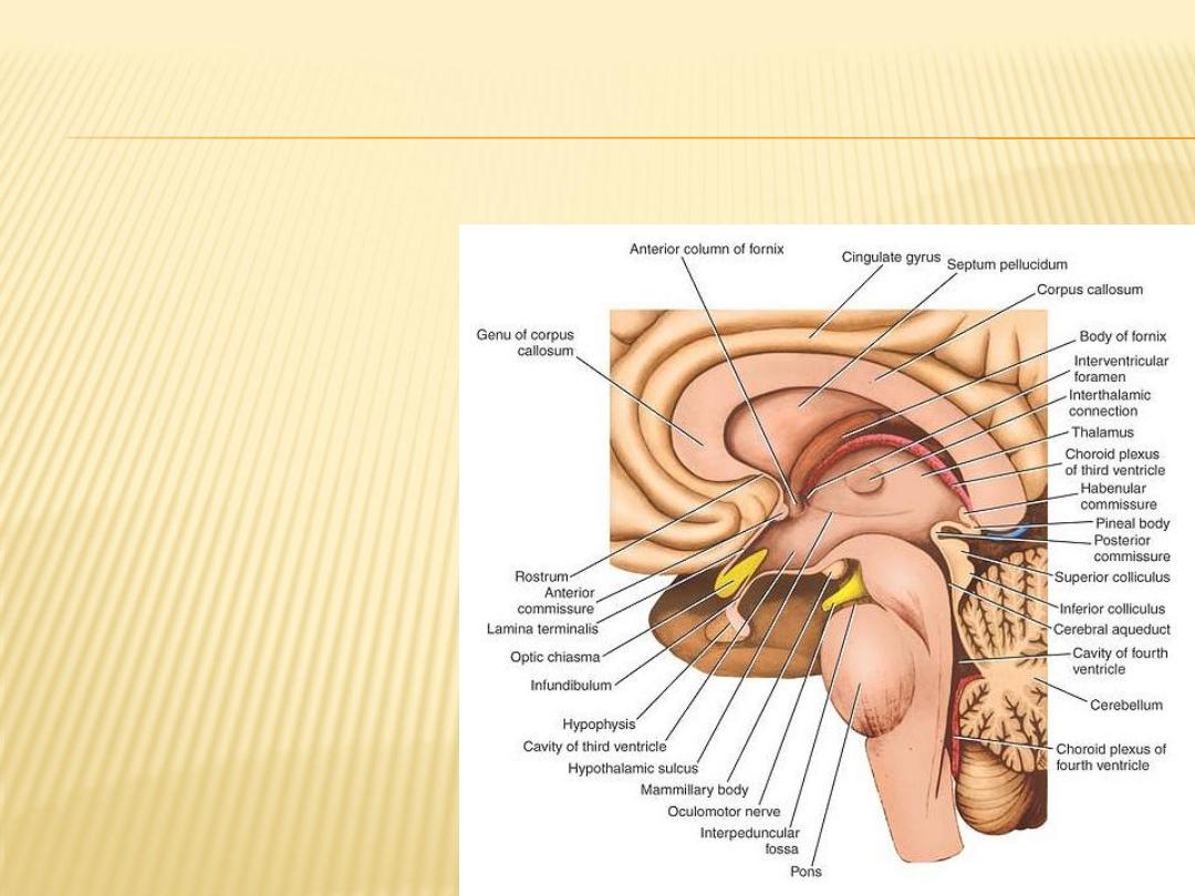
Third Ventricle
The third ventricle is a slitlike cleft between the two thalami

The choroid plexuses of third ventricle are formed from the tela choroidea
situated above the roof of the which projects downward on each side of the
midline, invaginating the ependymal roof
The blood supply of the tela choroidea and the choroid plexuses of the third
branches of the internal
choroidal
and lateral ventricles is derived from the
.
carotid and basilar arteries
The venous blood drains into the internal cerebral veins, which unite to form
the great cerebral vein. The great cerebral vein joins the inferior sagittal
sinus to form the straight sinus of the ventricle
Cerebral Aqueduct
The cerebral aqueduct (aqueduct of Sylvius), a narrow channel about ¾ of an
inch (1.8 cm) long, connects the third ventricle with the fourth ventricle It is
lined with ependyma and is surrounded by a layer of gray matter called the
central gray.
The direction of flow of cerebrospinal fluid is from the third to the fourth
ventricle. There is no choroid plexus in the cerebral aqueduct

Fourth Ventricle
The fourth ventricle is a tent-shaped cavity filled with cerebrospinal fluid. It
is situated anterior to the cerebellum and posterior to the pons and the
superior half of the medulla oblongata
It is lined with ependyma and is continuous above with the cerebral
aqueduct of the midbrain and below with the central canal of the medulla
oblongata and the spinal cordperior half of the medulla oblongata
Choroid Plexus of the Fourth Ventricle
The choroid plexus has a T shape; the vertical part of the T is double, it is
formed from the highly vascular tela choroidea.
The tela choroidea is a two-layered fold of pia mater that projects through the
roof of the ventricle and is covered by ependyma
The function of the choroid plexus is to produce cerebrospinal fluid

Sagittal section of the fourth ventricle showing the origin and circulation of
the cerebrospinal fluid

Central Canal of the Spinal Cord and Medulla Oblongata
into the fourth ventricle
superiorly
opens
, it extends through the inferior half of the medulla oblongata and
Inferiorly
through the entire length of the spinal cord
terminal
of the spinal cord, it expands to form the
medullaris
conus
In the
ventricle
. There
commissure
the gray
The central canal is surrounded by gray matter,
is no choroid plexus in the central canal

Subarachnoid Space
is the interval between the arachnoid mater and pia mater
The space is filled with cerebrospinal fluid and contains the large blood
vessels of the brain
This space is traversed by a network of fine trabeculae, formed of delicate
connective tissue
In certain situations around the base of the brain, the arachnoid does not
closely follow the surface of the brain. In such a case, the subarachnoid
space expands to form subarachnoid cisterns
the cerebellomedullary cistern, the pontine cistern, and the interpeduncular
cistern

Inferiorly, the subarachnoid space extends beyond the lower end of the
spinal cord and invests the cauda equina
The subarachnoid space ends below at the level of the interval between the
second and third sacral vertebrae


Cerebrospinal Fluid
is found in the ventricles of the brain and in the subarachnoid space around
the brain and spinal cord.
It has a volume of about 150 mL.
It is a clear, colorless fluid
It contain inorganic salts similar to those in the blood plasma, half of blood
glucose level, trace of albumin, few cells
Formation of CSF
1)
mainly in the choroid plexuses of the lateral, third, and fourth ventricles
2)
from the ependymal cells lining the ventricles
3)
the brain substance through the perivascular spaces

Microscopic structure of the choroid plexus showing the path taken by fluids in
the formation of cerebrospinal fluid

The Functions of the Cerebrospinal Fluid
1)
Cushions and protects the central nervous system from trauma
2)
Provides mechanical buoyancy and support for the brain
3)
Serves as a reservoir and assists in the regulation of the contents of the
skull
4)
Nourishes the central nervous system
5)
Removes metabolites from the central nervous system
6)
Serves as a pathway for pineal secretions to reach the pituitary gland

Circulation
1)
secretion from the choroid plexuses in the ventricles (mainly)
2)
lateral ventricles into the third ventricle through the interventricular
foramina
3)
passes into the fourth ventricle through the narrow cerebral aqueduct
4)
enters the subarachnoid space. Then moves through the
cerebellomedullary cistern and pontine cisterns and flows superiorly to
reach the inferior surface of the cerebrum and over the lateral aspect of
each cerebral hemisphere
5)
Some moves inferiorly in the subarachnoid space around the spinal cord
and cauda equina
6)
the absorption of is through arachnoid villi that project into the dural
venous sinuses, arachnoid villi tend to be grouped together to form
elevations known as arachnoid granulations

Circulation of the cerebrospinal fluid

Blood-Brain and Blood– Cerebrospinal Fluid Barriers
A barriers that isolate the CNS from the surrounding blood permitting to special
substances to pass from blood circulation to brain tissue
The permeability of the blood-brain barrier is differ from substance to another