
16
Lecture
Meninges and Blood
supply of Brain
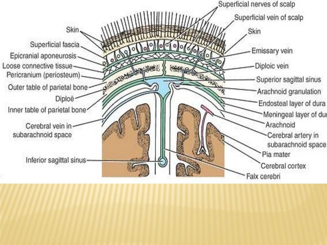
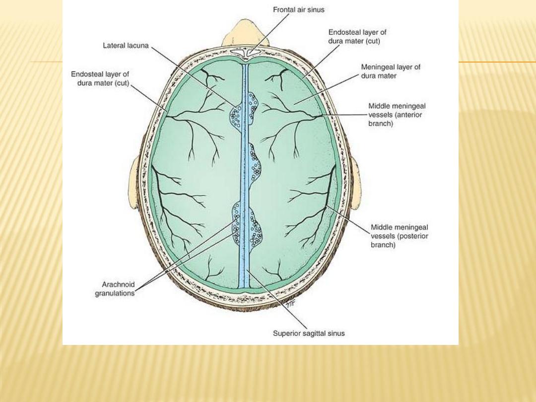
Coronal section of the upper part of the head showing the layers of the scalp,
sagittal suture of the skull, falx cerebri, venous sinuses, arachnoid
granulations, emissary veins, and relation of the cerebral blood vessels to
the subarachnoid space

The Meninges of the Brain
Dura Mater
as two layers:
1)
the endosteal layer
: is nothing more than the periosteum covering the
inner surface of the skull bones
At the foramen magnum, it does not become continuous with the Dura
mater of the spinal cord.
Around the margins of all the foramina in the skull, it becomes continuous
with the periosteum on the outside of the skull bones.
At the sutures, it is continuous with the sutural ligaments
It is most strongly adherent to the bones over the base of the skull
2)
meningeal layer
a dense, strong fibrous membrane covering the brain and is continuous
through the foramen magnum with the Dura mater of the spinal cord.
It provides tubular sheaths for the cranial nerves as they pass through the
foramina in the skull. Then the sheaths fuse with the epineurium of the
nerves outside the skull

Four septa derived from meningeal layer extended to intracranial are dividing
it into freely communicating spaces that lodge the subdivisions of the brain
1)
falx cerebri is a sickle-shaped fold of Dura mater that lies in the midline
between the two cerebral hemispheres
it has a narrow ant. end attached to the internal frontal crest and the crista
galli and a broad posterior part blends in the midline with the upper
surface of the tentorium cerebelli
superior sagittal sinus runs in its upper fixed margin, the inferior sagittal
sinus runs in its lower concave free margin, and the straight sinus runs
along its attachment to the tentorium cerebelli

2)
tentorium cerebelli is a crescent-shaped fold of Dura mater that roofs over
the posterior cranial fossa
tentorial notch is an ant opening for the passage of the midbrain with an
inner free border and an outer attached or fixed border.
The fixed border is attached to the posterior clinoid processes, the superior
borders of the petrous bones, and the margins of the grooves for the
transverse sinuses on the occipital bone
The falx cerebri and the falx cerebelli are attached to the upper and lower
surfaces of the tentorium, respectively
3)
The falx cerebelli, a small, sickle-shaped fold of Dura mater attached to the
internal occipital crest, projects forward between the two cerebellar
hemispheres. Its posterior fixed margin contains the occipital sinus
4)
diaphragma sellae is a small, circular fold of Dura mater that forms the roof
for the sella turcica
A small opening in its center allows passage of the stalk of the hypophysis
cerebri
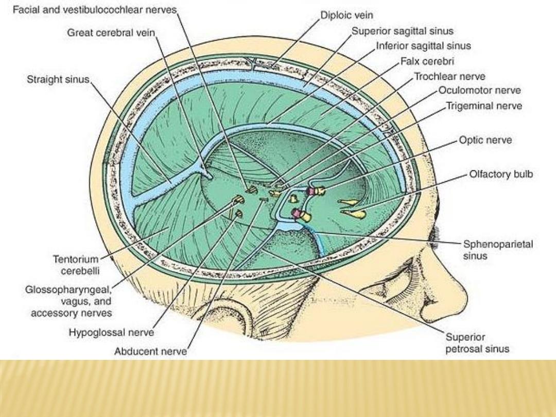
Interior of the skull showing the Dura mater and its contained
venous sinuses

Dural Nerve Supply
Branches of the trigeminal, vagus, and the first three cervical spinal
nerves and branches from the sympathetic trunk pass to the Dura
neuron endings sensitive to stretching, which produce the sensation of
headache
Dural Arterial Supply
Branches of internal carotid, maxillary, ascending pharyngeal,
occipital, and vertebral arteries
Clinically middle meningeal artery is the most important it arises
from the maxillary artery in the infratemporal fossa entering the
cranial cavity through the foramen spinosum and then lies between
the meningeal and endosteal layers of Dura, giving the ant and post
branches

Dural Venous Sinuses
The venous sinuses of the cranial cavity are situated between the layers of the
Dura mater
Their main function is to receive blood from the brain through the cerebral
veins and the cerebrospinal fluid from the subarachnoid space through the
arachnoid villi
blood in the Dural sinuses drains into the internal jugular veins in the neck
Emissary veins, which are also valve less, connect the Dural venous sinuses
with the diploic veins of the skull and with the veins of the scalp

Arachnoid Mater
a delicate, impermeable membrane covering the brain and lying between the
pia mater internally and the Dura mater externally.
It is separated from the Dura by the subdural space, filled by a film of fluid.
separated from the pia by the subarachnoid space, which is filled with
cerebrospinal fluid
in certain situations, the arachnoid and pia are widely separated to form the
subarachnoid cisternae (cisterna cerebellomedullaris and The cisterna
interpeduncularis)
Pia Mater
a vascular membrane covered by flattened mesothelial cells. It closely invests
the brain, covering the gyri and descending into the deepest sulci
The pia mater forms the tela choroidea of the roof of the third and fourth
ventricles of the brain, and it fuses with the ependyma to form the choroid
plexuses in the lateral, third, and fourth ventricles of the brain
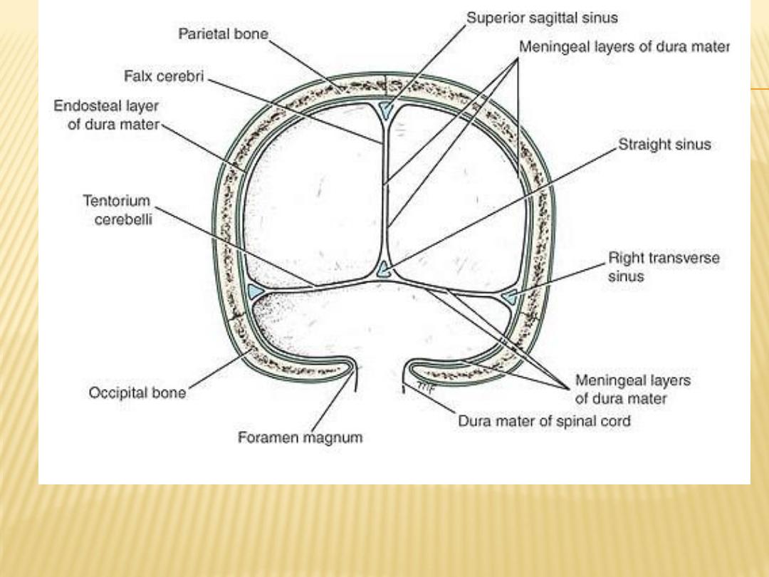
Falx cerebri and the tentorium cerebelli. Note the continuity between the
meningeal layer of Dura mater within the skull and the Dura mater of the spinal
cord at the foramen magnum
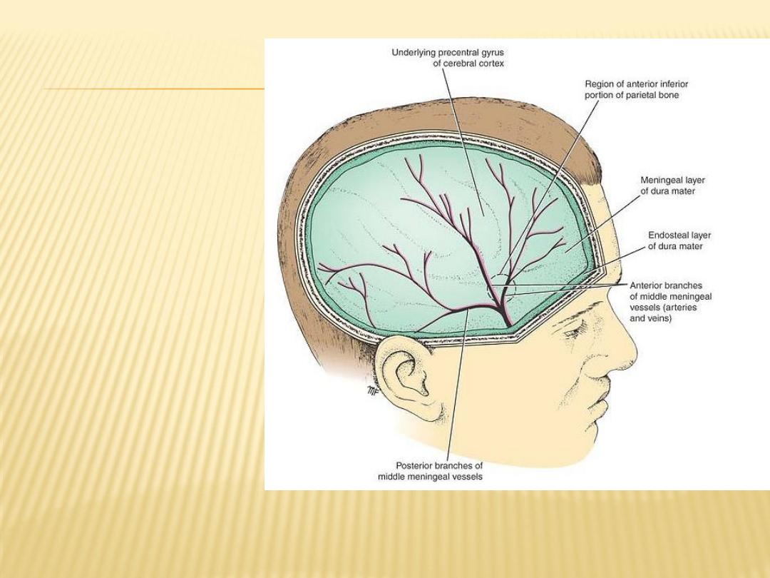
Right side of the head
showing the relation of
the middle meningeal
vessels to the layers of
the Dura mater and the
skull
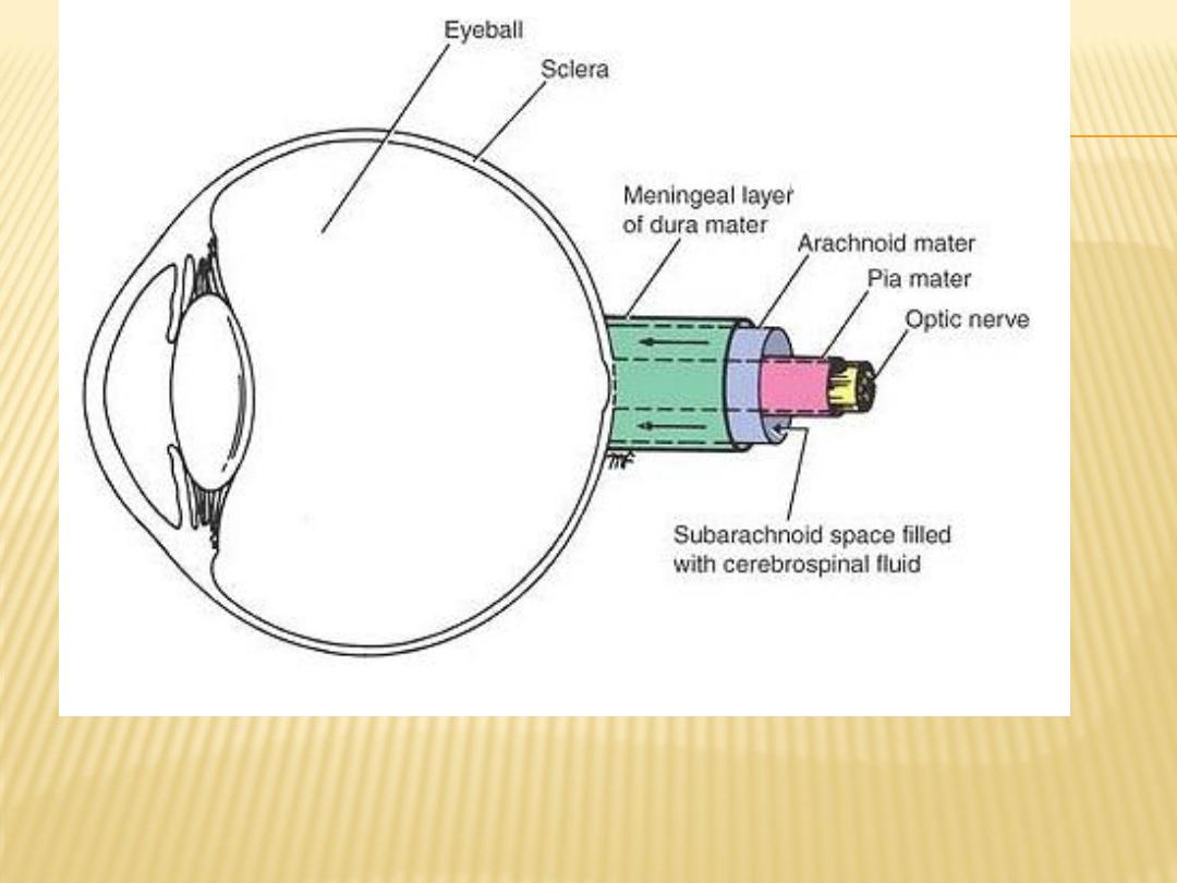
Sagittal section of the eyeball showing the attachment of the
meninges to the sclera. Note the extension of the subarachnoid
space around the optic nerve to the eyeball
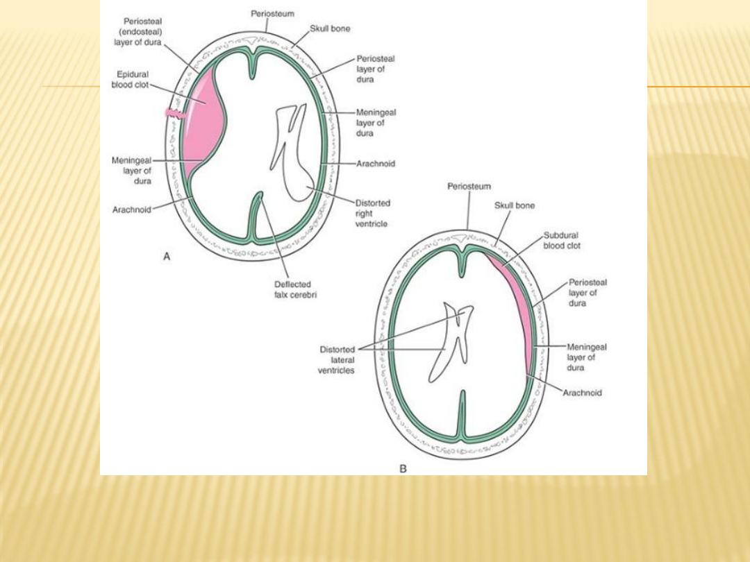
Diagrammatic representation of an epidural hemorrhage and a subdural
hemorrhage

Blood Supply of the Brain
Arteries of the Brain
The brain is supplied by the two internal carotid and the two vertebral
arteries.
The four arteries lie within the subarachnoid space, and their branches
anastomose on the inferior surface of the brain to form the circle of Willis
Internal Carotid Artery
Originate by the bifurcation of the common carotid artery, possesses a localized
dilatation, called the carotid sinus, enter the skull through the carotid canal
of the temporal bone
then runs forward through the cavernous sinus
by perforating the Dura mater and the arachnoid mater it reach the region of
the medial end of the lateral cerebral sulcus. Here, it divides into the
anterior and middle cerebral arteries
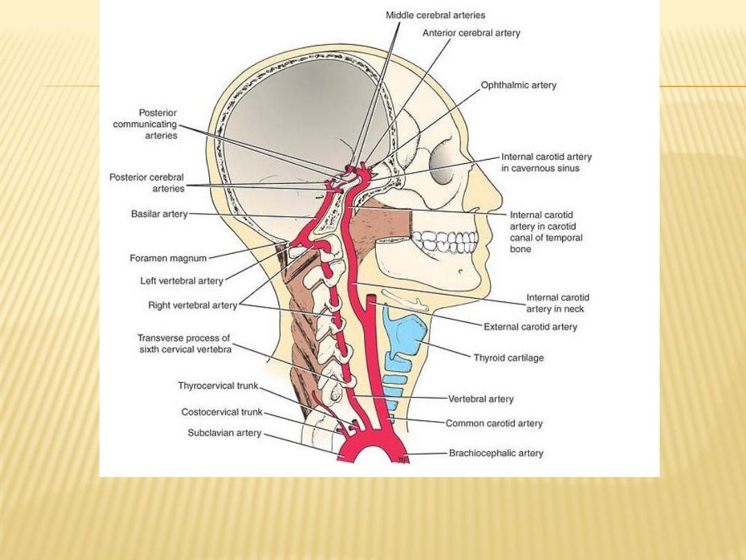
Origin and courses of the internal carotid and vertebral
arteries as they ascend the neck to enter the skull

Branches of the Cerebral Portion
The ophthalmic artery:
It enters the orbit through the optic canal
supply the frontal area of the scalp, the ethmoid and frontal sinuses, and the
dorsum of the nose
The posterior communicating artery:
forming part of the circle of Willis
The choroidal artery
The anterior cerebral artery:
it is joined to the anterior cerebral artery of the opposite side by the anterior
communicating artery
The middle cerebral artery:
Largest branch, Cortical branches supply the entire lateral surface of the
hemisphere, except for the narrow strip supplied by the anterior cerebral
artery, the occipital pole, and the inferolateral surface of the hemisphere,
which are supplied by the posterior cerebral artery

Vertebral Artery
a branch of the first part of the subclavian artery
passing through the foramina in the transverse processes of the upper six
cervical vertebrae
It enters the skull through the foramen magnum and pierces the Dura mater
and arachnoid to enter the subarachnoid space
At the lower border of the pons, it joins the vessel of the opposite side to form
the basilar artery
Branches of the Cranial Portion of Vertebral Artery
1)
The meningeal branches
2)
The posterior spinal artery
3)
The anterior spinal artery
4)
The posterior inferior cerebellar artery
5)
The medullary arteries

Basilar Artery
formed by the union of the two vertebral arteries
ascends in a groove on the anterior surface of the pons
At the upper border of the pons, it divides into the two posterior cerebral
arteries
Branches
1)
The pontine arteries supply the pons
2)
The labyrinthine accompanies the facial and the vestibulocochlear nerves
into the internal acoustic meatus and supplies the internal ear
3)
The anterior inferior cerebellar artery.
4)
The superior cerebellar artery
5)
The posterior cerebral artery form the cortical branches, central branches
and choroidal branch

Circle of Willis
It is formed by the anastomosis between the two internal carotid arteries and
the two vertebral arteries
lies in the interpeduncular fossa at the base of the brain
The anterior communicating, anterior cerebral, internal carotid, posterior
communicating, posterior cerebral, and basilar arteries all contribute to the
circle
The circle of Willis allows blood that enters by either internal carotid or
vertebral arteries to be distributed to any part of both cerebral hemispheres
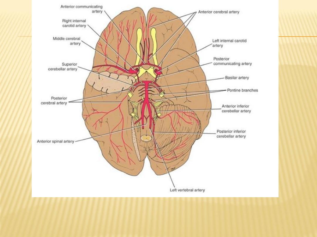
Arteries of the inferior surface of the brain. Note the formation of the
circle of Willis

Veins of the Brain
have no muscular tissue in their very thin walls, and they possess no valves
They drain into the cranial venous sinuses
External Cerebral Veins:
1)
superior cerebral veins
2)
superficial middle cerebral vein
3)
deep middle cerebral vein joined by the anterior cerebral and striate veins
to form the basal vein which in turn joins the great cerebral vein
Internal Cerebral Veins
two internal cerebral veins formed by the union of the thalamostriate vein and
the choroid vein they unite beneath the splenium of the corpus callosum to
form the great cerebral vein, which empties into the straight sinus