
amus; and (5) areas of the cerebral cortex.
medulla, pons, and mesencephalon of the brain; (3) the cerebellum; (4) the thal-
areas in (1) the spinal cord at all levels; (2) the reticular substance of the
some deep structures. This information enters the central nervous system
portion of the sensory system, which transmits
Figure 45–2 shows the
date.
for minutes, weeks, or years and determine bodily reactions at some future
from the brain, or memory of the experience can be stored in the brain
kinds of receptors. This sensory experience can either cause immediate reaction
receptors in the ears, tactile receptors on the surface of the body, or other
, whether visual receptors in the eyes, auditory
directions for performing specific nervous functions.
membranes of subsequent neurons). This forces the signal to travel in required
nervous system or peripheral body.
neuron. Then, this axon has many separate branches to other parts of the
fibers. Conversely, the output signal travels by way of a single axon leaving the
drites, but also on the cell body. For different types of neurons, there may be
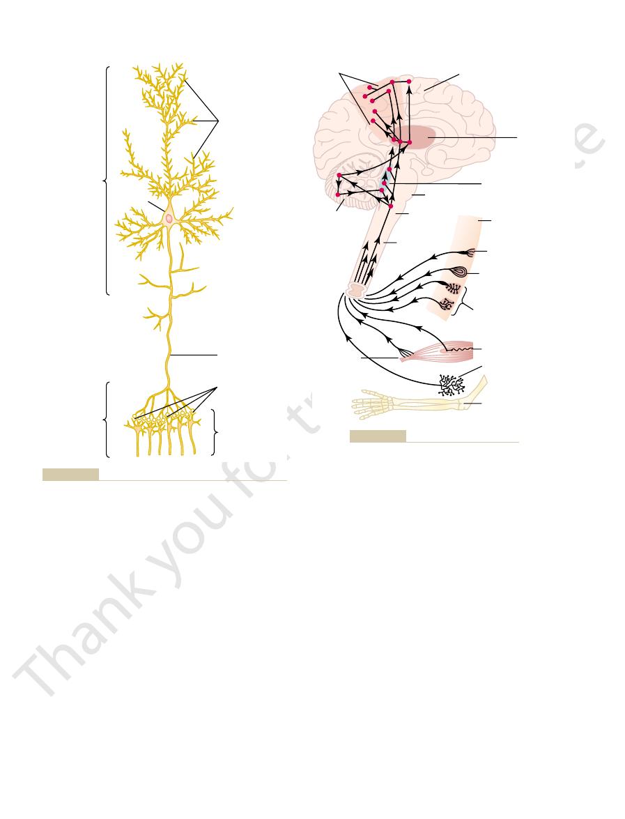
shows a typical neuron of a type found in the brain motor cortex. Incoming
The central nervous system contains more than 100 billion neurons. Figure 45–1
Functional Unit
Central Nervous System Neuron: The Basic
General Design of the Nervous System
through neuromuscular junctions.
nervous system, the reader should review Chapters 5 and 7, which present the
However, before beginning this discussion of the
body.
perform. It receives each minute literally millions of
The nervous system is unique in the vast complex-
Synapses, “Transmitter Substances”
System, Basic Functions of
C
H
A
P
T
E
R
4
5
555
Organization of the Nervous
ity of thought processes and control actions it can
bits of information from the different sensory
nerves and sensory organs and then integrates all
these to determine responses to be made by the
principles of membrane potentials and transmission of signals in nerves and
signals enter this neuron through synapses located mostly on the neuronal den-
only a few hundred or as many as 200,000 such synaptic connections from input
A special feature of most synapses is that the signal normally passes only in
the forward direction (from the axon of a preceding neuron to dendrites on cell
Sensory Part of the Nervous System—Sensory Receptors
Most activities of the nervous system are initiated by sensory experience
exciting sensory receptors
somatic
sensory information from the receptors of the entire body surface and from
through peripheral nerves and is conducted immediately to multiple sensory
portant. For instance, one is ordinarily unaware of the
occur. More than 99 per cent of all sensory informa-
“Integrative” Function of the
stimuli, and the higher regions with deliberate
automatic, instantaneous muscle responses to sensory
cific role, the lower regions concerned primarily with
motor cortex. Each of these areas plays its own spe-
(3) the basal ganglia; (4) the cerebellum; and (5) the
substance of the medulla, pons, and mesencephalon;
system, including (1) the spinal cord; (2) the reticular
Note in Figure 45–3 that the skeletal muscles can be
bodily systems; this is discussed in Chapter 60.
trolling smooth muscles, glands, and other internal
, for con-
system, called the
traction. Operating parallel to this axis is another
“skeletal” motor nerve axis
Figure 45–3 shows the
tated by the nerve signals.
of the nervous system, and the muscles and
body. These activities are collectively called
tion of smooth muscle in the internal organs, and
ate skeletal muscles throughout the body, (2) contrac-
system is to control the various bodily activities. This
The most important eventual role of the nervous
The Nervous System: A. General Principles and Sensory Physiology
556
Unit IX
Motor Part of the Nervous System—
Effectors
is achieved by controlling (1) contraction of appropri-
(3) secretion of active chemical substances by both
exocrine and endocrine glands in many parts of the
motor
functions
glands are called effectors because they are the actual
anatomical structures that perform the functions dic-
of
the nervous system for controlling skeletal muscle con-
autonomic nervous system
controlled from many levels of the central nervous
complex muscle movements controlled by the thought
processes of the brain.
Processing of Information—
Nervous System
One of the most important functions of the nervous
system is to process incoming information in such a
way that appropriate mental and motor responses will
tion is discarded by the brain as irrelevant and unim-
Brain
Spinal cord
Second-order
neurons
Axon
Synapses
Cell body
Dendrites
Anatomy and Physiology. Philadelphia: WB Saunders Co, 1987.)
tional parts. (Redrawn from Guyton AC: Basic Neuroscience:
Structure of a large neuron in the brain, showing its important func-
Figure 45–1
Golgi tendon
apparatus
Cerebellum
Motor cortex
Thalamus
Bulboreticular
formation
Pons
Somesthetic areas
Medulla
Spinal cord
Skin
Pain, cold,
warmth (Free
nerve ending)
Pressure
(Pacinian corpuscle)
(Expanded tip
receptor)
Touch
(Meissner's corpuscle)
Muscle spindle
Kinesthetic receptor
Joint
Muscle
Somatosensory axis of the nervous system.
Figure 45–2

, and (3) the
lower brain
, (2) the
spinal cord level
tional characteristics: (1) the
tionary development. From this heritage, three major
The human nervous system has inherited special func-
Nervous System Function
bodily responses.
ences with stored memories; the memories then help
mechanism for future “thinking.” That is, the thinking
system, they become part of the brain processing
memory process, but what is known about this and
We know little about the precise mechanisms by
tions are only memories of the sensations.
riencing the original sensations, although the percep-
not excited. This gives the person a perception of expe-
sequences of synapses, even when the sensory input is
large number of times, the synapses become so facili-
. After the
the next time, a process called
through sequences of synapses, these synapses become
That is, each time certain types of sensory signals pass
, and this, too, is a function of the synapses.
The storage of information is the process we call
, but even the basal regions of the
the thinking processes. Most storage occurs in the
response. But much of the information is stored for
signals, and often channeling these signals in many
weak signals while allowing strong signals to pass, but
synapses perform a selective action, often blocking
impulses, and others respond with only a few. Thus, the
times closing them. In addition, some postsynaptic
system can control synaptic transmission, sometimes
signals only with difficulty. Also,
neuron to the next with ease, whereas others transmit
system. Some synapses transmit signals from one
synaptic function. However, it is important to point out
Later in this chapter, we will discuss the details of
The synapse
the entire body away from the stove, and perhaps even
And other associated responses follow, such as moving
system. Thus, if a person places a hand on a hot stove,
responses. This channeling and processing of informa-
the mind, it is immediately channeled into proper inte-
But, when important sensory information excites
roundings is usually relegated to the subconscious.
field of vision, and even the perpetual noise of our sur-
attention is drawn only to an occasional object in one’s
well as of the seat pressure when sitting. Likewise,
parts of the body that are in contact with clothing, as
Organization of the Nervous System, Basic Functions of Synapses, “Transmitter Substances”
Chapter 45
557
grative and motor regions of the brain to cause desired
tion is called the integrative function of the nervous
the desired instantaneous response is to lift the hand.
shouting with pain.
Role of Synapses in Processing Information.
is the junction point from one neuron to the next.
here that synapses determine the directions that the
nervous signals will spread through the nervous
facilitatory and
inhibitory signals from other areas in the nervous
opening the synapses for transmission and at other
neurons respond with large numbers of output
at other times selecting and amplifying certain weak
directions rather than only one direction.
Storage of Information—Memory
Only a small fraction of even the most important
sensory information usually causes immediate motor
future control of motor activities and for use in
cerebral cortex
brain and the spinal cord can store small amounts of
information.
memory
more capable of transmitting the same type of signal
facilitation
sensory signals have passed through the synapses a
tated that signals generated within the brain itself can
also cause transmission of impulses through the same
which long-term facilitation of synapses occurs in the
other details of the sensory memory process are dis-
cussed in Chapter 57.
Once memories have been stored in the nervous
processes of the brain compare new sensory experi-
to select the important new sensory information and
to channel this into appropriate memory storage areas
for future use or into motor areas to cause immediate
Major Levels of Central
tional capabilities from each stage of human evolu-
levels of the central nervous system have specific func-
or subcortical level
higher
brain or cortical level.
Cerebellum
Alpha motor fiber
Motor
area
Motor nerve
to muscles
Thalamus
Putamen
Globus pallidus
Subthalamic nucleus
Bulboreticular formation
Gamma motor fiber
Caudate
nucleus
Muscle spindle
Figure 45–3
Skeletal motor nerve axis of the nervous system.

course of bodily activity.
ilarity to the nervous system. The fact that the basic
Figure 45–4 is a simple block diagram of a computer.
place.
forth, until complex sequences of thought or action take
sensation or motor activity, then to another, and so
This unit is analogous to the control mechanisms in our
, that determines the sequence of all operations.
sary to add still another unit, called the
as computers become even more complex, it is neces-
anisms of our higher nervous system. Furthermore,
been stored in memory in the computer, which is anal-
more complex computers, the output is determined both
similar to that of simple reflexes of the spinal cord. In
directly by the input signals, operating in a manner
In simple computers, the output signals are controlled
portion of the nervous system, and output circuits that
common with the nervous system. First, all computers
When computers were first developed, it soon became
Nervous System with
system performs specific functions. But it is the cortex
ery of the brain. Thus, each portion of the nervous
in the cerebral cortex, thus
In fact, it is the lower brain centers, not the cortex,
our thought processes, but it cannot function by itself.
Finally, the cerebral cortex is essential for most of
functions to determinative and precise operations.
lower brain centers are often imprecise. The vast store-
Without the cerebral cortex, the functions of the
memory storehouse. The cortex never functions alone
to do? The answer to this is complex, but it begins with
levels, one may ask, what is left for the cerebral cortex
response, reaction to pain, and reaction to pleasure,
emotional patterns, such as anger, excitement, sexual
cephalon, amygdala, and hypothalamus. And many
are controlled by areas in the medulla, pons, mesen-
mesencephalon. Feeding reflexes, such as salivation
and the reticular substance of the medulla, pons, and
medulla and pons. Control of equilibrium is a com-
ganglia. For instance, subconscious control of arterial
hypothalamus,
thalamus,
cerebellum,
and basal
of the brain—in the medulla, pons, mesencephalon,
Many, if not most, of what we call subconscious activ-
the cord centers to perform their functions.
the control centers of the cord, simply “commanding”
tion. In fact, the upper levels of the nervous system
vessels, gastrointestinal movements, or urinary excre-
against gravity, and (4) reflexes that control local blood
draw portions of the body from painful objects, (3)
cause (1) walking movements, (2) reflexes that with-
occur. For instance, neuronal circuits in the cord can
back to the body. This is far from the truth. Even after
the brain, or in the opposite direction from the brain
We often think of the spinal cord as being only a
The Nervous System: A. General Principles and Sensory Physiology
558
Unit IX
Spinal Cord Level
conduit for signals from the periphery of the body to
the spinal cord has been cut in the high neck region,
many highly organized spinal cord functions still
reflexes that stiffen the legs to support the body
often operate not by sending signals directly to the
periphery of the body but by sending signals to
Lower Brain or Subcortical Level
ities of the body are controlled in the lower areas
pressure and respiration is achieved mainly in the
bined function of the older portions of the cerebellum
and licking of the lips in response to the taste of food,
can still occur after destruction of much of the cere-
bral cortex.
Higher Brain or Cortical Level
After the preceding account of the many nervous
system functions that occur at the cord and lower brain
the fact that the cerebral cortex is an extremely large
but always in association with lower centers of the
nervous system.
house of cortical information usually converts these
that initiate wakefulness
opening its bank of memories to the thinking machin-
that opens a world of stored information for use by the
mind.
Comparison of the
a Computer
apparent that these machines have many features in
have input circuits that are comparable to the sensory
are comparable to the motor portion of the nervous
system.
by input signals and by information that has already
ogous to the more complex reflex and processing mech-
central process-
ing unit
brain that direct our attention first to one thought or
Even a rapid study of this diagram demonstrates its sim-
components of the general-purpose computer are anal-
ogous to those of the human nervous system demon-
strates that the brain is basically a computer that
continuously collects sensory information and uses this
along with stored information to compute the daily
Problem
Answer
Output
Result of
operations
Initial
data
Procedure
for solution
Central
processing unit
Computational
unit
Information
storage
Input
components and their interrelations.
Block diagram of a general-purpose computer, showing the basic
Figure 45–4

other neurons. Later, it will become evident that many
per cent on the soma. These presynaptic terminals are
dendrites and soma of the motor neuron, about 80 to
, which are great numbers of
cord; and the
, which extends from the
the neuron; a single
, which is the main body of
three major parts: the
the anterior horn of the spinal cord. It is composed of
Figure 45–5 shows a typical
Physiologic Anatomy of the Synapse
others.
tions of sensation, motor control, memory, and many
signals to be directed toward specific goals. Indeed, it
of the one-way conduction mechanism. It allows
Think for a moment about the extreme importance
trical synapses, which often transmit signals in either
at chemical synapses,
principle of one-way conduction
. This is the
mitter acts, called the
, to the neuron on which the trans-
that secretes the transmitter substance, called the
the signals in one direction: that is, from the neuron
most nervous system signals: they always transmit
“One-Way” Conduction at Chemical Synapses.
However, it is by way of gap junctions and other
cussed in Chapter 4. Only a few examples of gap junc-
cell to the interior of the next. Such junctions were dis-
one cell to the next. Most of these consist of small
, in contrast, are characterized by
(GABA), glycine, serotonin, and glutamate.
epinephrine, histamine, gamma-aminobutyric acid
the best known are acetylcholine, norepinephrine,
substances have been discovered thus far. Some of
some other way. More than 40 important transmitter
excite the neuron, inhibit it, or modify its sensitivity in
), and this transmitter in turn acts on recep-
. In these, the first neuron secretes at
chemical synapses
chemi-
There are two major types of synapses: (1) the
Types of Synapses
patterns of impulses in successive neurons. All these
repetitive impulses, or (3) may be integrated with
next, (2) may be changed from a single impulse into
another. However, in addition, each impulse (1) may
impulses,” through a succession of neurons, one after
form of nerve action potentials, called simply “nerve
Central Nervous System
Organization of the Nervous System, Basic Functions of Synapses, “Transmitter Substances”
Chapter 45
559
Synapses
Every medical student is aware that information is
transmitted in the central nervous system mainly in the
be blocked in its transmission from one neuron to the
impulses from other neurons to cause highly intricate
functions can be classified as synaptic functions of
neurons.
—Chemical
and Electrical
cal synapse and (2) the electrical synapse.
Almost all the synapses used for signal transmission
in the central nervous system of the human being are
its nerve ending synapse a chemical substance called a
neurotransmitter (or often called simply transmitter
substance
tor proteins in the membrane of the next neuron to
Electrical synapses
direct open fluid channels that conduct electricity from
protein tubular structures called gap junctions that
allow free movement of ions from the interior of one
tions have been found in the central nervous system.
similar junctions that action potentials are transmitted
from one smooth muscle fiber to the next in visceral
smooth muscle (Chapter 8) and from one cardiac
muscle cell to the next in cardiac muscle (Chapter 10).
Chemical
synapses have one exceedingly important characteris-
tic that makes them highly desirable for transmitting
presynaptic neuron
postsynaptic neuron
and it is quite different from conduction through elec-
direction.
is this specific transmission of signals to discrete and
highly focused areas both within the nervous system
and at the terminals of the peripheral nerves that
allows the nervous system to perform its myriad func-
anterior motor neuron in
soma
axon
soma into a peripheral nerve that leaves the spinal
dendrites
branching projections of the soma that extend as
much as 1 millimeter into the surrounding areas of the
cord.
As many as 10,000 to 200,000 minute synaptic knobs
called presynaptic terminals lie on the surfaces of the
95 per cent of them on the dendrites and only 5 to 20
the ends of nerve fibrils that originate from many
Dendrites
Axon
Soma
the neuronal soma and dendrites. Note also the single axon.
Typical anterior motor neuron, showing presynaptic terminals on
Figure 45–5

specific cellular functions.
serve as “second messengers” to increase or decrease
the postsynaptic neuron. These substances in turn
cytoplasm and activates one or more substances inside
“second messenger” activator
ion channel
types: (1) an
synaptic neuron. The ionophore in turn is one of two
two important components: (1) a
Figure 45–6. The molecules of these receptors have
, also shown in
The membrane of the postsynaptic neuron contains
“Receptor Proteins”
on the Postsynaptic Neuron—Function of
Action of the Transmitter Substance
action potentials.
cules of acetylcholine are present in each vesicle, and
mitter acetylcholine, between 2000 and 10,000 mole-
potential. For those vesicles that store the neurotrans-
brane, allowing a few transmitter vesicles to release
. This binding in turn
membrane, called
nal, it is believed that they bind with special protein
When the calcium ions enter the presynaptic termi-
following.
release is not known, but it is believed to be the
to the number of calcium ions that enter. The precise
of calcium ions to flow into the terminal. The quantity
potential depolarizes the presynaptic membrane,
. When an action
voltage-gated calcium channels
. It contains large numbers
The membrane of the presynaptic terminal is called
Presynaptic Terminals—Role of Calcium Ions
Causes Transmitter Release from the
the neuronal receptor characteristics.
or inhibition of the postsynaptic neuron, depending on
naptic neuronal membrane, and this leads to excitation
The released transmitter in turn causes an immediate
tic terminal, depolarization of its membrane causes
When an action potential spreads over a presynap-
new transmitter substance.
chondria provide adenosine triphosphate (ATP),
. The mito-
, inhibits if the
into the synaptic cleft, either
that, when released
. The transmitter vesicles
mitochondria
inhibitory function of the synapse: the
usually of 200 to 300 angstroms. The terminal has
neuronal soma by a
membrane surface of a postsomatic neuron. The presy-
synapse, showing a single presynaptic terminal on the
Figure 45–6 illustrates the basic structure of a
, or
boutons, end-feet
oval knobs and, therefore, are sometimes called
anatomical forms, but most resemble small round or
Presynaptic Terminals.
and, therefore, perform many different functions.
from only a few to as many as 200,000. These differ-
number of presynaptic terminals, which may range
ters; (3) the length and size of the axon; and (4) the
cell body; (2) the length, size, and number of dendrites,
postsynaptic neuron. But other presynaptic terminals
—that is,
The Nervous System: A. General Principles and Sensory Physiology
560
Unit IX
of these presynaptic terminals are excitatory
they secrete a transmitter substance that excites the
are inhibitory—they secrete a transmitter substance
that inhibits the postsynaptic neuron.
Neurons in other parts of the cord and brain differ
from the anterior motor neuron in (1) the size of the
ranging in length from almost zero to many centime-
ences make neurons in different parts of the nervous
system react differently to incoming synaptic signals
Electron microscopic studies of
the presynaptic terminals show that they have varied
termi-
nal knobs,
synaptic knobs.
naptic terminal is separated from the postsynaptic
synaptic cleft having a width
two internal structures important to the excitatory or
transmitter vesi-
cles and the
contain the transmitter substance
excites or inhibits the
postsynaptic neuron—excites if the neuronal mem-
brane contains excitatory receptors
membrane contains inhibitory receptors
which in turn supplies the energy for synethesizing
a small number of vesicles to empty into the cleft.
change in permeability characteristics of the postsy-
Mechanism by Which an Action Potential
the presynaptic membrane
of
these calcium channels open and allow large numbers
of transmitter substance that is then released from
the terminal into the synaptic cleft is directly related
mechanism by which the calcium ions cause this
molecules on the inside surface of the presynaptic
release sites
causes the release sites to open through the mem-
their transmitter into the cleft after each single action
there are enough vesicles in the presynaptic terminal
to transmit from a few hundred to more than 10,000
large numbers of receptor proteins
binding component
that protrudes outward from the membrane into the
synaptic cleft—here it binds the neurotransmitter
coming from the presynaptic terminal—and (2) an
ionophore component that passes all the way through
the postsynaptic membrane to the interior of the post-
that allows passage of speci-
fied types of ions through the membrane or (2) a
that is not an ion channel
but instead is a molecule that protrudes into the cell
Transmitter vesicles
Mitochondria
Synaptic cleft
(200-300
angstroms)
Presynaptic
terminal
Soma of neuron
Receptor
proteins
Figure 45–6
Physiologic anatomy of the synapse.
often stays open for a prolonged time, in contrast
opened in response to the G-protein; this channel
. Shown in the upper
Opening specific ion channels through the
can occur. They are as follows:
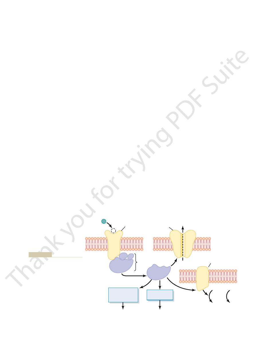
of neuron. Shown in Figure 45–7 are four changes that
nent performs one or more of multiple functions,
Inside the cytoplasm, the separated alpha compo-
the cytoplasm of the cell.
receptor protein. On activation by a nerve impulse, the
the G-protein, and beta (
G-protein in turn consists of three components: an
that protrudes into the interior of the cell. The
upper left corner a membrane receptor protein. A
. Figure 45–7 shows in the
systems. One of the most common types uses a group
There are several types of second messenger
inside the postsynaptic neuronal cell itself, and then it
by activating a “second messenger” chemical system
present. However, in many instances, prolonged post-
mitter substance is gone. The ion channels are not suit-
Many functions of the nervous system—for instance,
for very rapid control of postsynaptic neurons.
longer present, the channel closes equally rapidly. The
a millisecond; when the transmitter substance is no
channel, the channel usually opens within a fraction of
When a transmitter substance activates an ion
inhibits the neuron. Therefore, transmitter substances
allows negative electrical charges to enter, which
. Conversely, opening anion channels
turn excite this neuron. Therefore, a transmitter sub-
and allow positively charged sodium ions to enter, the
We will learn later that when cation channels open
pass.
sodium, potassium, and calcium cations are blocked,
channels and on through to the opposite side, whereas
become large enough, chloride ions pass into the
, when the channel diameters
anion channels
For the
prevent their passage.
repel chloride ions and other anions
that of the hydrated sodium ion. But those same neg-
lined with negative charges. These charges attract the
cation channels
The
of other anions.
anion channels
calcium ions as well, and (2)
when opened, but sometimes allow potassium and/or
channels
ronal membrane are usually of two types: (1)
The ion channels in the postsynaptic neu-
Organization of the Nervous System, Basic Functions of Synapses, “Transmitter Substances”
Chapter 45
561
Ion Channels.
cation
that most often allow sodium ions to pass
that allow
mainly chloride ions to pass but also minute quantities
that conduct sodium ions are
positively charged sodium ions into the channel when
the channel diameter increases to a size larger than
ative charges
and
mainly because their hydrated ions are too large to
positive electrical charges of the sodium ions will in
stance that opens cation channels is called an excita-
tory transmitter
that open these channels are called inhibitory
transmitters.
opening and closing of ion channels provide a means
“Second Messenger” System in the Postsynaptic Neuron.
the process of memory—require prolonged changes in
neurons for seconds to months after the initial trans-
able for causing prolonged postsynaptic neuronal
changes because these channels close within millisec-
onds after the transmitter substance is no longer
synaptic neuronal excitation or inhibition is achieved
is the second messenger that causes the prolonged
effect.
of proteins called G-proteins
G-protein is attached to the portion of the receptor
alpha (
a) component that is the activator portion of
b) and gamma (g) compo-
nents that are attached to the alpha component and
also to the inside of the cell membrane adjacent to the
alpha portion of the G-protein separates from the beta
and gamma portions and then is free to move within
depending on the specific characteristic of each type
1.
postsynaptic cell membrane
right of the figure is a potassium channel that is
to rapid closure of directly activated ion channels
that do not use the second messenger system.
Transmitter substance
G-protein
Opens
channel
K
+
Membrane
enzyme
Potassium
channel
Receptor
protein
1
2
3
4
Activates one or
more intracellular
enzymes
Activates gene
transcription
Specific cellular
chemical activators
Proteins and
structural changes
Activates
enzymes
ATP
cAMP
or
g
b
a
a
GTP
cGMP
tion in the second neuron.
the neuron’s membrane; 3, activat-
membrane of the second neuron;
G-protein are shown, including 1,
quent possible effects of the
neuron’s cytoplasm. Four subse-
a “G-protein” into the second
second neuron by first releasing
an initial neuron can activate a
which a transmitter substance from
Figure 45–7
“Second messenger” system by
opening an ion channel in the
2, activating an enzyme system in
ing an intracellular enzyme system;
and/or 4, causing gene transcrip-

synapses.
closure of certain ion channels, and possibly even long-
numbers of neuronal receptors, long-term opening or
prolonged actions, such as long-term changes in
The neuropeptides, in contrast, usually cause more
to the brain and of motor signals back to the muscles.
nervous system, such as transmission of sensory signals
The small-molecule, rapidly acting transmitters are
much more slowly acting.
The other is made up of a large number of
small-molecule, rapidly acting transmitters
give two groups of synaptic transmitters. One group
Many of them are listed in Tables 45–1 and 45–2, which
or postulated to function as synaptic transmitters.
as Synaptic Transmitters
number of excitatory receptors.
inside the neuron; this is inhibitory.
the exterior, which causes increased negativity
. This allows positive ions to diffuse to
inhibitory.
and increasing the negativity inside, which is
inside, thereby carrying negative charges inward
. This allows
Opening of chloride ion channels through the
number of inhibitory membrane receptors.
some instances, to increase the number of
to excite cell activity or, in
Various changes in the internal metabolism of the
positive than normal, which is excitatory.
to the outside. In either instance, the effect is to
. This decreases the
potassium channels, or both
Depressed conduction through chloride or
excitation. It is by far the most widely used means
. This raises the
numbers of positive electrical charges to flow to the
Opening of sodium channels to allow large
inhibition include the following.
The different molecular and membrane mechanisms
additional dimension to nervous function, allowing
inhibition. The importance of having inhibitory as well
excitation of the postsynaptic neuron, and others cause
Some postsynaptic receptors, when activated, cause
Excitatory or Inhibitory Receptors in the
tics of different neuronal pathways. We will return to
G-protein type or of other types, is extremely impor-
systems within the neuron, whether they be of the
processes.
neurons do occur, especially in long-term memory
or its structure. Indeed, it is well known that
neuron, thereby changing its metabolic machinery
This is one of the
intracellular enzymes. In turn the enzymes can
The G-protein can directly activate one or more
structure itself, which in turn alters long-term
results, including long-term changes in cell
therefore, can initiate any one of many chemical
cyclic AMP or cyclic GMP can activate highly
. Recall that either
The Nervous System: A. General Principles and Sensory Physiology
562
Unit IX
2. Activation of cyclic adenosine monophosphate
(cAMP) or cyclic guanosine monophosphate
(cGMP) in the neuronal cell
specific metabolic machinery in the neuron and,
excitability of the neuron.
3. Activation of one or more intracellular enzymes.
cause any one of many specific chemical functions
in the cell.
4. Activation of gene transcription.
most important effects of activation of the second
messenger systems because gene transcription
can cause formation of new proteins within the
structural changes of appropriately activated
It is clear that activation of second messenger
tant for changing the long-term response characteris-
this subject in more detail in Chapter 57 when we
discuss memory functions of the nervous system.
Postsynaptic Membrane
as excitatory types of receptors is that this gives an
restraint of nervous action as well as excitation.
used by the different receptors to cause excitation or
Excitation
1.
interior of the postsynaptic cell
intracellular membrane potential in the positive
direction up toward the threshold level for
for causing excitation.
2.
diffusion of negatively charged chloride ions to
the inside of the postsynaptic neuron or decreases
the diffusion of positively charged potassium ions
make the internal membrane potential more
3.
postsynaptic neuron
excitatory membrane receptors or decrease the
Inhibition
1.
postsynaptic neuronal membrane
rapid diffusion of negatively charged chloride ions
from outside the postsynaptic neuron to the
2. Increase in conductance of potassium ions out of
the neuron
3. Activation of receptor enzymes that inhibit cellular
metabolic functions that increase the number of
inhibitory synaptic receptors or decrease the
Chemical Substances That Function
More than 50 chemical substances have been proved
comprises
.
neuropep-
tides of much larger molecular size that are usually
the ones that cause most acute responses of the
term changes in numbers of synapses or sizes of
Table 45–1
Gamma-aminobutyric acid (GABA)
Class III: Amino Acids
Class II: The Amines
Small-Molecule, Rapidly Acting Transmitters
Class I
Acetylcholine
Norepinephrine
Epinephrine
Dopamine
Serotonin
Histamine
Glycine
Glutamate
Aspartate
Class IV
Nitric oxide (NO)

ganglia, and many areas of the cortex. It is believed
nerve terminals in the spinal cord, cerebellum, basal
GABA
transmitter.
spinal cord. It is believed to always act as an inhibitory
is mainly in the striatal region of the basal ganglia. The
the substantia nigra. The termination of these neurons
inhibits others.
nervous system, where it excites some organs but
receptors instead. Norepinephrine is also secreted by
tory receptors, but in a few areas, it activates inhibitory
these areas, norepinephrine probably activates excita-
such as increasing the level of wakefulness. In most of
pons send nerve fibers to widespread areas of the brain
stem and hypothalamus. Specifically, norepinephrine-
vagus nerves.
nerve endings, such as inhibition of the heart by the
has an excitatory effect; however, it is known to have
thetic nervous system. In most instances, acetylcholine
neurons of the parasympathetic nervous system, and
the autonomic nervous system, (5) the postganglionic
skeletal muscles, (4) the preganglionic neurons of
basal ganglia, (3) the motor neurons that innervate the
cortex, (2) several different types of neurons in the
Acetylcholine
transmitters are the following.
The most important of the small-molecule
Transmitters.
acetylcholine.
cles are recycled; choline is actively transported back
then again, inside the presynaptic terminal, the vesi-
reticulum that fills the space of the synaptic cleft. And
, which is present in the proteoglycan
cholinesterase
ronal signal transmission, the acetylcholine is rapidly
vesicles. When the vesicles later release the acetyl-
. Then it is transported into its specific
choline
in the presynaptic terminal from acetyl coenzyme A
stated earlier. This transmitter substance is synthesized
vesicle.
off to form a new vesicle. And the new vesicular
membrane. However, within seconds to minutes, the
to release their transmitter substance, the vesicle mem-
are continually recycled and used over and over again.
The vesi-
Recycling of the Small-Molecule Types of Vesicles.
sium or chloride conductance, which causes inhibition.
ductance, which causes excitation, or to increase potas-
ion channels; an example is to increase sodium con-
within another millisecond or less. Most often the
described earlier. The subsequent action of the small-
transmitter into the synaptic cleft. This usually occurs
naptic terminal, a few vesicles at a time release their
Then, each time an action potential reaches the presy-
are synthesized in the cytosol of the presynaptic ter-
In most cases, the small-molecule types of transmitters
Small-Molecule, Rapidly Acting Transmitters
Organization of the Nervous System, Basic Functions of Synapses, “Transmitter Substances”
Chapter 45
563
minal and are absorbed by means of active transport
into the many transmitter vesicles in the terminal.
within a millisecond or less by the mechanism
molecule type of transmitter on the membrane recep-
tors of the postsynaptic neuron usually also occurs
effect is to increase or decrease conductance through
cles that store and release small-molecule transmitters
After they fuse with the synaptic membrane and open
brane at first simply becomes part of the synaptic
vesicle portion of the membrane invaginates back to
the inside of the presynaptic terminal and pinches
membrane still contains appropriate enzyme proteins
or transport proteins required for synthesizing and/or
concentrating new transmitter substance inside the
Acetylcholine is a typical small-molecule transmit-
ter that obeys the principles of synthesis and release
and choline in the presence of the enzyme
acetyltransferase
choline into the synaptic cleft during synaptic neu-
split again to acetate and choline by the enzyme
into the terminal to be used again for synthesis of new
Characteristics of Some of the More Important Small-Molecule
is secreted by neurons in many areas
of the nervous system but specifically by (1) the ter-
minals of the large pyramidal cells from the motor
(6) some of the postganglionic neurons of the sympa-
inhibitory effects at some peripheral parasympathetic
Norepinephrine is secreted by the terminals of many
neurons whose cell bodies are located in the brain
secreting neurons located in the locus ceruleus in the
to help control overall activity and mood of the mind,
most postganglionic neurons of the sympathetic
Dopamine is secreted by neurons that originate in
effect of dopamine is usually inhibition.
Glycine is secreted mainly at synapses in the
(gamma-aminobutyric acid) is secreted by
always to cause inhibition.
Neuropeptide, Slowly Acting Transmitters or Growth
Table 45–2
From other tissues
Vasoactive intestinal polypeptide (VIP)
Cholecystokinin
Vasopressin
Thyrotropin
-Melanocyte-stimulating hormone
Adrenocorticotropic hormone (ACTH)
Thyrotropin-releasing hormone
Factors
Hypothalamic-releasing hormones
Luteinizing hormone–releasing hormone
Somatostatin (growth hormone inhibitory factor)
Pituitary peptides
b-Endorphin
a
Prolactin
Luteinizing hormone
Growth hormone
Oxytocin
Peptides that act on gut and brain
Leucine enkephalin
Methionine enkephalin
Substance P
Gastrin
Nerve growth factor
Brain-derived neurotropic factor
Neurotensin
Insulin
Glucagon
Angiotensin II
Bradykinin
Carnosine
Sleep peptides
Calcitonin

the neuron less excitable. This is the basis for the two
the membrane of the neuron more excitable, whereas
of the degree of excitability of the neuron. That is,
skeletal muscle fibers; the lower voltage is important
livolts. This is somewhat less negative than the
45–8 shows the soma of a spinal motor neuron, indi-
Figure
Resting Membrane Potential of the Neuronal Soma.
to these neurons. Except for quantitative differences,
anterior horns of the spinal cord. Therefore, the events
The electrical events in neuronal excitation have been
only beginning to develop.
days, but others perhaps for months or years. Our
inhibitory receptors. Some of these effects last for
vation of specific genes in the cell nucleus, and/or
ery of cells, prolonged changes in activation or deacti-
channels, prolonged changes in the metabolic machin-
they often cause much more prolonged actions. Some
as potent as the small-molecule transmitters. Another
ters. This is partly compensated for by the fact that the
neuropeptides, much smaller quantities of them are
However, the vesicle is autolyzed and is not reused.
the same manner as for small-molecule transmitters.
Finally, these vesicles release their transmitter at the
at the slow rate of only a few centimeters per day.
of the axon cytoplasm, traveling
into the cytoplasm. Then the transmitter vesicles are
Second, the Golgi apparatus packages the neuropep-
matically split into smaller fragments, some of which
occur: First, the neuropeptide-forming protein is enzy-
quently inside the Golgi apparatus, where two changes
The protein molecules then enter the spaces inside
cules by ribosomes in the neuronal cell body.
cytosol of the presynaptic terminals. Instead, they are
ters. The neuropeptides are not synthesized in the
The neuropeptides are an entirely different class of
Neuropeptides
seconds, minutes, or perhaps even longer.
neuron, it usually does not greatly alter the membrane
postsynaptic neurons nearby. In the postsynaptic
released in vesicular packets. Next, it diffuses into
needed, and it then diffuses out of the presynaptic
mitters. Instead, it is synthesized almost instantly as
postsynaptic neuron. It is not preformed and stored in
standing. Nitric oxide is different from other small-
ior and for memory. Therefore, this transmitter system
Nitric oxide
mood of the person, perhaps even to cause sleep.
cord, and an inhibitor action in the higher regions of
horns of the spinal cord and to the hypothalamus.
brain and spinal cord areas, especially to the dorsal
bral cortex. It probably always causes excitation.
nervous system, as well as in many areas of the cere-
The Nervous System: A. General Principles and Sensory Physiology
564
Unit IX
Glutamate is secreted by the presynaptic terminals
in many of the sensory pathways entering the central
Serotonin is secreted by nuclei that originate in the
median raphe of the brain stem and project to many
Serotonin acts as an inhibitor of pain pathways in the
the nervous system is believed to help control the
is especially secreted by nerve terminals
in areas of the brain responsible for long-term behav-
might in the future explain some behavior and
memory functions that thus far have defied under-
molecule transmitters in its mechanism of formation
in the presynaptic terminal and in its actions on the
vesicles in the presynaptic terminal as are other trans-
terminals over a period of seconds rather than being
potential but instead changes intracellular metabolic
functions that modify neuronal excitability for
transmitters that are synthesized differently and
whose actions are usually slow and in other ways quite
different from those of the small-molecule transmit-
synthesized as integral parts of large-protein mole-
the endoplasmic reticulum of the cell body and subse-
are either the neuropeptide itself or a precursor of it.
tide into minute transmitter vesicles that are released
transported all the way to the tips of the nerve fibers
by axonal streaming
neuronal terminals in response to action potentials in
Because of this laborious method of forming the
usually released than of the small-molecule transmit-
neuropeptides are generally a thousand or more times
important characteristic of the neuropeptides is that
of these actions include prolonged closure of calcium
prolonged alterations in numbers of excitatory or
knowledge of the functions of the neuropeptides is
Electrical Events During
Neuronal Excitation
studied especially in the large motor neurons of the
described in the next few sections pertain essentially
they apply to most other neurons of the nervous
system as well.
cating a resting membrane potential of about
-65 mil-
-90
millivolts found in large peripheral nerve fibers and in
because it allows both positive and negative control
decreasing the voltage to a less negative value makes
increasing this voltage to a more negative value makes
Dendrite
Axon
Axon hillock
14 mEq/L
(Pumps)
?
Pump
120 mEq/L
8 mEq/L
-
65
mV
Na
+
: 142 mEq/L
K
+
: 4.5 mEq/L
Cl
-
: 107 mEq/L
neuronal somal membrane; origin of the intrasomal membrane
Distribution of sodium, potassium, and chloride ions across the
Figure 45–8
potential.

inside the neuron, which is more negative than the
This calculates to a Nernst potential of
120 mEq/L inside the neuron and 4.5 mEq/L outside.
For potassium ions, the concentration gradient is
to the exterior by the sodium pump, thus maintaining
61 millivolts. Therefore, those sodium ions
volts, not
However, the actual membrane potential is
61 millivolts.
on the interior, the membrane potential that will
Figure 45–8, 142 mEq/L on the exterior and 14 mEq/L
For the sodium concentration difference shown in
arate ions: sodium, potassium, and chloride.
Now, let us calculate the Nernst potential that will
) for negative ions.
. The potential will be negative
for that ion; the equation
the potential is of proper polarity and magnitude. A
differences to membrane potentials. It will be recalled
than outside.
ride ions, forcing them outward through the pores until
65 millivolts in the neuron. That is, this
be a weak chloride pump. Yet most of the reason
. It also shows that the membrane
low concentra-
chloride ion
Figure 45–8 shows the
pump) that pumps potassium to the interior.
(4.5 mEq/L). It shows that
low in the extracellular fluid
(120 mEq/L) but
The figure also shows that
This sodium concentration gradient is caused by a
(14 mEq/L).
low inside the neuron
(142 mEq/L) but
chloride ions. At the top, the
neuronal function: sodium ions, potassium ions, and
Figure 45–8 also shows the concen-
Concentration Differences of Ions Across the Neuronal
inhibition—as explained in detail in the next sections.
Organization of the Nervous System, Basic Functions of Synapses, “Transmitter Substances”
Chapter 45
565
modes of function of the neuron—either excitation or
Somal Membrane.
tration differences across the neuronal somal mem-
brane of the three ions that are most important for
sodium ion concentra-
tion is shown to be high in the extracellular fluid
strong somal membrane sodium pump that continually
pumps sodium out of the neuron.
potassium ion concentra-
tion is high inside the neuronal soma
there is a potassium pump (the other half of the Na
+
-
K
+
to be of high con-
centration in the extracellular fluid but
tion inside the neuron
is quite permeable to chloride ions and that there may
for the low concentration of chloride ions inside the
neuron is the
-
negative voltage repels the negatively charged chlo-
the concentration is much less inside the membrane
Let us recall at this point what we learned in Chap-
ters 4 and 5 about the relation of ionic concentration
that an electrical potential across the cell membrane
can oppose movement of ions through a membrane if
potential that exactly opposes movement of an ion is
called the Nernst potential
for this is the following:
where EMF is the Nernst potential in millivolts on the
inside of the membrane
(
-) for positive ions and positive (+
exactly oppose the movement of each of the three sep-
exactly oppose sodium ion movement through the
sodium channels calculates to be
+
-65 milli-
+
that leak to the interior are immediately pumped back
the
-65 millivolt negative potential inside the neuron.
-86 millivolts
61
log
= ±
¥
EMF mV
Concentration inside
Concentration outside
(
)
Ê
Ë
ˆ
¯
postsynaptic neuron, thus exciting it. (In this case, the
itive direction, it will elicit an action potential in the
that is, to a less negative value
45 millivolts. This positive
resting membrane potential. Thus, in Figure 45
The rapid in
membrane.
neuron, sodium ions diffuse rapidly to the inside of the
. Because of the large sodium concentration
to increase the membrane’s permeability
brane. This transmitter acts on the membrane excita-
Figure 45
65 millivolts.
everywhere in the soma is
resting on its surface. The resting membrane potential
Figure 45
Excitatory Postsynaptic Potential.
see in subsequent sections of this chapter.
entering the neuron from multiple sources, as we shall
an action potential). This is an important principle,
soma (that is, as long as the neuron is not transmitting
another part. Therefore, any change in potential in any
ters), causing almost no resistance to conduction of
of the neuron. Furthermore, the diameter of
highly conductive electrolytic solution, the
The interior of the neuronal soma contains a
Uniform Distribution of Electrical Potential Inside the
channels.
Keep these three Nernst potentials in mind and
chloride pump.
are moved back to the exterior, perhaps by an active
to the interior of the neuron, but those few that do leak
volts. Therefore, chloride ions tend to leak very slightly
millivolts inside the neuron, which is only
and 8 mEq/L inside, yields a Nernst potential of
Finally, the chloride ion gradient, 107 mEq/L outside
interior.
outside of the neuron, but this is opposed by continual
net tendency for potassium ions to diffuse to the
intracellular potassium ion concentration, there is a
65 that actually exists. Therefore, because of the high
-
pumping of these potassium ions back to the
-70
slightly more
negative than the actual measured value of
-65 milli-
remember the direction in which the different ions
tend to diffuse because this information is important
in understanding both excitation and inhibition of the
neuron by synapse activation or inactivation of ion
Soma.
intracellu-
lar fluid
the neuronal soma is large (from 10 to 80 microme-
electric current from one part of the somal interior to
part of the intrasomal fluid causes an almost exactly
equal change in potential at all other points inside the
because it plays a major role in “summation” of signals
Effect of Synaptic Excitation on the Postsynaptic Membrane—
–9A shows the
resting neuron with an unexcited presynaptic terminal
-
–9B shows a presynaptic terminal that has
secreted an excitatory transmitter into the cleft
between the terminal and the neuronal somal mem-
tory receptor
to Na
+
gradient and large electrical negativity inside the
flux of positively charged sodium ions
to the interior neutralizes part of the negativity of the
–9B, the
resting membrane potential has increased in the posi-
tive direction from
-65 to -
increase in voltage above the normal resting neuronal
potential—
—is called
the excitatory postsynaptic potential (or EPSP),
because if this potential rises high enough in the pos-

synapse.
5 millivolts, which
70 millivolts. This mem-
ux out of the cell, with the membrane potential
potential caused by activation of inhibitory synapses,
Figure 45
inhibitory postsynaptic potential (IPSP)
potential. Therefore, an increase in negativity beyond
. This
lular negativity, which is called
more negative than usual. Thus, both chloride in
charged potassium ions to move to the exterior, and
negative than normal, approaching the
uid to the interior, which
brane. Therefore, opening the chloride channels
This potential is more negative than the
70 millivolts.
potential for chloride ions. We calculated the Nernst
of chloride ions. Now, to understand how the
open mainly chloride channels,
The inhibitory synapses
Inhibitory Postsynaptic Potential.
Effect of Inhibitory Synapses on the Postsynaptic Membrane—
65 millivolts.
that is, 20 millivolts more pos-
45 millivolts, which represents an
cannot generate action potentials at all. Thus, in Figure
they, like the neuronal soma, have very few voltage-
the dendrites, too, but not into all of them, because
the soma. In some instances, it travels backward into
Once the action potential begins, it travels periph-
20 millivolts. This is in contrast to the
can the soma. The EPSP that will elicit an action
channels as does the soma and, therefore, can gener-
versely,
sodium channels to elicit an action potential. Con-
sodium channels in its membrane, which makes it dif-
where the axon leaves the neuronal soma. The main
does not begin adjacent to the excitatory synapses.
potential in the neuron. However, the action potential
When
cussed in detail in the next sections.
, which is dis-
at the same time or in rapid succession. This
45 millivolts. An increase of this
However, we must issue a word of warning. Dis-
itive than the resting value.)
that is, 20 millivolts more pos-
The Nervous System: A. General Principles and Sensory Physiology
566
Unit IX
EPSP is
+20 millivolts—
charge of a single presynaptic terminal can never
increase the neuronal potential from
-65 millivolts
all the way up to
-
magnitude requires simultaneous discharge of many
terminals—about 40 to 80 for the usual anterior motor
neuron—
occurs by a process called summation
Generation of Action Potentials in the Initial Segment of the
Axon Leaving the Neuron—Threshold for Excitation.
the EPSP rises high enough in the positive direction,
there comes a point at which this initiates an action
Instead, it begins in the initial segment of the axon
reason for this point of origin of the action potential
is that the soma has relatively few voltage-gated
ficult for the EPSP to open the required number of
the membrane of the initial segment has seven
times as great a concentration of voltage-gated sodium
ate an action potential with much greater ease than
potential in the axon initial segment is between
+10
and
+
+30 or +40
millivolts or more required on the soma.
erally along the axon and usually also backward over
gated sodium channels and therefore frequently
45–9B, the threshold for excitation of the neuron is
shown to be about
-
EPSP of
+20 millivolts—
itive than the normal resting neuronal potential of
-
Electrical Events During Neuronal
Inhibition
allowing easy passage
inhibitory synapses inhibit the postsynaptic neuron,
we must recall what we learned about the Nernst
potential for chloride ions to be about
-
-65 millivolts
normally present inside the resting neuronal mem-
will allow negatively charged chloride ions to move
from the extracellular fl
will make the interior membrane potential more
-70 millivolt
level.
Opening potassium channels will allow positively
this will also make the interior membrane potential
flux
and potassium efflux increase the degree of intracel-
hyperpolarization
inhibits the neuron because the membrane potential is
even more negative than the normal intracellular
the normal resting membrane potential level is called
an
.
–9C shows the effect on the membrane
allowing chloride influx into the cell and/or potassium
effl
decreasing from its normal value of
-65 millivolts to
the more negative value of
-
brane potential is 5 millivolts more negative than
normal and is therefore an IPSP of
-
inhibits transmission of the nerve signal through the
70 mV
45 mV
65 mV
Resting Neuron
Spread of
action potential
Excited Neuron
Inhibitory
A
B
C
Initial segment
of axon
Excitatory
Na
+
influx
Cl
-
influx
-
-
Inhibited Neuron
K
+
efflux
-
potassium ion efflux, chloride ion influx, or both.
ative intraneuronal membrane potential (
, with a more neg-
, Neuron in an
with a less negative intraneuronal potential (
, Neuron in an
neuronal potential of
, with a normal intra-
Resting neuron
Three states of a neuron.
Figure 45–9
A,
-65 millivolts. B
excited state,
-45 millivolts) caused
by sodium influx. C
inhibited state
-70 millivolts) caused by
becomes more positive by 0.5 to 1.0 millivolt. When
charges simultaneously, the total intrasomal potential
body. Therefore, for each excitatory synapse that dis-
exactly equally. This is true because of the very high
tation does occur. The reason for this is the following:
that is, they can add to one another until neuronal exci-
areas of the neuron, their effects can still
terminals are usually stimulated at the same time.
threshold for excitation. However, many presynaptic
EPSP usually no greater than 0.5 to 1 millivolt, instead
The reason for this is that suf
Threshold for Firing
“Spatial Summation” in Neurons—
substances.
seconds, minutes, or hours. This is especially true for
dies away in about 15 milliseconds.
normal, thereby creating the IPSP. This potential also
for 1 to 2 milliseconds, and this decreases the intra-
the membrane to potassium or chloride ions, or both,
is, the inhibitory synapse increases the permeability of
Precisely the opposite effect occurs for an IPSP; that
10. This potential then slowly declines
of Figure 45
millivolts, thus creating the excitatory postsynaptic
this very short time, enough sodium ions diffuse
meable to sodium ions for 1 to 2 milliseconds. During
neuron, the neuronal membrane becomes highly per-
When an excitatory synapse excites the anterior motor
of this phenomenon more fully in subsequent chapters.
of signals in sensory tracts. We discuss the importance
another, which minimizes sideways spread and mixing
pathways in the nervous system. In fact, adjacent
arrives.
bril. The
opening anion channels, allowing large numbers of
aminobutyric acid). This has a speci
inhibitory transmitter substance is GABA (gamma-
on the postsynaptic neuron. In most instances, the
way.
, occurs in the following
signal ever reaches the synapse. This type of inhibition,
, another type of inhibition
operating at the neuronal membrane, which is called
Presynaptic Inhibition
Organization of the Nervous System, Basic Functions of Synapses, “Transmitter Substances”
Chapter 45
567
In addition to inhibition caused by inhibitory synapses
postsynaptic inhibition
often occurs at the presynaptic terminals before the
called presynaptic inhibition
Presynaptic inhibition is caused by release of an
inhibitory substance onto the outsides of the presy-
naptic nerve fibrils before their own endings terminate
fic effect of
chloride ions to diffuse into the terminal fi
negative charges of these ions inhibit synaptic trans-
mission because they cancel much of the excitatory
effect of the positively charged sodium ions that also
enter the terminal fibrils when an action potential
Presynaptic inhibition occurs in many of the sensory
sensory nerve fibers often mutually inhibit one
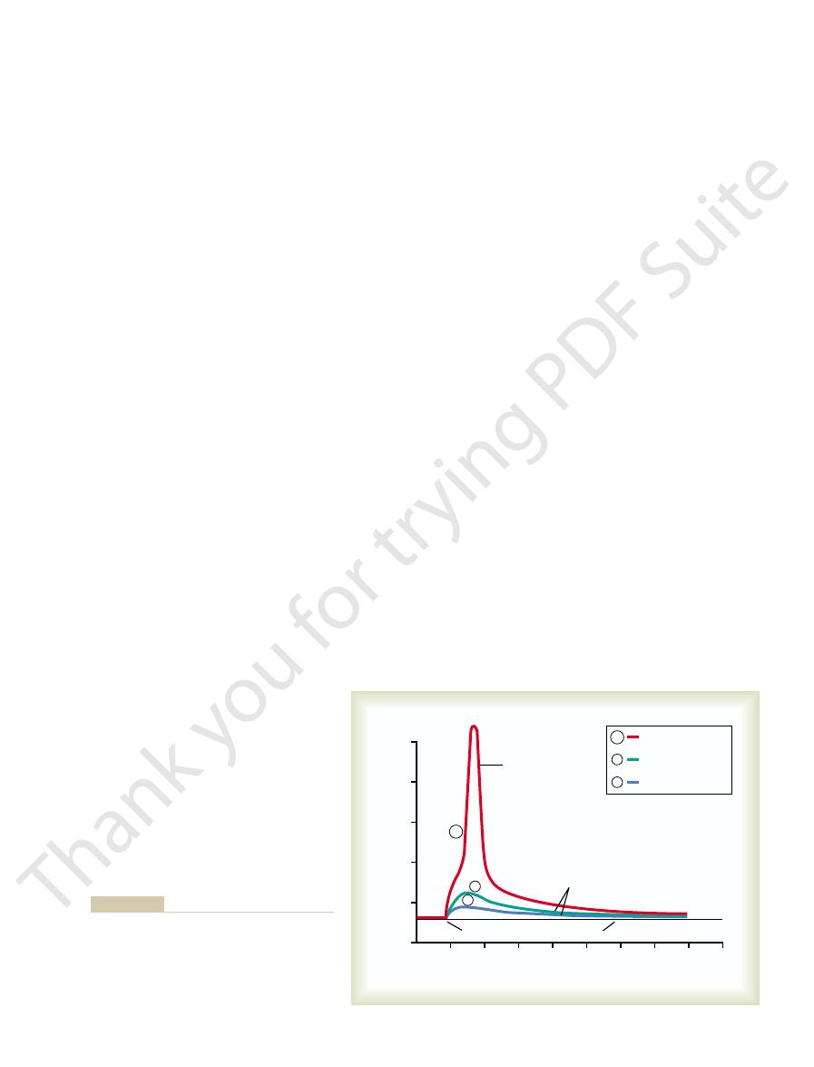
Time Course of Postsynaptic Potentials
rapidly to the interior of the postsynaptic motor
neuron to increase its intraneuronal potential by a few
potential (EPSP) shown by the blue and green curves
–
over the next 15 milliseconds because this is the time
required for the excess positive charges to leak out of
the excited neuron and to re-establish the normal
resting membrane potential.
neuronal potential to a more negative value than
Other types of transmitter substances can excite or
inhibit the postsynaptic neuron for much longer
periods—for hundreds of milliseconds or even for
some of the neuropeptide types of transmitter
Excitation of a single presynaptic terminal on the
surface of a neuron almost never excites the neuron.
ficient transmitter sub-
stance is released by a single terminal to cause an
of the 10 to 20 millivolts normally required to reach
Even though these terminals are spread over wide
summate;
It was pointed out earlier that a change in potential at
any single point within the soma will cause the poten-
tial to change everywhere inside the soma almost
electrical conductivity inside the large neuronal cell
12
14
16
– 80
2
10
0
4
6
8
Action potential
Excitatory postsynaptic
potential
Resting membrane potential
16 synapses firing
16
8
4
8 synapses firing
4 synapses firing
Millivolts
Milliseconds
–60
–40
–20
+20
0
16
8
4
to threshold for excitation and cause a superim-
cause sufficient summated potential to elicit an
Excitatory postsynaptic potentials, showing that
Figure 45–10
simultaneous firing of only a few synapses will not
action potential, but that simultaneous firing of
many synapses will raise the summated potential
posed action potential.

membrane potentials at these points. However, a
that is, note the
tip ends; note the high levels of excitatory postsynap-
drites to the left, there are excitatory effects near the
stimulating the dendrites of a neuron. On the two den-
In Figure 45
Decrement of Electrotonic Conduction in the Den-
teristics, as follows.
generation of action potentials. Stimulation (or inhibi-
the dendrites to the soma. Transmission of electrotonic
occur. Yet they do transmit
few voltage-gated sodium channels, and their thresh-
Can Transmit Signals Within the Same Neuron by Electrotonic
Most Dendrites Cannot Transmit Action Potentials—But They
vided by signals transmitted by way of the dendrites.
fore, the preponderant share of the excitation is pro-
20 per cent terminating on the neuronal soma. There-
neuron terminate on dendrites, in contrast to only 5 to
bers.
This provides a vast opportunity for summation of
ronal soma. And these dendrites can receive signals
The den-
for Exciting Neurons
Special Functions of Dendrites
other sources.
very easily. Diffuse signals in the nervous system often
quently, another excitatory signal entering the neuron
ring level. Conse-
than normal, but not yet at the
. That is, its
happens, the neuron is said to be
ring by the postsynaptic neuron. When this
“Facilitation” of Neurons
less than threshold value for excitation, thus turning
excited by an EPSP, an inhibitory signal from another
tially nullify each other. Thus, if a neuron is being
time, these two effects can either completely or par-
Simultaneous Summation of Inhibitory and Excitatory Postsy-
This type of summation is
summate.
that is, they can
if they occur rapidly enough, can add to one another;
greater the postsynaptic potential becomes. Thus, suc-
level, and the more rapid the rate of stimulation, the
Therefore, a second opening of the same channels can
for at most a millisecond or so. But the changed post-
res, the released
“Temporal Summation”
This effect of summing simultaneous postsynaptic
ring threshold had been reached, and an action
stimulation of 16 synapses. In this last instance, the
nally, a still higher EPSP was caused by
by simultaneous stimulation of 4 synapses; the next
gure was caused
10. The
axon. This is demonstrated in Figure 45
the EPSP becomes great enough, the
The Nervous System: A. General Principles and Sensory Physiology
568
Unit IX
threshold for
firing will be reached and an action potential will
develop spontaneously in the initial segment of the
–
bottom postsynaptic potential in the fi
higher potential was caused by stimulation of 8
synapses; fi
fi
potential was generated in the axon.
potentials by activating multiple terminals on widely
spaced areas of the neuronal membrane is called
spatial summation.
Each time a presynaptic terminal fi
transmitter substance opens the membrane channels
synaptic potential lasts up to 15 milliseconds after the
synaptic membrane channels have already closed.
increase the postsynaptic potential to a still greater
cessive discharges from a single presynaptic terminal,
“
”
called temporal summation.
naptic Potentials.
If an IPSP is tending to decrease the
membrane potential to a more negative value while an
EPSP is tending to increase the potential at the same
source can often reduce the postsynaptic potential to
off the activity of the neuron.
Often the summated postsynaptic potential is excita-
tory but has not risen high enough to reach the thresh-
old for fi
facilitated
membrane potential is nearer the threshold for firing
fi
from some other source can then excite the neuron
do facilitate large groups of neurons so that they can
respond quickly and easily to signals arriving from
Large Spatial Field of Excitation of the Dendrites.
drites of the anterior motor neurons often extend 500
to 1000 micrometers in all directions from the neu-
from a large spatial area around the motor neuron.
signals from many separate presynaptic nerve fi
It is also important that between 80 and 95 per cent
of all the presynaptic terminals of the anterior motor
Conduction.
Most dendrites fail to transmit action
potentials because their membranes have relatively
olds for excitation are too high for action potentials to
electrotonic current down
current means direct spread of electrical current by ion
conduction in the fluids of the dendrites but without
tion) of the neuron by this current has special charac-
drites—Greater Excitatory (or Inhibitory) Effect by
Synapses Located Near the Soma.
–11,
multiple excitatory and inhibitory synapses are shown
tic potentials at these ends—
less neg-
ative
60 mV
-
40
-
40
-
30
-
30
-
10
-
20
-
20
-
20
-
35
-
50
-
60
-
50
-
60
-
70
-
75
-
I
I
I
I
I
I
-
40
-
50
-
75
-
70
-
60
E
E
E
E
E
E
E
E
E
E
A powerful effect of inhibitory synapses at the initial segment of
bition (I) of dendritic excitation in the dendrite that is uppermost.
(E) electrotonic potentials in the two dendrites to the left and inhi-
drites, showing, especially, decremental conduction of excitatory
Stimulation of a neuron by presynaptic terminals located on den-
Figure 45–11
the axon is also shown.

ination, one can readily understand the importance of
maximum frequencies of discharge. With a little imag-
thresholds for excitation, and have widely differing
ferent neurons respond differently, have different
posing an inhibitory state on the neuron. Thus, dif-
ring can even be stopped, by superim-
decreased, or
increasing their excitatory state. The frequency can be
state is above the threshold level. Their frequency of
frequency.
charge, whereas neuron 3 has the highest maximum
neuron 2 has the lowest maximum frequency of dis-
neuron 3 has a high threshold. But note also that
neuron 1 has a low threshold for excitation, whereas
neurons to varying levels of excitatory state. Note that
level. Figure 45
the threshold for excitation, the neuron will
When the excitatory state of a neuron rises above
inhibitory state.
there is more inhibition than excitation, then it is said
. Conversely, if
than inhibition of the neuron at any given instant, then
the neuron. If there is a higher degree of excitation
The
“Excitatory State.”
Neuron to Rate of Firing
Relation of State of Excitation of the
segment. This location provides especially powerful
potentials in the same way that the soma can. Also
trotonic conduction toward the soma. Thus, dendrites
inhibitory synapses acting on the same dendrite. These
synaptic potential, but nearer the soma are two
stimulated by both excitatory and inhibitory synapses.
11 is shown to be
uppermost dendrite of Figure 45
The
Therefore, those synapses that lie near the soma have
of the neuron, the greater will be the decrement, and
The farther the excitatory synapse is from the soma
through the membrane. This decrease in membrane
soma, a large share of the potential is lost by leakage
fore, before the excitatory potentials can reach the
to electric current. There-
ions, making them
dendrites are long, and their membranes are thin and
lost before it reaches the soma. The reason is that the
Organization of the Nervous System, Basic Functions of Synapses, “Transmitter Substances”
Chapter 45
569
large share of the excitatory postsynaptic potential is
at least partially permeable to potassium and chloride
“leaky”
potential as it spreads electrotonically along dendrites
toward the soma is called decremental conduction.
the less will be excitatory signal reaching the soma.
far more effect in causing neuron excitation or inhibi-
tion than those that lie far away from the soma.
Summation of Excitation and Inhibition in Dendrites.
–
At the tip of the dendrite is a strong excitatory post-
inhibitory synapses provide a hyperpolarizing voltage
that completely nullifies the excitatory effect and
indeed transmits a small amount of inhibition by elec-
can summate excitatory and inhibitory postsynaptic
shown in the figure are several inhibitory synapses
located directly on the axon hillock and initial axon
inhibition because it has the direct effect of increasing
the threshold for excitation at the very point where the
action potential is normally generated.
“excitatory state” of a neuron is
defined as the summated degree of excitatory drive to
it is said that there is an excitatory state
that there is an
fire repet-
itively as long as the excitatory state remains at that
–12 shows responses of three types of
Some neurons in the central nervous system fire
continuously because even the normal excitatory
firing can usually be increased still more by further
fi
having different neurons with these many types of
response characteristics to perform the widely varying
functions of the nervous system.
10
15
20
25
30
35
Frequency of discharge per second
Excitatory state (arbitrary units)
0
5
Threshold
Neuron 1
Neuron 2
Neuron 3
0
600
500
400
300
200
100
neurons to different levels of excitatory state.
Response characteristics of different types of
Figure 45–12

Science, 4th ed. New York: McGraw-Hill, 2000.
Kandel ER, Schwartz JH, Jessell TM: Principles of Neural
logue between genes and synapses. Science 294:1030, 2001.
Kandel ER: The molecular biology of memory storage: a dia-
Churchill Livingstone, 2003.
Haines DE, Lancon JA: Review of Neuroscience. New York:
Churchill Livingstone, 1997.
Haines DE: Fundamental Neuroscience. New York:
signaling. Arch Neurol 61:641, 2004.
Girault JA, Greengard P: The neurobiology of dopamine
rosci 5:135, 2004.
receptors and control of transmitter release. Nat Rev Neu-
Engelman HS, MacDermott AB: Presynaptic inotropic
Ann N Y Acad Sci 994:175, 2003.
actions of peripheral hormones on melanocortin neurons.
Cowley MA, Cone RD, Enriori P, et al: Electrophysiological
the garbage, they recycle. Neuron 40:667, 2003.
Brasnjo G, Otis TS: Glycine transporters not only take out
Rev Neurosci 26:105, 2003.
Boehning D, Snyder SH: Novel neural modulators. Annu
mysteries that remain). Ann N Y Acad Sci 929:11, 2001.
Albright TD, Jessell TM, Kandell ER, Posner MI: Progress
From the measure of delay time, one can then estimate
into a pool of neurons and the consequent output volley.
synaptic delay.
simultaneously, is about 0.5 millisecond. This is called
required for all these events to take place, even when
elicit an action potential. The
the receptor to increase the membrane permeability,
transmitter on the membrane receptor, (4) action of
postsynaptic neuronal membrane, (3) action of the
naptic terminal, (2) diffusion of the transmitter to the
from a presynaptic neuron to a postsynaptic neuron, a
Synaptic Delay.
agents.
membranes, making them less responsive to excitatory
soluble, it has been reasoned that some of them might
tonic muscle spasms.
go into rapidly repetitive discharge, resulting in severe
whelming, and the neurons become so excited that they
inhibitory effect of glycine in the spinal cord. Therefore,
, especially the
the neurons; instead, it
increase excitability of neurons. However, it does not
threshold for excitation of neurons.
neuronal excitability, presumably by reducing the
found in coffee, tea, and cocoa, respectively, all
, which are
, and
others are known to decrease excitability. For instance,
known to increase the excitability of neurons, and
Many drugs are
Effect of Drugs on Synaptic Transmission.
the person becomes unconscious.
temporarily interrupted, because within 3 to 7 seconds,
neurons. This is observed when the brain
supply of oxygen. Cessation of oxygen for only a few
Effect of Hypoxia on Synaptic Transmission.
uremic acidosis, coma virtually always develops.
comatose state. For instance, in very severe diabetic or
Conversely,
momentarily, but even this short time can often precip-
to overbreathe. The overbreathing blows off carbon
neurons. This can be demonstrated especially well by
For instance, a
greatly increases neuronal excitability.
uids.
Effect of Acidosis or Alkalosis on Synaptic Transmission.
factors as well: (1) progressive inactivation of many of
few seconds to a few minutes of rapid stimulation. Part
tials, and the transmitter can be exhausted in only a
stance in the presynaptic terminals. The excitatory ter-
The mechanism of fatigue is mainly exhaustion or
anism against excess neuronal activity. This is dis-
Thus, the development of fatigue is a protective mech-
nally subdued so that the seizure ceases.
lose this excess excitability after awhile. For example,
system become overexcited, fatigue causes them to
Fatigue is an exceedingly important characteristic of
sively less in succeeding milliseconds or seconds. This
rst very great, but the
synapses are repetitively stimulated at a rapid rate, the
When excitatory
Fatigue of Synaptic Transmission.
of Synaptic Transmission
The Nervous System: A. General Principles and Sensory Physiology
570
Unit IX
Some Special Characteristics
number of discharges by the postsynaptic neuron is at
fi
firing rate becomes progres-
is called fatigue of synaptic transmission.
synaptic function because when areas of the nervous
fatigue is probably the most important means by which
the excess excitability of the brain during an epileptic
seizure is fi
cussed further in the description of reverberating
neuronal circuits in Chapter 46.
partial exhaustion of the stores of transmitter sub-
minals on many neurons can store enough excitatory
transmitter to cause only about 10,000 action poten-
of the fatigue process probably results from two other
the postsynaptic membrane receptors and (2) slow
development of abnormal concentrations of ions
inside the postsynaptic neuronal cell.
Most
neurons are highly responsive to changes in pH of
the surrounding interstitial fl
Normally, alkalosis
rise in arterial blood pH from the 7.4 norm to 7.8 to
8.0 often causes cerebral epileptic seizures because of
increased excitability of some or all of the cerebral
asking a person who is predisposed to epileptic seizures
dioxide and therefore elevates the pH of the blood
itate an epileptic attack.
acidosis greatly depresses neuronal activ-
ity; a fall in pH from 7.4 to below 7.0 usually causes a
Neuronal
excitability is also highly dependent on an adequate
seconds can cause complete inexcitability of some
’s blood flow is
caffeine, theophylline
theobromine
increase
Strychnine is one of the best known of all agents that
do this by reducing the threshold for excitation of
inhibits the action of some nor-
mally inhibitory transmitter substances
the effects of the excitatory transmitters become over-
Most anesthetics increase the neuronal membrane
threshold for excitation and thereby decrease synaptic
transmission at many points in the nervous system.
Because many of the anesthetics are especially lipid-
change the physical characteristics of the neuronal
During transmission of a neuronal signal
certain amount of time is consumed in the process of
(1) discharge of the transmitter substance by the presy-
and (5) inward diffusion of sodium to raise the excita-
tory postsynaptic potential to a high enough level to
minimal period of time
large numbers of excitatory synapses are stimulated
the
Neurophysiologists can measure the
minimal delay time between an input volley of impulses
the number of series neurons in the circuit.
References
in the neural sciences in the century after Cajal (and the

Annu Rev Physiol 64:355, 2002.
Zucker RS, Regehr WG: Short-term synaptic plasticity.
26:147, 2003.
in the control of action potential output. Trends Neurosci
Williams SR, Stuart GJ: Role of dendritic synapse location
dopaminergic systems. Ann N Y Acad Sci 1003:53, 2003.
West AR, Floresco SB, Charara A, et al: Electrophysiologi-
spines. Trends Neurosci 27:77, 2004.
Tsay D, Yuste RL: On the electrical function of dendritic
development and cessation. Neuroscience 123:299, 2004.
Timofeev I, Steriade M: Neocortical seizures: initiation,
197, 2004.
Stern JE: Nitric oxide and homeostatic control: an intercel-
maintaining the tone. Trends Neurosci 27:262, 2004.
Tonically active GABA A receptors: modulating gain and
Semyanov A, Walker MC, Kullmann DM, Silver RA:
ity. Ann N Y Acad Sci 978:359, 2002.
making of a complex spike: ionic composition and plastic-
Schmolesky MT, Weber JT, De Zeeuw CI, Hansel C: The
overview. Ann N Y Acad Sci 998:1, 2003.
Ruff RL: Neurophysiology of the neuromuscular junction:
Annu Rev Physiol 65:453, 2003.
cation currents: from molecules to physiological function.
Robinson RB, Siegelbaum SA: Hyperpolarization-activated
nels: a functional patchwork.Trends Neurosci 26:683, 2003.
Reid CA, Bekkers JM, Clements JD: Presynaptic Ca
function. Prog Neurobiol 64:51, 2001.
Prast H, Philippu A: Nitric oxide as modulator of neuronal
function of dendritic spines. Annu Rev Physiol 64:313,
Nimchinsky EA, Sabatin BL, Svoboda K: Structure and
Physiol Rev 80:315, 2000.
Nicholls DG, Budd SL: Mitochondria and neuronal survival.
rosci 26:573, 2003.
role in activity-dependent synaptogenesis. Trends Neu-
Muller D, Nikonenko I: Dynamic presynaptic varicosities: a
3:362, 2002.
tions of active dendritic conductances. Nat Rev Neurosci
Migliore M, Shepherd GM: Emerging rules for the distribu-
Nat Rev Neurosci 1:181, 2000.
Magee JC: Dendritic integration of excitatory synaptic input.
Organization of the Nervous System, Basic Functions of Synapses, “Transmitter Substances”
Chapter 45
571
2002.
2
+
chan-
lular signaling molecule contributing to autonomic and
neuroendocrine integration? Prog Biophys Mol Biol 84:
cal interactions between striatal glutamatergic and