stituent amino acids.
hydrolysis. That is, the proteolytic enzymes return hydrogen and hydroxyl ions
bound together by condensation, and digestion occurs by the reverse effect:
succeeding one; thus, the successive amino acids in the protein chain are also
. At each linkage, a hydroxyl ion has been
and thereby split the fatty acid molecules away from the glycerol. Here again,
Digestion of the triglycerides consists of the reverse process: the fat-
molecule. During condensation, three molecules
erides (neutral fats), which are combinations of three
hydrolysis,
from each other. This process, called
bohydrates are converted into monosaccharides. Specific enzymes in the diges-
When carbohydrates are digested, the above process is reversed and the car-
combine with each other at these sites of removal, and the hydrogen and
OH) has been removed from the next one. The two monosaccharides then
) has been removed from one of the monosaccharides, and a hydroxyl ion
. This means that a hydrogen ion
monosaccha-
, which are combinations of
disaccharides
polysaccharides
Digestion of the Various Foods by Hydrolysis
products as well as water, electrolytes, and other substances are absorbed.
pounds for absorption and, second, the mechanisms by which the digestive end
which carbohydrates, fats, and proteins are digested into small enough com-
fore, this chapter discusses, first, the processes by
as nutrients without preliminary digestion. There-
trointestinal mucosa and, for this reason, are useless
. They generally cannot be
, and
The major foods on which the body lives (with the
Gastrointestinal Tract
Digestion and Absorption in the
C
H
A
P
T
E
R
6
5
808
exception of small quantities of substances such as
vitamins and minerals) can be classified as carbo-
hydrates, fats
proteins
absorbed in their natural forms through the gas-
Hydrolysis of Carbohydrates.
Almost all the carbohydrates of the diet are either
large
or
rides bound to one another by condensation
(H
+
(
-
hydroxyl ions combine to form water (H
2
O).
tive juices of the gastrointestinal tract return the hydrogen and hydroxyl ions
from water to the polysaccharides and thereby separate the monosaccharides
is the following (in which
R
≤-R¢ is a disaccharide):
Hydrolysis of Fats.
Almost the entire fat portion of the diet consists of triglyc-
fatty acid molecules con-
densed with a single glycerol
of water are removed.
digesting enzymes return three molecules of water to the triglyceride molecule
the digestive process is one of hydrolysis.
Hydrolysis of Proteins.
Proteins are formed from multiple amino acids that are
bound together by peptide linkages
removed from one amino acid and a hydrogen ion has been removed from the
from water molecules to the protein molecules to split them into their con-
R -R
H O
R OH R H
æ
¢¢
+ ¢
¢¢ ¢ +
æ
Æ
ææ
2
digestive
enzyme
marized in Figure 65
The major steps in carbohydrate digestion are sum-
products of carbohydrate digestion, and galactose and
In the ordinary diet, which contains far more
digestion are all monosaccharides. They are all water
. Thus, the
multiple molecules
glucose.
. Sucrose splits into a molecule of
these enterocytes.
, so that the
the intestinal microvilli brush border
rides. These enzymes are located
glucose polymers, into their constituent monosaccha-
charides lactose, sucrose, and maltose, plus other small
), which are capable of splitting the disac-
, and
enterocytes lining the villi of the small intestine
The
In general, the carbohydrates are almost totally con-
mixes with pancreatic juice, virtually all the carbohy-
Therefore, within 15 to 30 minutes after the chyme
tion, like saliva, contains a large quantity of
Digestion by Pancreatic Amylase.
secretions, as much as 30 to 40 per cent of the starches
on the average, before food and its accompanying
pH of the medium falls below about 4.0. Nevertheless,
secretions. Then activity of the salivary amylase is
However, starch digestion sometimes continues in
remains in the mouth only a short time, so that prob-
1. However, the food
molecules, as shown in Figure 65
mainly by the parotid glands. This enzyme hydrolyzes
food is chewed, it is mixed with saliva, which contains
When
be considered a food for humans.
human digestive tract. Consequently, cellulose cannot
which is a carbohydrate. However, no enzymes
The diet also contains a large amount of cellulose,
, and minor quantities of
pyruvic acid
potatoes and the different types of grains. Other car-
present in almost all nonanimal foods, particularly in
, which are large polysaccharides
starches
in milk; and
, which is a disaccharide found
, which is the disaccharide known popularly
of carbohydrates exist in the normal human diet. They
Digestion of Carbohydrates
All the digestive enzymes are proteins. Their secre-
is involved. The
because, in the case of all three major types of food,
Therefore, the chemistry of digestion is simple
Digestion and Absorption in the Gastrointestinal Tract
Chapter 65
809
the same basic process of hydrolysis
only difference lies in the types of enzymes required
to promote the hydrolysis reactions for each type of
food.
tion by the different gastrointestinal glands was dis-
cussed in Chapter 64.
Carbohydrate Foods of the Diet.
Only three major sources
are sucrose
as cane sugar; lactose
bohydrates ingested to a slight extent are amylose,
glycogen, alcohol, lactic acid,
, pectins, dex-
trins
carbohydrate derivatives
in meats.
capable of hydrolyzing cellulose are secreted in the
Digestion of Carbohydrates in the Mouth and Stomach.
the digestive enzyme ptyalin (an
a-amylase) secreted
starch into the disaccharide maltose and other small
polymers of glucose that contain three to nine glucose
–
ably not more than 5 per cent of all the starches will
have become hydrolyzed by the time the food is
swallowed.
the body and fundus of the stomach for as long as 1
hour before the food becomes mixed with the stomach
blocked by acid of the gastric secretions because the
amylase is essentially nonactive as an enzyme once the
saliva do become completely mixed with the gastric
will have been hydrolyzed mainly to form maltose.
Digestion of Carbohydrates in the Small Intestine
Pancreatic secre-
a-amylase
that is almost identical in its function with the
a-amylase of saliva but is several times as powerful.
empties from the stomach into the duodenum and
drates will have become digested.
verted into maltose and/or other very small glucose
polymers before passing beyond the duodenum or
upper jejunum.
Hydrolysis of Disaccharides and Small Glucose Polymers
into Monosaccharides by Intestinal Epithelial Enzymes.
contain four enzymes (lactase, sucrase, maltase
a-dextrinase
in the enterocytes cov-
ering
disaccharides are digested as they come in contact with
Lactose splits into a molecule of galactose and a
molecule of glucose
fructose and a molecule of
Maltose and other
small glucose polymers all split into
of glucose
final products of carbohydrate
soluble and are absorbed immediately into the portal
blood.
starches than all other carbohydrates combined,
glucose represents more than 80 per cent of the final
fructose each seldom more than 10 per cent.
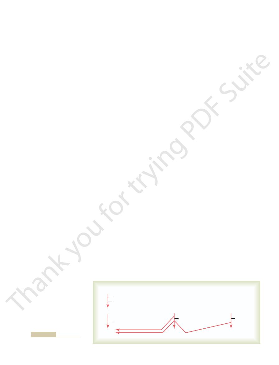
–1.
Maltose and 3 to 9 glucose polymers
Lactose
Sucrose
Ptyalin (saliva)–20–40%
Starches
Glucose
Fructose
Galactose
Sucrase
(intestine)
Lactase
(intestine)
Pancreatic amylase–50–80%
Maltase and
a
-dextrinase
(intestine)
Figure 65–1
Digestion of carbohydrates.
minutes, virtually all the last dipeptides and tripeptides
ing types of linkages between amino acids. Within
Finally, inside the cytosol of the enterocyte are mul-
enterocyte.
into amino acids. Both the amino acids plus the dipep-
. They succeed in splitting the remaining larger
Two types of peptidase enzymes are especially
uids.
exterior, where they come in contact with the intestinal
projecting from the surface of each cell. In the mem-
microvilli
brush border
mainly in the duodenum and jejunum. These cells have
enterocytes that line the villi of the small intestine,
The last digestive stage of
tripeptides.
pancreatic juices. Most remain as dipeptides and
that partially hold meats together.
, which then digests elastin
, in turn, is con-
ends of the polypeptides.
cules into small polypeptides; carboxypolypeptidase
, as shown in Figure
, and
carboxy-
chymotrypsin
stomach, the partial breakdown products of the
in the duodenum and jejunum, under the in
protein digestion occurs in the upper small intestine,
linkages.
The dietary proteins are chemically
Digestion of Proteins
810
Unit XII
Gastrointestinal Physiology
Proteins of the Diet.
long chains of amino acids bound together by peptide
A typical linkage is the following:
Digestion of Proteins by Pancreatic Secretions.
Most
fluence
of proteolytic enzymes from pancreatic secretion.
Immediately on entering the small intestine from the
protein foods are attacked by major proteolytic
pancreatic enzymes: trypsin,
,
polypeptidase
proelastase
65–2.
Both trypsin and chymotrypsin split protein mole-
then cleaves individual amino acids from the carboxyl
Proelastase
verted into elastase
fibers
Only a small percentage of the proteins are digested
all the way to their constituent amino acids by the
Digestion of Peptides by Peptidases in the Enterocytes That
Line the Small Intestinal Villi.
the proteins in the intestinal lumen is achieved by the
a
that consists of hundreds of
brane of each of these microvilli are multiple pepti-
dases that protrude through the membranes to the
fl
important, aminopolypeptidase and several dipepti-
dases
polypeptides into tripeptides and dipeptides and a few
tides and tripeptides are easily transported through
the microvillar membrane to the interior of the
tiple other peptidases that are specific for the remain-
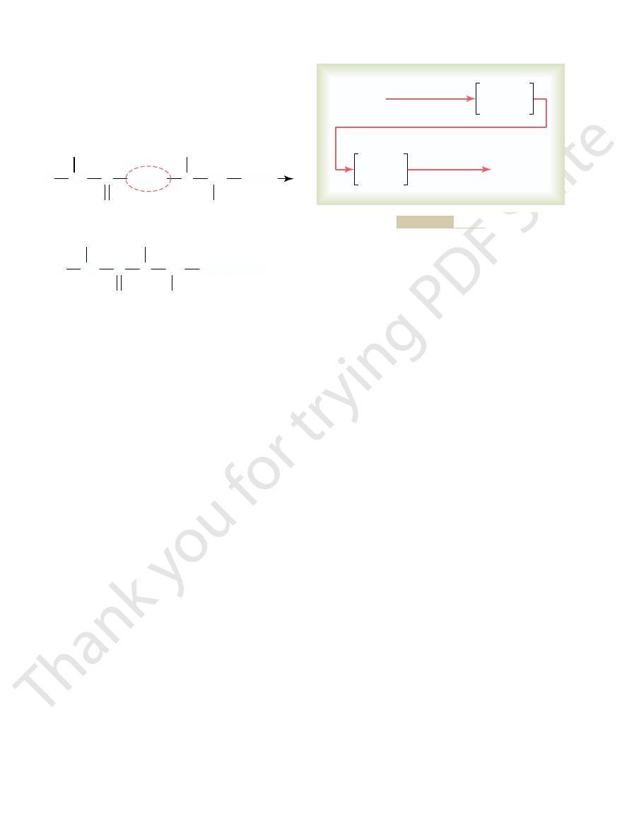
Trypsin, chymotrypsin, carboxypolypeptidase,
Pepsin
Peptidases
Amino acids
Polypeptides
+
Amino acids
Proteoses
Peptones
Polypeptides
Proteins
proelastase
Digestion of proteins.
Figure 65–2
CH
COOH
CH
COOH
C
N
O
OH
+ H
NH
2
CH
R
R
H
C
N
O
+ H
2
O
NH
2
CH
R
R
H
acids.
tides. This splitting of proteins occurs as a result of
the protein to proteoses, peptones, and a few polypep-
process of protein digestion, usually providing only 10
2, pepsin only initiates the
As shown in Figure 65
the other digestive enzymes and, therefore, may be
juices, the ingested meats are less well penetrated by
quently, in persons who lack pepsin in the stomach
bers be digested. Conse-
meats and digest the other meat proteins, it is
cellular connective tissue of meats; therefore, for the
enzymes. Collagen is a major constituent of the inter-
, an albuminoid
range of acidity for pepsin activity.
then averages around 2.0 to 3.0, a highly favorable
the nonoxyntic glandular cells of the stomach, the pH
glands at a pH of about 0.8, but by the time it is mixed
a large quantity of hydrochloric acid. This hydrochlo-
As explained in Chapter 64, the gastric glands secrete
action on protein, the stomach juices must be acidic.
Consequently, for this enzyme to cause digestive
of 2.0 to 3.0 and is inactive at a pH above about 5.0.
peptic enzyme of the stomach, is most active at a pH
, the important
Pepsin
acids. The physical and chemical characteristics of dif-
The characteristics of each protein are determined
by the types of amino acids in the protein molecule
and by the sequential arrangements of these amino
ferent proteins important in human tissues are dis-
cussed in Chapter 69.
Digestion of Proteins in the Stomach.
ric acid is secreted by the parietal (oxyntic) cells in the
with the stomach contents and with secretions from
One of the important features of pepsin digestion is
its ability to digest the protein collagen
type of protein that is affected little by other digestive
digestive enzymes of the digestive tract to penetrate
first nec-
essary that the collagen fi
poorly digested.
–
to 20 per cent of the total protein digestion to convert
hydrolysis at the peptide linkages between amino
End Products of Fat Digestion.
, but this is usually not
lipase, known as
the enterocytes of the small intestine contain still more
minute all triglycerides that it can reach. In addition,
tities in pancreatic juice, enough to digest within 1
, present in enormous quan-
Digestion of Triglycerides by Pancreatic Lipase.
for digestion of fats.
Consequently, it can be readily understood how impor-
and can attack the fat globules only on their surfaces.
The lipase enzymes are water-soluble compounds
cation process.
occurred is less than 1 micrometer, this represents an
manyfold. Because the average diameter of the fat
intestine, the total surface area of the fat increases
used in household cleaners for removing grease.
tation with the water in the small bowel. This action is
salts and lecithin, especially the lecithin, in the bile is
is great. Consequently, a major function of the bile
tion, can be broken up into many very minute particles
uid, on agita-
uid is low, this nonmiscible
When the interfacial tension of a globule of non-
uids, which greatly decreases the
jecting. The polar projections, in turn, are soluble in the
layer of the fat globules, with the polar portions pro-
highly soluble in fat. Therefore, the fat-soluble por-
lecithin molecules are highly soluble in water, whereas
cation of the fat. The polar parts (the points
, are extremely important for
. Both of these,
However, bile does contain a large quantity of
the liver that does not contain any digestive enzymes.
, the secretion from
Then, most of the emulsi
, and it begins
digestive enzymes can act on the globule surfaces. This
globules into very small sizes so that the water-soluble
The
Emulsification of Fat by Bile Acids and Lecithin.
intestine as follows.
Instead, essentially all fat digestion occurs in the small
swallowed with the saliva. This amount of digestion is
in the stomach
cholesterol is considered, from a dietary point of view,
fats and is metabolized similarly to fats. Therefore,
chemical characteristics of fats; plus, it is derived from
fatty acid, but it does exhibit some of the physical and
terol, however, is a sterol compound that contains no
therefore can be considered fats themselves. Choles-
pholipids, cholesterol, and cholesterol esters. The phos-
of animal origin but much, much less so in food of
3. Neutral fat is a major constituent in food
Figure 65
nucleus and three fatty acid side chains, as shown in
diet are the neutral fats, also known as
disturbances, as discussed in Chapter 34.
rare absorption of whole protein molecules. Even
with only rare absorption of peptides and very, very
products that are absorbed are individual amino acids,
the enterocyte and thence into the blood.
acids; these then pass on through to the other side of
Digestion and Absorption in the Gastrointestinal Tract
Chapter 65
811
are digested to the final stage to form single amino
More than 99 per cent of the final protein digestive
these very few absorbed molecules of whole protein
can sometimes cause serious allergic or immunologic
Digestion of Fats
Fats of the Diet.
By far the most abundant fats of the
triglycerides,
each molecule of which is composed of a glycerol
–
plant origin.
In the usual diet are also small quantities of phos-
pholipids and cholesterol esters contain fatty acid and
a fat.
Digestion of Fats in the Intestine.
A small amount of
triglycerides is digested
by lingual lipase
that is secreted by lingual glands in the mouth and
less than 10 per cent and generally unimportant.
first step in fat digestion is physically to break the fat
process is called emulsification of the fat
by agitation in the stomach to mix the fat with the
products of stomach digestion.
fication occurs in the duo-
denum under the influence of bile
bile salts
as well as the phospholipid lecithin
but especially the lecithin
emulsifi
where ionization occurs in water) of the bile salts and
most of the remaining portions of their molecules are
tions of these liver secretions dissolve in the surface
surrounding watery fl
interfacial tension of the fat and makes it soluble as
well.
miscible fl
fl
far more easily than it can when the interfacial tension
to make the fat globules readily fragmentable by agi-
the same as that of many detergents that are widely
Each time the diameters of the fat globules are sig-
nificantly decreased as a result of agitation in the small
particles in the intestine after emulsification has
increase of as much as 1000-fold in total surface areas
of the fats caused by the emulsifi
tant this detergent function of bile salts and lecithin is
By far the
most important enzyme for digestion of the triglyc-
erides is pancreatic lipase
enteric lipase
needed.
Most of the triglyc-
erides of the diet are split by pancreatic lipase into
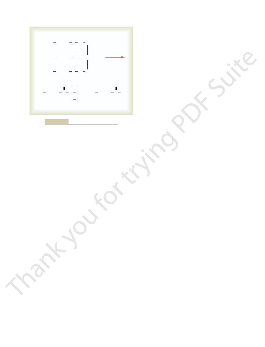
O
CH + 2H
(2-Monoglyceride)
(Stearic acid)
HO
CH
HO
CH
(Tristearin)
O
CH
O
CH
CH
3
(CH
2
)
16
C
O
2
CH
3
(CH
2
)
16
C
O
2
CH
3
(CH
2
)
16
C
O
Lipase
CH
3
(CH
2
)
16
(CH
2
)
16
C
O
O
C
O
OH
2
2
2
O
CH + 2CH
3
Hydrolysis of neutral fat catalyzed by lipase.
Figure 65–3
small intestinal mucosa, showing many folds called
Figure 65
absorbed in small quantities.
such as alcohol and some drugs like aspirin, can be
tions. Only a few highly lipid-soluble substances,
type of absorptive membrane, and also because the
The stomach is a poor absorptive area of the gas-
day.
absorbed in the small intestine, leaving only 1.5 liters
a total of 8 to 9 liters. All but about 1.5 liters of this is
trointestinal secretions (about 7 liters). This comes to
The total quantity of
Anatomical Basis of Absorption
branes discussed in detail in Chapter 4. The following
Gastrointestinal Absorption
absorbed without this function of the micelles.
free fatty acids. Indeed, essentially no cholesterol is
The bile salt micelles play the same role in
hydrolyze the cholesterol ester, and
cholesterol ester hydrolase
acid within their molecules. Both the cholesterol esters
molecule of fatty acid. Phospholipids also contain fatty
lesterol in the diet is in the form of cholesterol esters,
process.
absorbed into the blood, as discussed later, but the bile
There the monoglycerides and free fatty acids are
the brush borders of the intestinal epithelial cells.
of which would otherwise be relatively insoluble, to
to carry the monoglycerides and free fatty acids, both
The bile salt micelles also act as a transport medium
groups are negatively charged, they allow the entire
cover the surface of the micelle. Because these polar
a small fat globule in the middle of a resulting micelle,
sterol nucleus encompasses the fat digestate, forming
and a polar group that is highly water-soluble. The
salt. These develop because each bile salt molecule is
small spherical, cylindrical globules 3 to 6 nanometers
, which are
water, have the propensity to form
Bile salts, when in high enough concentration in
occurs in the following way.
these end products of digestion are formed. This
ing fats quickly blocks further digestion. But the bile
reversible process; therefore, accumulation of mono-
The hydrolysis of triglycerides is a highly
Figure 65
, as shown in
812
Unit XII
Gastrointestinal Physiology
free fatty acids and 2-monoglycerides
–4.
Role of Bile Salts to Accelerate Fat Digestion—Formation of
Micelles.
glycerides and free fatty acids in the vicinity of digest-
salts play the additional important role of removing
the monoglycerides and free fatty acids from the vicin-
ity of the digesting fat globules almost as rapidly as
micelles
in diameter composed of 20 to 40 molecules of bile
composed of a sterol nucleus that is highly fat-soluble
with polar groups of bile salts projecting outward to
micelle globule to dissolve in the water of the diges-
tive fluids and to remain in stable solution until the fat
is absorbed into the blood.
salts themselves are released back into the chyme to
be used again and again for this “ferrying”
Digestion of Cholesterol Esters and Phospholipids.
Most cho-
which are combinations of free cholesterol and one
and the phospholipids are hydrolyzed by two other
lipases in the pancreatic secretion that free the fatty
acids—the enzyme
to
phospholipase A
2
to hydrolyze the phospholipid.
“ferry-
ing” free cholesterol and phospholipid molecule diges-
tates that they play in “ferrying” monoglycerides and
Basic Principles of
It is suggested that the reader review the basic princi-
ples of transport of substances through cell mem-
paragraphs present specialized applications of these
transport processes during gastrointestinal absorption.
fluid that must be absorbed each
day by the intestines is equal to the ingested fluid
(about 1.5 liters) plus that secreted in the various gas-
to pass through the ileocecal valve into the colon each
trointestinal tract because it lacks the typical villus
junctions between the epithelial cells are tight junc-
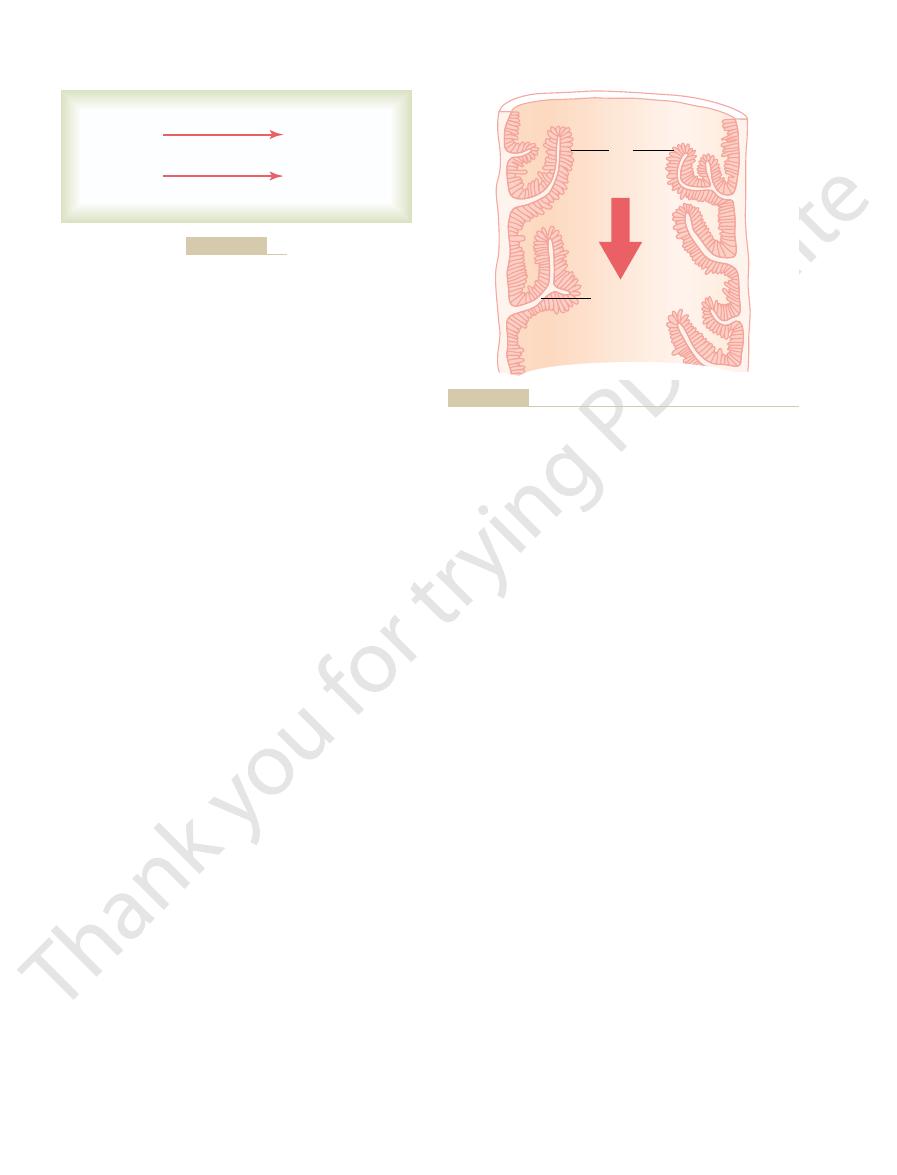
Absorptive Surface of the Small Intestinal Mucosa Villi.
–5 demonstrates the absorptive surface of the
(Bile + Agitation)
Pancreatic lipase
Emulsified fat
Fatty acids and
2-monoglycerides
Fat
Emulsified fat
Figure 65–4
Digestion of fats.
Villi
Food
movement
Food
movement
Valvulae
conniventes
conniventes covered by villi.
Figure 65–5
Longitudinal section of the small intestine, showing the valvulae

of several hundred grams of carbohydrates, 100 or
movement of the microvilli, keeping them constantly
been entrapped. Small amounts of substances are
are pinched-off portions of infolded enterocyte mem-
, which
pinocytic vesicles
Figure 65
shows a cross section of the villus, and
Figure 65
general organization of the villus, emphasizing (1) the
Figure 65
area of the mucosa perhaps 1000-fold, making a
villi, and the microvilli increases the total absorptive
Thus, the combination of the folds of Kerckring, the
7. This increases the surface
micrograph in Figure 65
chyme; these microvilli are shown in the electron
microvilli
brush border,
Finally, each intestinal epithelial cell on each villus
less profuse in the distal small intestine. The presence
that they touch in most areas, but their distribution is
6. The villi
5 and in individual detail in Figure 65
on the surfaces of the valvulae conniventes in Figure
millimeter from the surface of the mucosa, as shown
. These project about 1
developed in the duodenum and jejunum, where they
about threefold. These folds extend circularly most of
), which
folds of Kerckring
Digestion and Absorption in the Gastrointestinal Tract
Chapter 65
813
valvulae conniventes (or
increase the surface area of the absorptive mucosa
the way around the intestine and are especially well
often protrude as much as 8 millimeters into the
lumen.
Also located on the epithelial surface of the small
intestine all the way down to the ileocecal valve are
literally millions of small villi
65–
–
lie so close to one another in the upper small intestine
of villi on the mucosal surface enhances the total
absorptive area another 10-fold.
is characterized by a
consisting of as
many as 1000
1 micrometer in length and 0.1
micrometer in diameter protruding into the intestinal
–
area exposed to the intestinal materials at least
another 20-fold.
tremendous total area of 250 or more square meters
for the entire small intestine—about the surface area
of a tennis court.
–6A shows in longitudinal section the
advantageous arrangement of the vascular system for
absorption of fluid and dissolved material into the
portal blood and (2) the arrangement of the “central
lacteal” lymph vessel for absorption into the lymph.
–6B
–7 shows many small
brane forming vesicles of absorbed fluids that have
absorbed by this physical process of pinocytosis.
Extending from the epithelial cell body into each
microvillus of the brush border are multiple actin fila-
ments that contract rhythmically to cause continual
exposed to new quantities of intestinal fluid.
Absorption in the
Small Intestine
Absorption from the small intestine each day consists
Central
lacteal
Central
lacteal
Venules
Basement
membrane
A
B
Brush
border
Arteriole
Capillaries
Vein
Artery
Blood
capillaries
border at the other ends of these
Cross section showing a base-
Figure 65–6
Functional organization of the
villus. A, Longitudinal section.
B,
ment membrane beneath the
epithelial cells and a brush
cells.
Dr. William Lockwood.)
reticulum lying immediately beneath the brush border. (Courtesy
Brush border of a gastrointestinal epithelial cell, showing also
Figure 65–7
absorbed pinocytic vesicles, mitochondria, and endoplasmic
chloride in the feces and also little water loss. Thus, the
This effect of aldosterone is especially important in
substances.
absorption of chloride ions, water, and some other
by the intestinal epithelium. And the increased sodium
the adrenal glands. Within 1 to 3 hours this aldosterone
person becomes dehydrated, large amounts of aldos-
When a
villus.
nally, into the circulating blood of the
cells themselves. And osmotic movement of water
of the epithelial cells, but much also occurs through the
in the paracellular space. Much of this osmosis occurs
spaces. This occurs because a large osmotic gradient
The next step in the transport
Osmosis of the Water.
cells into the paracellular spaces.
into the epithelial cell cytoplasm. This provides still
(that is, about equal to that in plasma), sodium moves
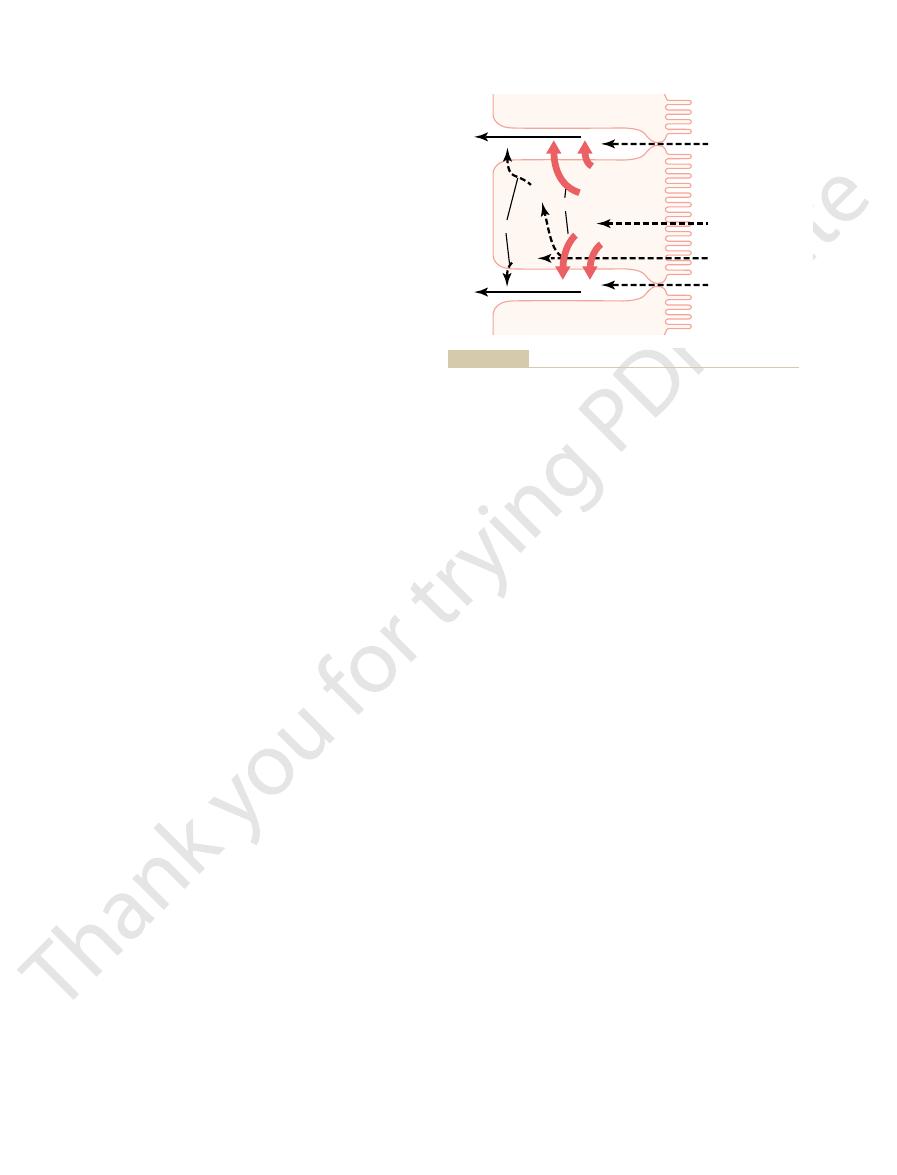
8. Because the sodium con-
as also shown in Figure 65
tion inside the cell to a low value (about 50 mEq/L),
of the sodium ions.
fact, the negatively charged chloride ions are mainly
the sodium is absorbed along with chloride ions; in
enzymes in the cell membrane (see Chapter 4). Part of
port: it requires energy, and the energy process is cat-
8. This
strated by the heavy red arrows in Figure 65
these cells into paracellular spaces. This is demon-
The motive power for sodium absorption is pro-
mechanism, discussed in Chapter 4, are also essentially
8. The principles of this
intestine is shown in Figure 65
The basic mechanism of sodium absorption from the
discussions.
and amino acids, as we shall see in subsequent
absorbed through the intestinal mucosa. Sodium also
however, less than 0.5 per cent of the intestinal sodium
depleted to lethal levels within hours. Normally,
tions are lost to the exterior, as in extreme diarrhea,
Whenever signi
one seventh of all the sodium present in the body.
35 grams of sodium each day, which is equal to about
sodium into the feces, the intestines must absorb 25 to
sodium each day. Therefore, to prevent net loss of
In addition, the average person eats 5 to 8 grams of
are secreted in the intestinal secretions each day.
Twenty to 30 grams of sodium
Active Transport of Sodium.
Absorption of Ions
minutes, suf
charged from the stomach into the duodenum. Within
from plasma into the chyme. This
Conversely, water can also be transported in the
of the villi almost entirely by osmosis.
Therefore, when the chyme is dilute enough, water is
more, this diffusion obeys the usual laws of osmosis.
. Further-
Water is transported through the
Absorption of Water
tional water and ions, although very few nutrients.
water per day. The
700 grams of proteins per day, and 20 or more liters of
bohydrates per day, 500 grams of fat per day, 500 to
greater than this: as much as several kilograms of car-
to 100 grams of ions, and 7 to 8 liters of water. The
more grams of fat, 50 to 100 grams of amino acids, 50
814
Unit XII
Gastrointestinal Physiology
absorptive capacity of the normal small intestine is far
large intestine can absorb still addi-
Isosmotic Absorption.
intestinal membrane entirely by diffusion
absorbed through the intestinal mucosa into the blood
opposite direction—
occurs especially when hyperosmotic solutions are dis-
ficient water usually will be transferred by
osmosis to make the chyme isosmotic with the plasma.
ficant amounts of intestinal secre-
the sodium reserves of the body can sometimes be
is lost in the feces each day because it is rapidly
plays an important role in helping to absorb sugars
–
the same as for absorption of sodium from the
gallbladder and renal tubules as discussed in Chapter
27.
vided by active transport of sodium from inside the
epithelial cells through the basal and side walls of
–
active transport obeys the usual laws of active trans-
alyzed by appropriate adenosine triphosphatase
passively “dragged” by the positive electrical charges
Active transport of sodium through the basolateral
membranes of the cell reduces the sodium concentra-
–
centration in the chyme is normally about 142 mEq/L
down this steep electrochemical gradient from the
chyme through the brush border of the epithelial cell
more sodium ions to be transported by the epithelial
process is osmosis of water into the paracellular
has been created by the elevated concentration of ions
through the tight junctions between the apical borders
creates flow of fluid into and through the paracellular
spaces and, fi
Aldosterone Greatly Enhances Sodium Absorption.
terone almost always are secreted by the cortices of
causes increased activation of the enzyme and trans-
port mechanisms for all aspects of sodium absorption
absorption in turn causes secondary increases in
the colon because it allows virtually no loss of sodium
H
2
O
H
2
O
H
2
O
H
2
O
H
2
O
H
2
O
Na
Active transport
Diffusion
Osmosis
Na
(50 mEq/L)
Na (142 mEq/L)
H
2
O
Na
through the epithelial membrane.
Absorption of sodium through the intestinal epithelium. Note also
Figure 65–8
osmotic absorption of water—that is, water “follows” sodium

sugar.
, the galactose derived from milk and the fructose
most abundant carbohydrate food, the starches. The
of carbohydrate calories absorbed. The reason for
, usually accounting for more than 80 per cent
none as larger carbohydrate compounds. By far
absorbed in the form of monosaccharides; only a small
Absorption of Nutrients
body.
tion of sodium ions. Fortunately, only small quantities
amounts; for example, maximum absorption of
absorbed with ease and in great quantities. Conversely,
intestinal mucosa. In general, the monovalent ions are
phosphate,
Potassium
globin, are discussed in Chapter 32.
need for iron, especially for the formation of hemo-
intestine. The principles of iron absorption and regu-
Chapter 79.
calcium absorption. These effects are discussed in
Parathyroid hormone activates vitamin D, and the
by the parathyroid glands, and another is
the body for calcium. One important factor controlling
num, and the amount of calcium ion absorption is very
make up for the loss.
whole body that might ensue. In most instances, the life
in combating the disease, but too much of a good thing
uid along with the salt. All this excess
extreme osmosis of water from the blood, thus provid-
ride ions. Finally, all this extra sodium chloride causes
from inside the cell into the intestinal crypts. In turn, this
chloride channels, allowing chloride ions to
monophosphate, which opens tremendous numbers of
This stimulates formation of excess cyclic adenosine
a subunit of cholera toxin into the epithelial cells.
uid alone.
Within 1 to 5 days, many severely affected patients die
each day.
be reabsorbed, thus sometimes causing loss of 5 to 10
The toxins of cholera and of some other types of diar-
intestinal digestates.
the folds, thus providing
and water into the intestinal lumen. This secretion in
deep folds, the epithelial cells secrete sodium chloride
the luminal surfaces of the intestines. While still in the
new epithelial cells. These in turn spread outward over
the Large Intestine Epithelium in Some Types of Diarrhea.
Extreme Secretion of Chloride Ions, Sodium Ions, and Water from
in the large intestine.
in exchange for absorption of chloride ions. This is
The epithelial cells on the surfaces of the villi in the
and Large Intestine—Simultaneous Absorption
Secretion of Bicarbonate Ions in the Ileum
kidneys.
active absorption of bicarbonate ions.
subsequently expired through the lungs. Thus, this is
remains as part of the chyme in the intestines, but the
ciates to form water and carbon dioxide. The water
), which then disso-
of the gut in exchange for some of the sodium. These
as follows: When sodium ions are absorbed, moderate
bile.The bicarbonate ion is absorbed in an indirect way
the sodium ions.
the epithelial cells. Then chloride ions move along this
that is, absorption of sodium ions through the epithe-
the upper part of the small intestine, chloride ion
tubules, which also serves to conserve sodium chloride
Digestion and Absorption in the Gastrointestinal Tract
Chapter 65
815
function of aldosterone in the intestinal tract is the
same as that achieved by aldosterone in the renal
and water in the body when a person becomes
dehydrated.
Absorption of Chloride Ions in the Duodenum and Jejunum.
In
absorption is rapid and occurs mainly by diffusion—
lium creates electronegativity in the chyme and
electropositivity in the paracellular spaces between
electrical gradient to “follow”
Absorption of Bicarbonate Ions in the Duodenum and Jejunum.
Often large quantities of bicarbonate ions must be
reabsorbed from the upper small intestine because
large amounts of bicarbonate ions have been secreted
into the duodenum in both pancreatic secretion and
amounts of hydrogen ions are secreted into the lumen
hydrogen ions in turn combine with the bicarbonate
ions to form carbonic acid (H
2
CO
3
carbon dioxide is readily absorbed into the blood and
so-called “
” It is
the same mechanism that occurs in the tubules of the
of Chloride Ions
ileum as well as on all surfaces of the large intestine
have a special capability of secreting bicarbonate ions
important because it provides alkaline bicarbonate
ions that neutralize acid products formed by bacteria
Deep
in the spaces between the intestinal epithelial folds are
immature epithelial cells that continually divide to form
turn is reabsorbed by the older epithelial cells outside
flow of water for absorbing
rheal bacteria can stimulate the fold secretion so greatly
that this secretion often becomes much greater than can
liters of water and sodium chloride as diarrhea
from this loss of fl
Extreme diarrheal secretion is initiated by entry of
flow rapidly
is believed to activate a sodium pump that pumps
sodium ions into the crypts to go along with the chlo-
ing rapid flow of fl
fluid washes away most of the bacteria and is of value
can be lethal because of serious dehydration of the
of a cholera victim can be saved by administration of
tremendous amounts of sodium chloride solution to
Absorption of Other Ions.
Calcium ions are actively
absorbed into the blood especially from the duode-
exactly controlled to supply exactly the daily need of
calcium absorption is parathyroid hormone secreted
vitamin D.
activated vitamin D in turn greatly enhances
Iron ions are also actively absorbed from the small
lation of its absorption in proportion to the body’s
, magnesium,
and probably still
other ions can also be actively absorbed through the
bivalent ions are normally absorbed in only small
calcium ions is only 1/50 as great as the normal absorp-
of the bivalent ions are normally required daily by the
Absorption of Carbohydrates
Essentially all the carbohydrates in the food are
fraction are absorbed as disaccharides and almost
the most abundant of the absorbed monosaccharides
is glucose
this is that glucose is the final digestion product of our
remaining 20 per cent of absorbed monosaccharides
are composed almost entirely of galactose and fruc-
tose
as one of the monosaccharides digested from cane

This allows direct diffusion of these short-chain fatty
are more water-soluble and mostly are not recon-
erides and absorbed by way of the lymphatics. The
as those from butterfat, are absorbed directly into the
quantities of short- and medium-chain fatty acids, such
epithelial cell, to
chylomicrons
endoplasmic reticulum; here, they are mainly used to
After entering the epithelial cell, the fatty acids and
only 40 to 50 per cent can be absorbed.
the fat is absorbed; in the absence of the bile micelles,
of an abundance of bile micelles, about 97 per cent of
is highly important for fat absorption. In the presence
function that
Thus, the micelles perform a
monoglycerides and fatty acids.
leaves the bile micelles still in the chyme, where they
are also soluble in the epithelial cell membrane. This
the epithelial cells, which is possible because the lipids
recesses among the moving, agitating microvilli. Here,
chyme. In this form, the monoglycerides and free fatty
of their highly charged exterior, they are soluble in
are only 3 to 6 nanometers in diameter, and because
bile micelles
acids, both of these digestive end products
Earlier in this chapter, it was pointed out that when
amino acids and peptides.
This multiplicity of transport proteins is required
the luminal membranes of intestinal epithelial cells.
ported, by facilitated diffusion.
of the amino acids and peptides.
and pulls the amino acid or peptide along with it. This
occur. After binding, the sodium ion then moves down
sodium co-transport of glucose occurs. That is, most
The energy for most of this transport is supplied by a
of dipeptides, tripeptides, and a few free amino acids.
after digestion, are absorbed through the luminal
As explained earlier in the chapter, most proteins,
Absorption of Proteins
one half that of glucose or galactose.
with sodium, its overall rate of transport is only about
into the blood. Because fructose is not co-transported
phosphorylated, then converted to glucose, and
Much of the fructose, on entering the cell, becomes
by the sodium co-transport mechanism. Instead, fruc-
glucose. Conversely, fructose transport does not occur
membranes.
To summarize, it is the initial active transport of
epithelial cell, other transport proteins and enzymes
with it the glucose at the same time. Once inside the
transported together to the interior of the cell. Thus,
simultaneously with the same transport protein, and
glucose. Fortunately, intestinal glucose also combines
, but the trans-
. That is, a sodium
Second, decrease of sodium inside the cells causes
thereby depleting sodium inside the epithelial cells.
through the intestinal membrane. First is active trans-
There are two stages in the transport of
The reason is that glucose absorption occurs in a co-
tinal membrane, virtually no glucose can be absorbed.
Glucose Is Transported by a Sodium Co-Transport Mechanism.
absorption of glucose.
an active transport process. Let us
Virtually all the monosaccharides are absorbed by
816
Unit XII
Gastrointestinal Physiology
first discuss the
In the absence of sodium transport through the intes-
transport mode with active transport of sodium.
sodium
port of sodium ions through the basolateral mem-
branes of the intestinal epithelial cells into the blood,
sodium from the intestinal lumen to move through the
brush border of the epithelial cells to the cell interiors
by a process of facilitated diffusion
ion combines with a transport protein
port protein will not transport the sodium to the inte-
rior of the cell until the protein itself also combines
with some other appropriate substance such as
then both the sodium ion and glucose molecule are
the low concentration of sodium inside the cell liter-
ally “drags” sodium to the interior of the cell and along
cause facilitated diffusion of the glucose through the
cell’s basolateral membrane into the paracellular
space and from there into the blood.
sodium through the basolateral membranes of the
intestinal epithelial cells that provides the eventual
motive force for moving glucose also through the
Absorption of Other Monosaccharides.
Galactose is trans-
ported by almost exactly the same mechanism as
tose is transported by facilitated diffusion all the way
through the intestinal epithelium but not coupled with
sodium transport.
finally
transported in the form of glucose the rest of the way
membranes of the intestinal epithelial cells in the form
sodium co-transport mechanism in the same way that
peptide or amino acid molecules bind in the cell’s
microvillus membrane with a specific transport protein
that requires sodium binding before transport can
its electrochemical gradient to the interior of the cell
is called co-transport (or secondary active transport)
A few amino acids do
not require this sodium co-transport mechanism but
instead are transported by special membrane trans-
port proteins in the same way that fructose is trans-
At least five types of transport proteins for trans-
porting amino acids and peptides have been found in
because of the diverse binding properties of different
Absorption of Fats
fats are digested to form monoglycerides and free fatty
first become
dissolved in the central lipid portions of
.
Because the molecular dimensions of these micelles
acids are carried to the surfaces of the microvilli of the
intestinal cell brush border and then penetrate into the
both the monoglycerides and fatty acids diffuse imme-
diately out of the micelles and into the interior of
function again and again to help absorb still more
“ferrying”
monoglycerides are taken up by the cell’s smooth
form new triglycerides that are subsequently released
in the form of
through the base of the
flow upward through the thoracic
lymph duct and empty into the circulating blood.
Direct Absorption of Fatty Acids into the Portal Blood.
Small
portal blood rather than being converted into triglyc-
cause of this difference between short- and long-chain
fatty acid absorption is that the short-chain fatty acids
verted into triglycerides by the endoplasmic reticulum.
acids from the intestinal epithelial cells directly into
the capillary blood of the intestinal villi.

Membr Biol 184:233, 2001.
amino acids: from apical cytosol to villus capillaries. J
Pappenheimer JR: Intestinal absorption of hexoses and
mammals. Physiol Rev 80:1633, 2000.
Pacha J: Development of intestinal transport function in
64:635, 2002.
Meier PJ, Stieger B: Bile salt transporters. Annu Rev Physiol
Physiol Rev 82:245, 2002.
malian colon: mechanisms and implications for disease.
Kunzelmann K, Mall M: Electrolyte transport in the mam-
Gastroenterology 126:322, 2004.
salt transporters in normal physiology and liver disease.
Kullak-Ublick GA, Stieger B, Meier PJ: Enterohepatic bile
Mosby, 2001.
Johnson LR: Gastrointestinal Physiology, 6th ed. St. Louis:
Physiol 283:G833, 2002.
intestinal epithelial cells. Am J Physiol Gastrointest Liver
antigen processing and Toll-like receptor signaling in
cking. V. Polarized compartmentalization of
Hershberg RM: The epithelial cell cytoskeleton and intra-
915:327, 2000.
of altered colonic sodium transport. Ann N Y Acad Sci
Greig E, Sandle GI: Diarrhea in ulcerative colitis. The role
diarrhea. J Clin Invest 111:931, 2003.
Field M: Intestinal ion transport and the pathophysiology of
port. Physiol Rev 77:257, 1997.
Ferraris RP, Diamond J: Regulation of intestinal sugar trans-
troenterol Hepatol 18:479, 2003.
barrier: an interface between health and disease. J Gas-
Farhadi A, Banan A, Fields J, Keshavarzian A: Intestinal
peptide transport. Annu Rev Physiol 66:361, 2004.
Daniel H: Molecular and integrative physiology of intestinal
Rev Physiol 62:535, 2000.
epithelium: molecular basis and regulatory aspects. Annu
Barrett KE, Keely SJ: Chloride secretion by the intestinal
Am J Physiol Gastrointest Liver Physiol 285:G779, 2003.
oligopeptide transporter (Pept-1) in health and disease.
Adibi SA: Regulation of expression of the intestinal
hydrogen sulfide.
, and
food eaten. The actual odoriferous products include
ucts vary from one person to another, depending on
principally by products of bacterial action; these prod-
, derivatives of bilirubin. The odor is caused
cells. The brown color of feces is caused by
tive juices, such as bile pigment and sloughed epithelial
, and 30 per cent
, 10 to 20 per cent
The feces normally are about
The bacteria-formed vitamin K is especially important
cially carbon dioxide, hydrogen gas, and methane.
in the colon, espe-
avin, and
, thiamine, ribo
ity are vitamin K, vitamin B
ble importance in human beings.
cant, although it is of negligi-
nutrition for the body. In herbivorous animals, this
cellulose, in this way providing a few calories of extra
They are capable of digesting small amounts of
colon bacilli, are present even normally in the absorbing
Numerous bacteria, especially
uid each day,
rhea. As noted earlier in the chapter, toxins from
this amount, the excess appears in the feces as diar-
uid and electrolytes each day. When the total quan-
The
which in turn causes absorption of water.
end products of bacterial action in the large intestine.
described. The bicarbonate helps neutralize the acidic
small intestine, the mucosa of the large intestine
In addition, as occurs in the distal portion of the
port capability.
can occur in the small intestine. This is especially true
that is,
junctions, thus allowing the large intestinal mucosa to
than those of the small intestine. This prevents signif-
tion as well. The tight junctions between the epithelial
sodium, and the electrical potential gradient created
intestine, has a high capability for active absorption of
mucosa of the large intestine, like that of the small
The
Absorption and Secretion of Electrolytes and Water.
, whereas the distal
in the proximal one half of the colon, giving this
be lost in the feces.
essentially all the ions are absorbed, leaving only 1 to
uid to be excreted in the feces. Also,
absorbed in the colon, usually leaving less than 100
the ileocecal valve into the large intestine each day.
Intestine: Formation of Feces
Absorption in the Large
Digestion and Absorption in the Gastrointestinal Tract
Chapter 65
817
About 1500 milliliters of chyme normally pass through
Most of the water and electrolytes in this chyme are
milliliters of fl
5 milliequivalents each of sodium and chloride ions to
Most of the absorption in the large intestine occurs
portion the name absorbing colon
colon functions principally for feces storage until a
propitious time for feces excretion and is therefore
called the storage colon.
by absorption of the sodium causes chloride absorp-
cells of the large intestinal epithelium are much tighter
icant amounts of back-diffusion of ions through these
absorb sodium ions far more completely—
against a much higher concentration gradient—than
when large quantities of aldosterone are available
because aldosterone greatly enhances sodium trans-
secretes bicarbonate ions while it simultaneously
absorbs an equal number of chloride ions in an
exchange transport process that has already been
Absorption of sodium and chloride ions creates an
osmotic gradient across the large intestinal mucosa,
Maximum Absorption Capacity of the Large Intestine.
large intestine can absorb a maximum of 5 to 8 liters
of fl
tity entering the large intestine through the ileocecal
valve or by way of large intestine secretion exceeds
cholera or certain other bacterial infections often
cause the crypts in the terminal ileum and in the large
intestine to secrete 10 or more liters of fl
leading to severe and sometimes lethal diarrhea.
Bacterial Action in the Colon.
colon.
source of energy is signifi
Other substances formed as a result of bacterial activ-
12
fl
various gases that contribute to flatus
because the amount of this vitamin in the daily ingested
foods is normally insufficient to maintain adequate
blood coagulation.
Composition of the Feces.
three-fourths water and one-fourth solid matter that
itself is composed of about 30 per cent dead bacteria, 10
to 20 per cent fat
inorganic matter, 2
to 3 per cent protein
undigested
roughage from the food and dried constituents of diges-
stercobilin
and urobilin
each person’s colonic bacterial flora and on the type of
indole, skatole, mercaptans
References
cellular traffi

polysaccharides. Physiol Rev 81:1031, 2001.
colonic function: roles of resistant starch and nonstarch
Topping DL, Clifton PM: Short-chain fatty acids and human
of nutrients. Physiol Rev 78:393, 1998.
Stevens CE, Hume ID: Contributions of microbes in verte-
absorption of water-soluble vitamins. Annu Rev Physiol
Said HM: Recent advances in carrier-mediated intestinal
enzymes. Physiol Rev 82:1, 2002.
Rothman S, Liebow C, Isenman L: Conservation of digestive
Rev Physiol 64:877, 2002.
action between genetic and environmental factors. Annu
sodium channel and the control of sodium balance: inter-
Rossier BC, Pradervand S, Schild L, Hummler E: Epithelial
62:939, 2000.
glucose absorption in the intestine. Annu Rev Physiol
Reuss L: One-hundred years of inquiry: the mechanism of
calcium-transporting epithelia. News Physiol Sci 18:158,
Peng JB, Brown EM, Hediger MA: Apical entry channels in
818
Unit XII
Gastrointestinal Physiology
2003.
2004;66:419-46.
brate gastrointestinal tract to production and conservation