
Chapter 7
The Gut Tube
and the Body
Cavities
A tube on top of a tube
• During 3
rd
& 4
th
weeks: neural tube dorsally + gut tube ventrally=
tube on top of a tube
Formation of the body cavity
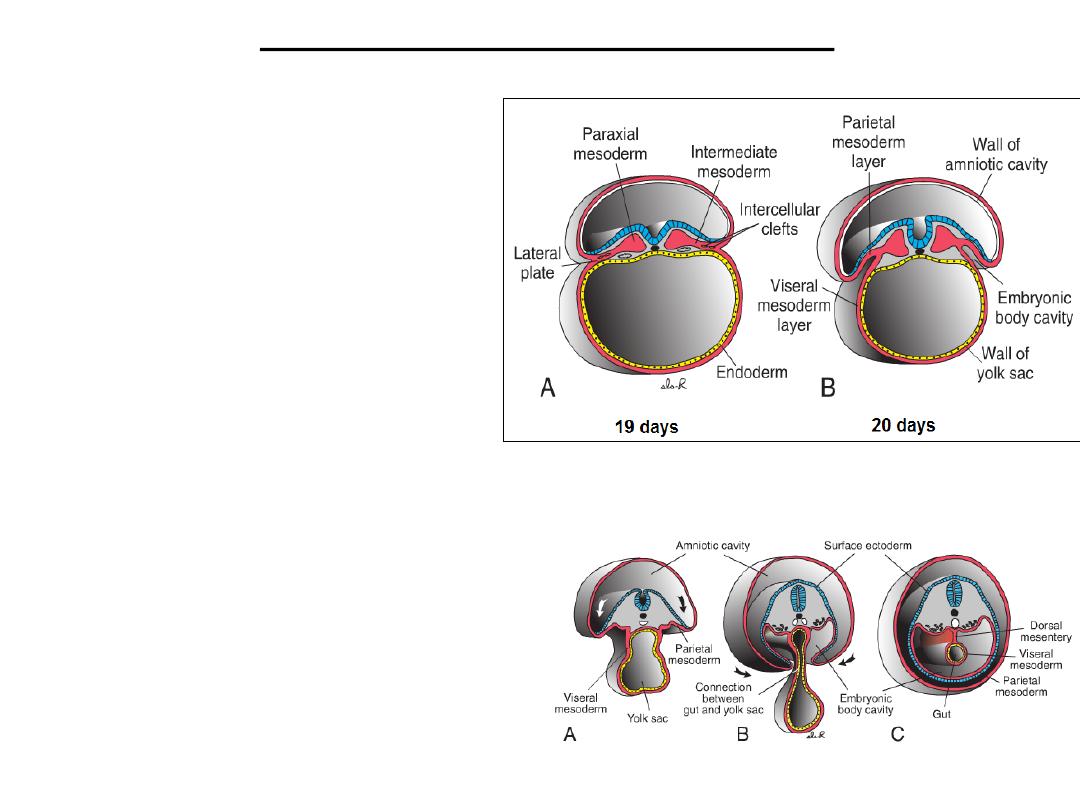
• At the end of the third week,
• Intraembryonic mesoderm
differentiates into:
• Paraxial mesoderm , that forms
somitomeres and somites;
• Intermediate mesoderm , that
contributes to the urogenital
system; and
• Lateral plate mesoderm that is
involved in forming the body cavity
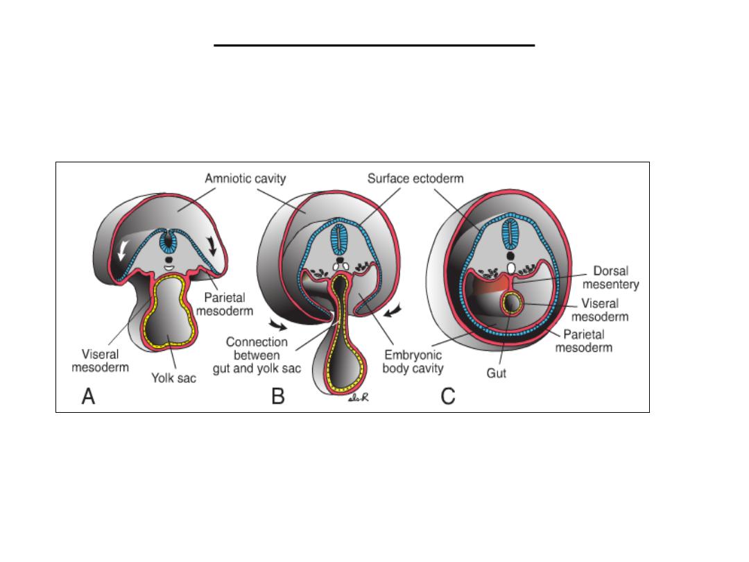
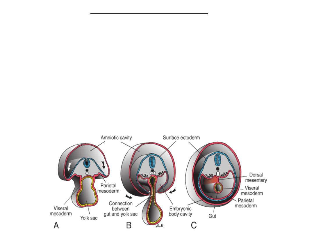
• Lateral plate mesoderm split into two:
• (a) the parietal (somatic) layer
adjacent to the surface ectoderm
• (b) the visceral (splanchnic) layer
adjacent to endoderm forming the gut
tube
• The space created between the two
layers of lateral plate mesoderm
constitutes the primitive body cavity .
Formation of body wall
• During the fourth week, the sides of the embryo begin to grow ventrally forming
two lateral body wall folds (lateral folding).
• These folds consist of the parietal layer of lateral plate mesoderm, overlying
ectoderm, and cells from adjacent somites that migrate into this mesoderm across
the lateral somitic frontier.
• As these folds progress, the endoderm layer also folds ventrally and closes to
form the gut tube.
• By the end of the fourth week, the lateral body wall folds meet in the midline
and fuse to close the ventral body wall.
Formation of body wall
• This closure is aided by head and tail folds that cause the embryo to curve into
the fetal position (cephalocaudal folding).
Lateral folding
Cephalocaudal folding
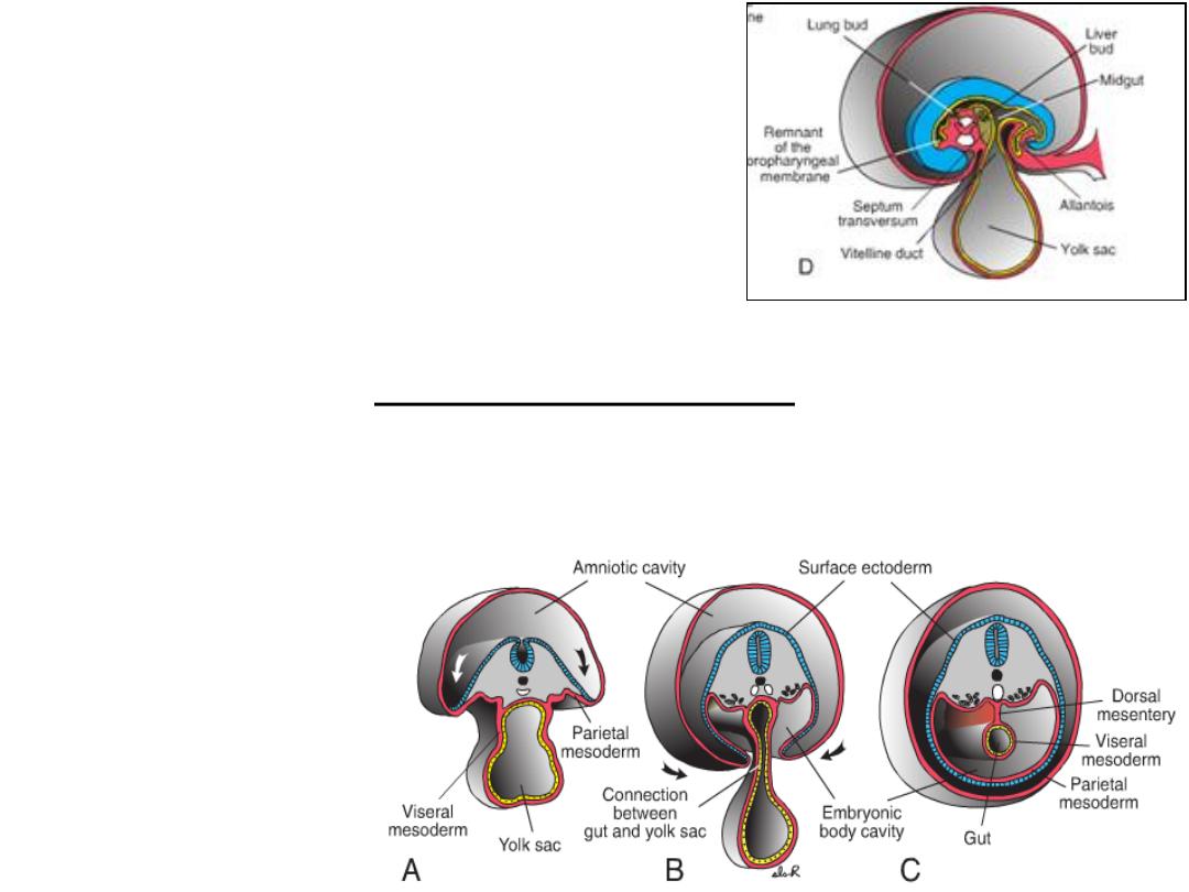
• Closure of the ventral body wall is complete
except in the region of the connecting stalk.
• Similarly, closure of the gut tube is complete
except for a connection from the midgut region
to the yolk sac that forms the vitelline (yolk
sac) duct. This duct is incorporated into the
umbilical cord, becomes very narrow, and
degenerates later.
Serous membranes
• Parietal layer of lateral plate mesoderm forms the parietal layer of the serous
membranes lining the outside of the peritoneal, pleural , and pericardial cavities .
• visceral layer of
lateral plate
mesoderm forms the
visceral layer of the
serous membranes
covering the
abdominal organs,
lungs, and heart.
Dorsal & ventral mesentry
Visceral and parietal layers are
continuous with each other as
the dorsal mesentery, which
suspends the gut tube from the
posterior body wall into the
peritoneal cavity. Dorsal
mesentery extends
continuously from the caudal
limit of the foregut to the end
of the hindgut.
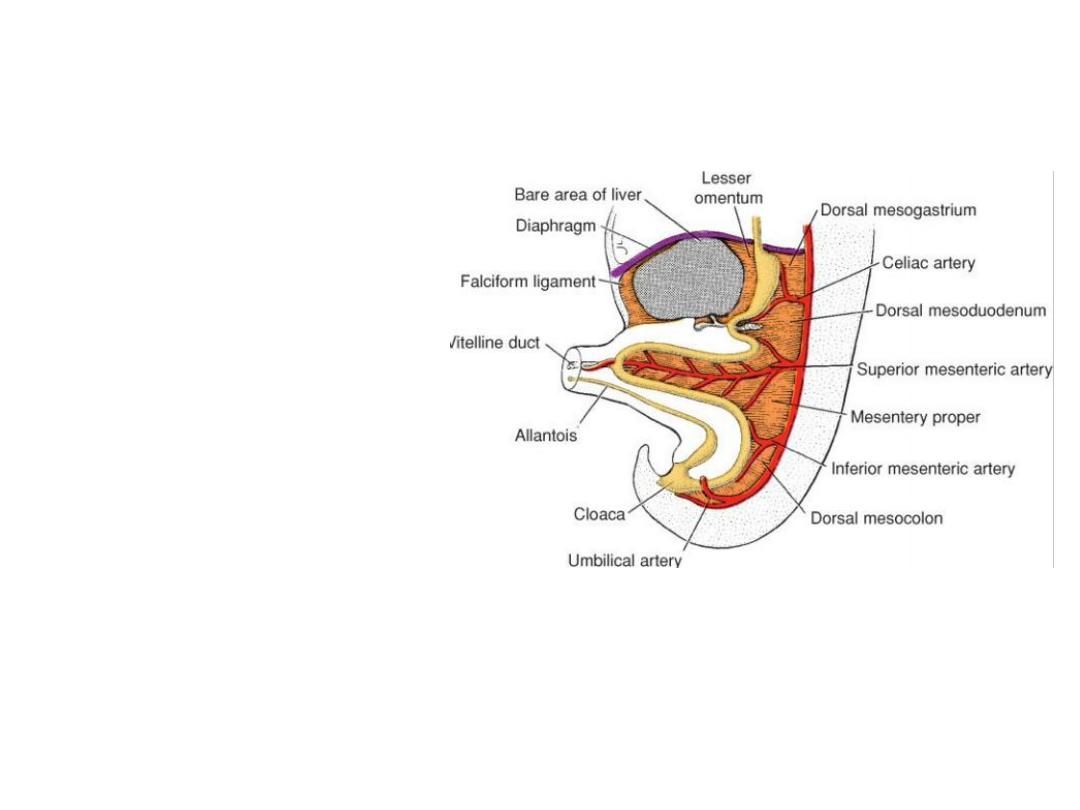
Ventral mesentery: exists only
from the caudal foregut to the
upper portion of the duodenum
and results from thinning of
mesoderm of the septum
transversum.
These mesenteries are double
layers of peritoneum that
provide a pathway for blood
vessels, nerves, and lymphatics
to the organs.
Clinical Correlates
Ventral Body Wall Defects :
• Occur in the thorax, abdomen, and pelvis and involve the heart (ectopia cordis) ,
abdominal viscera (gastroschisis), and/or urogenital organs (bladder or cloacal
exstrophy), depending upon the location and size of the abnormality.
• The malformations are due to a failure of the ventral body wall to close.
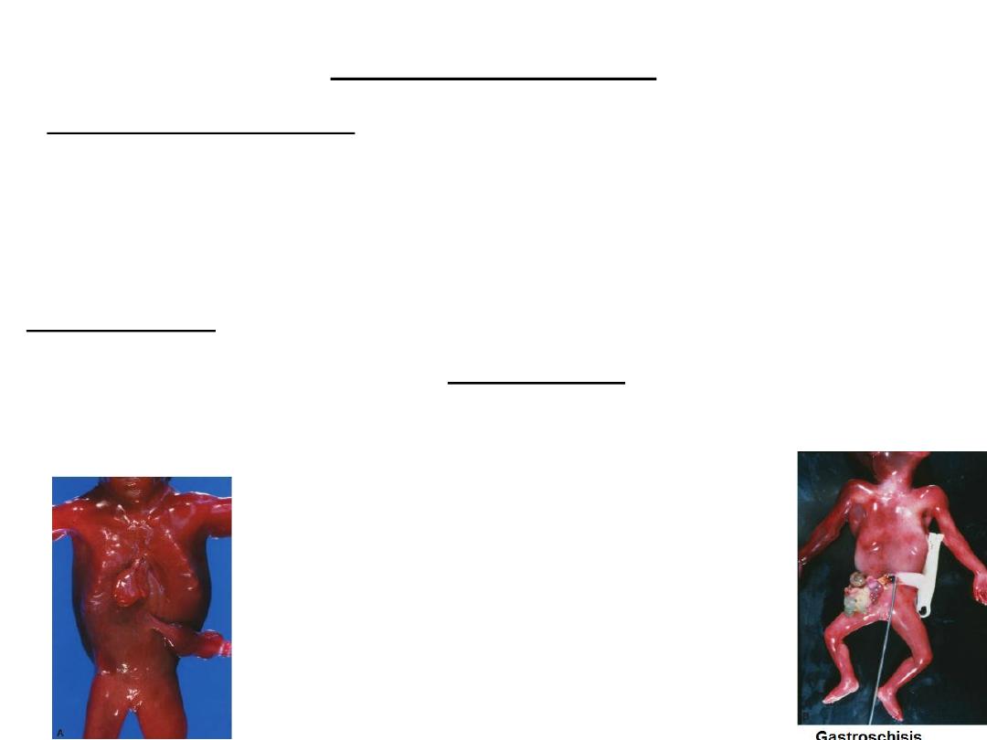
Ectopia cordis
Body wall folds fail to close the
midline in the thoracic region
causing the heart to lie outside
the body cavity.
Gastroschisis
•
Occurs when body wall closure
fails in the abdominal region.
•
As a result, intestinal loops
herniate into the amniotic cavity
through the defect, which usually
lies to the right of the umbilicus.
•
The malformation is not
associated with chromosome
abnormalities.
•
is associated with elevated alpha
fetoprotein (AFP) concentrations.
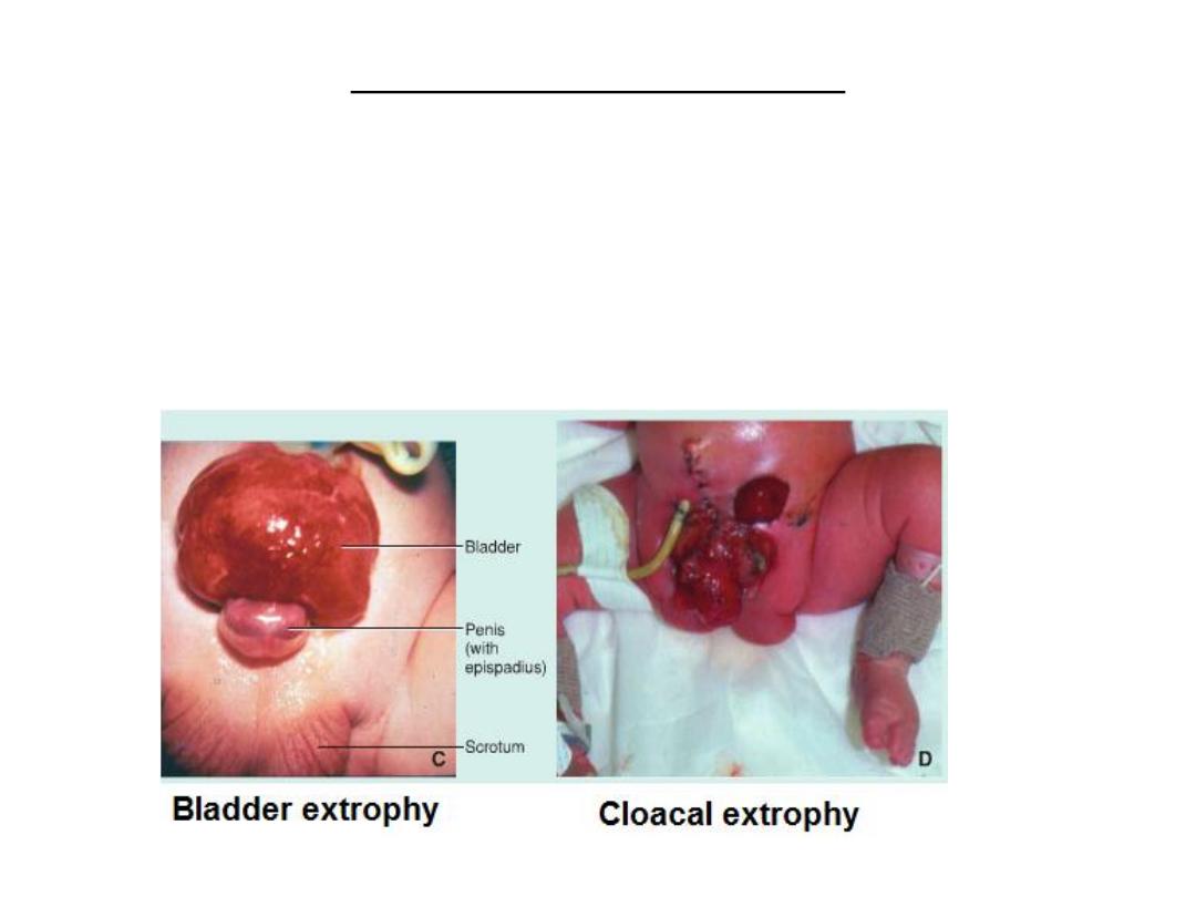
Bladder and cloacal exstrophy
• results from abnormal body wall closure in the pelvic region.
• Bladder exstrophy represents a less severe closure defect in this region
and only the bladder is exposed; in males, the penis may be involved and
epispadius [a split in the dorsum of the penis; is common).
• Cloacal exstrophy results from a more severe failure of body wall closure
in the pelvis such that the bladder and rectum, which are derived from the
cloaca, are exposed.
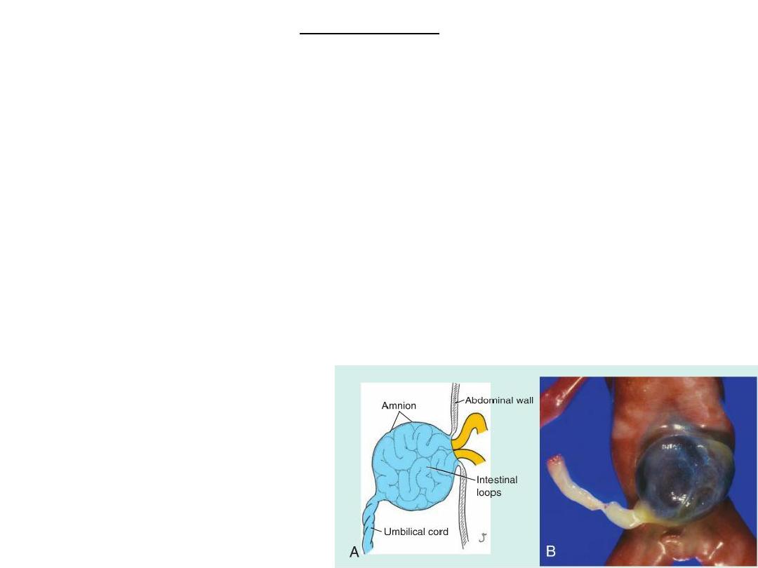
Omphalocele
•
A ventral body wall defect
•
Does not arise from a failure in body wall closure.
•
Originates when portions of the gut tube (the midgut), that normally herniates into the
umbilical cord during the 6th to 10th weeks (physiological umbilical herniation) , fails to
return to the abdominal cavity. Subsequently, loops of bowel, and other viscera, including
the liver may herniate into the defect. Since the umbilical cord is covered by a reflection of
the amnion, the defect is covered by this epithelial layer. (In contrast, loops of bowel in
gastroschisis are not covered by amnion because they herniate through the abdominal wall
directly into the amniotic cavity.)
•
Omphalocele is associated with high mortality rates and severe malformations, including
cardiac abnormalities and neural tube defects.
• Chromosome abnormalities are
present in 15% of cases.
• Omphaloceles is associated with
elevated alpha fetoprotein (afp)
concentrations.
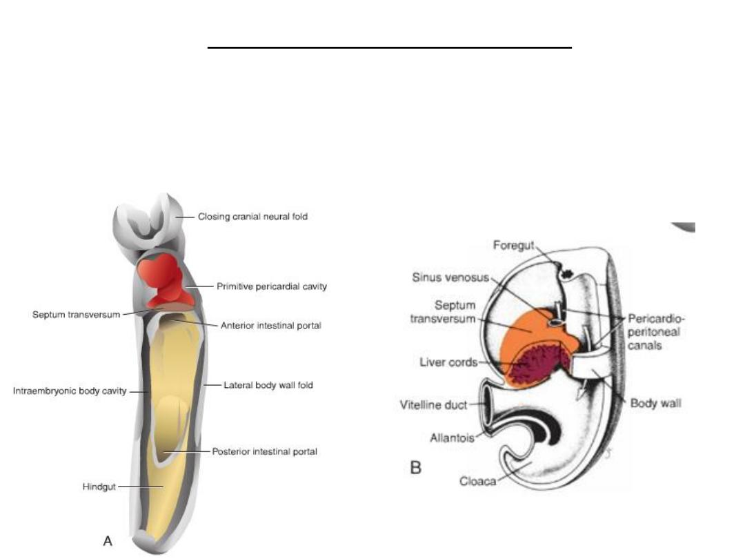
Diaphragm and thoracic cavity
The septum transversum is a thick
plate of mesodermal tissue occupying
the space between t he thoracic cavity
and the stalk of the yolk sac.
This septum does not separate the
thoracic and abdominal cavities
completely but leaves large openings, the
pericardio-peritoneal canals, on each side
of the foregut.

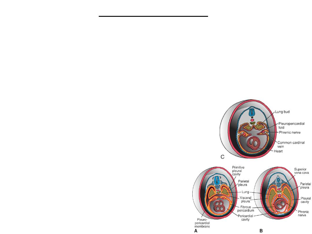
• When lung buds begin to grow, they expand within the pericardio-peritoneal canals.
• As a result of the rapid growth of the lungs, the pericardioperitoneal canals become too
small, & the lungs begin to expand into the mesenchyme of the body wall.
• Ventral and lateral expansion is posterior to the pleuroperi-cardial folds.
• With expansion of the lungs, mesoderm of the body wall splits into 2 components:
(a) the definitive wall of the thorax and
(b) the Pleuro-pericardial membranes, which are extensions of the pleuro-
pericardial folds that contain the common cardinal veins and phrenic nerves .
• Descent of the heart and positional changes of the sinus venosus shift the common
cardinal veins toward the midline, and the pleuropericardial membranes are drawn
out in mesentery-like fashion.
• Finally, they fuse with each other and with the root of the lungs, and the thoracic
cavity is divided into the definitive pericardial cavity and two pleural cavities.
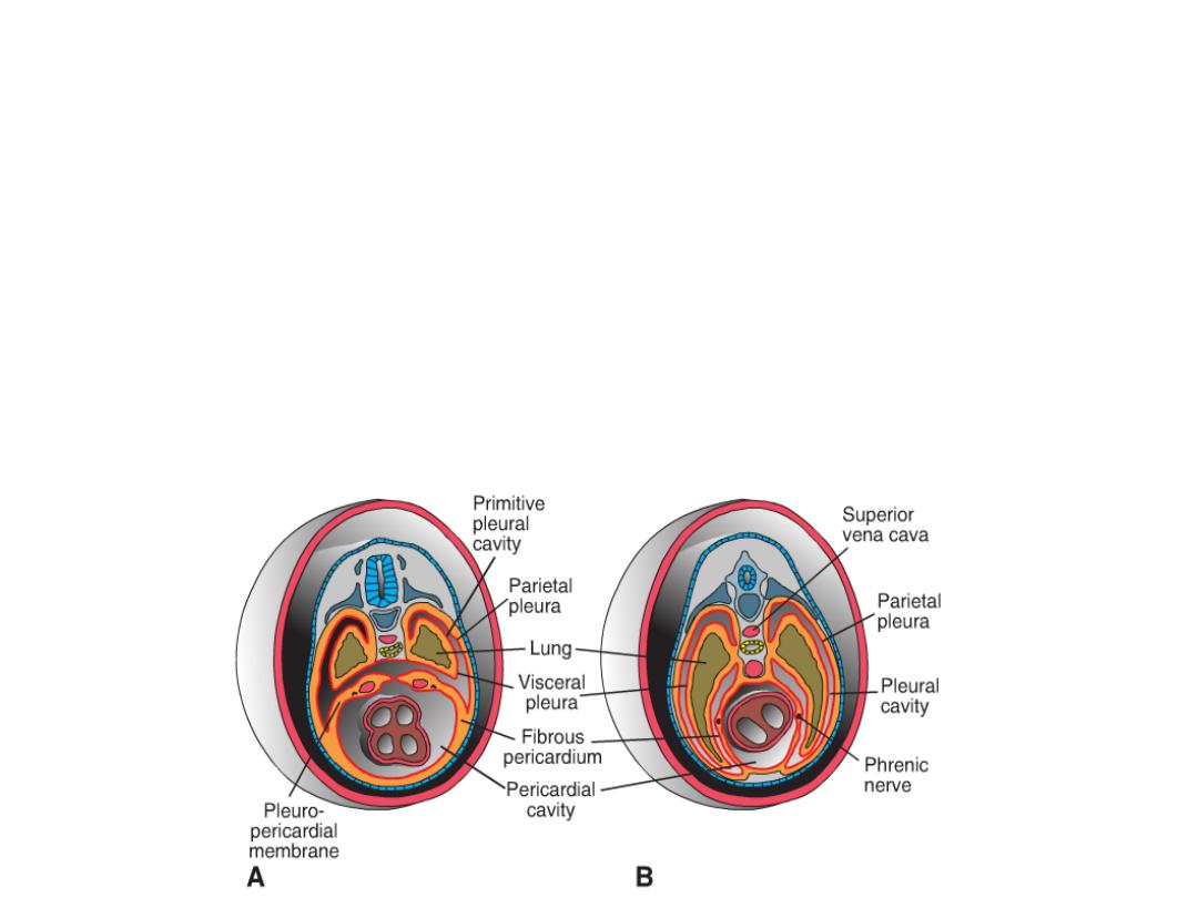
• In the adult, the pleuropericardial membranes form the fibrous pericardium.
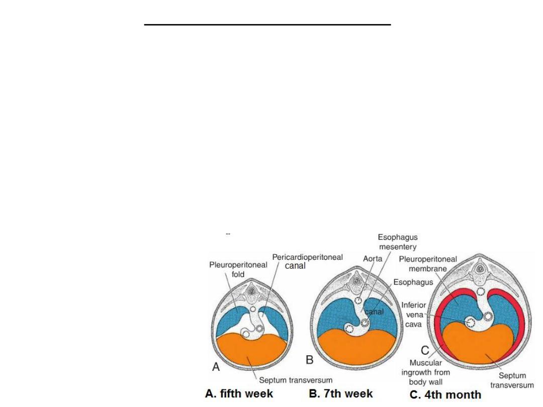
Formation of the diaphragm
• Formation of septum transversum
• During further development, the pericardioperitoneal canals are closed by growth
of the pleuroperitoneal folds which will form the pleuroperitoneal membranes.
• Further expansion of the pleural cavities relative to mesenchyme of the body wall
adds a peripheral rim to the pleuroperitoneal membranes. Once this rim is
established, myoblasts originating from somites at cervical segments three to
five(C 3-5) penetrate the membranes to form the muscular part of the diaphragm.
• Thus, the diaphragm is derived from the following structures:
the septum transversum,
which forms the central
tendon of the diaphragm;
the two pleuroperitoneal
membranes;
muscular components from
somites at cervical
segments three to five; and
the mesentery of the
esophagus, in which the
crura of the diaphragm
develop.
The phrenic nerves
•
During the fourth week, the septum transversum lies opposite cervical somites, and nerve
components of the 3, 4, and 5 cervical segments of the spinal cord grow into the septum.
•
At first, the nerves, known as phrenic nerves , pass into the septum through the
pleuropericardial folds. This explains why further expansion of the lungs and descent of the
septum shift the phrenic nerves that innervate the diaphragm into the fibrous pericardium
.
•
Although the septum transversum lies opposite cervical segments during the fourth week,
by the sixth week, the developing diaphragm is at the level of thoracic somites.
• The repositioning of the diaphragm is caused
by rapid growth of the dorsal part of the
embryo (vertebral column), compared with that
of the ventral part. By the beginning of the third
month, some of the dorsal bands of the
diaphragm originate at the level of the first
lumbar vertebra.
• The phrenic nerves supply the diaphragm with
its motor and sensory innervation .
• Since the most peripheral part of the
diaphragm is derived from mesenchyme of the
thoracic wall, it is generally accepted that some
of the lower intercostal (thoracic) nerves
contribute sensory fibers to the peripheral part of
the diaphragm.
Clinical Correlates
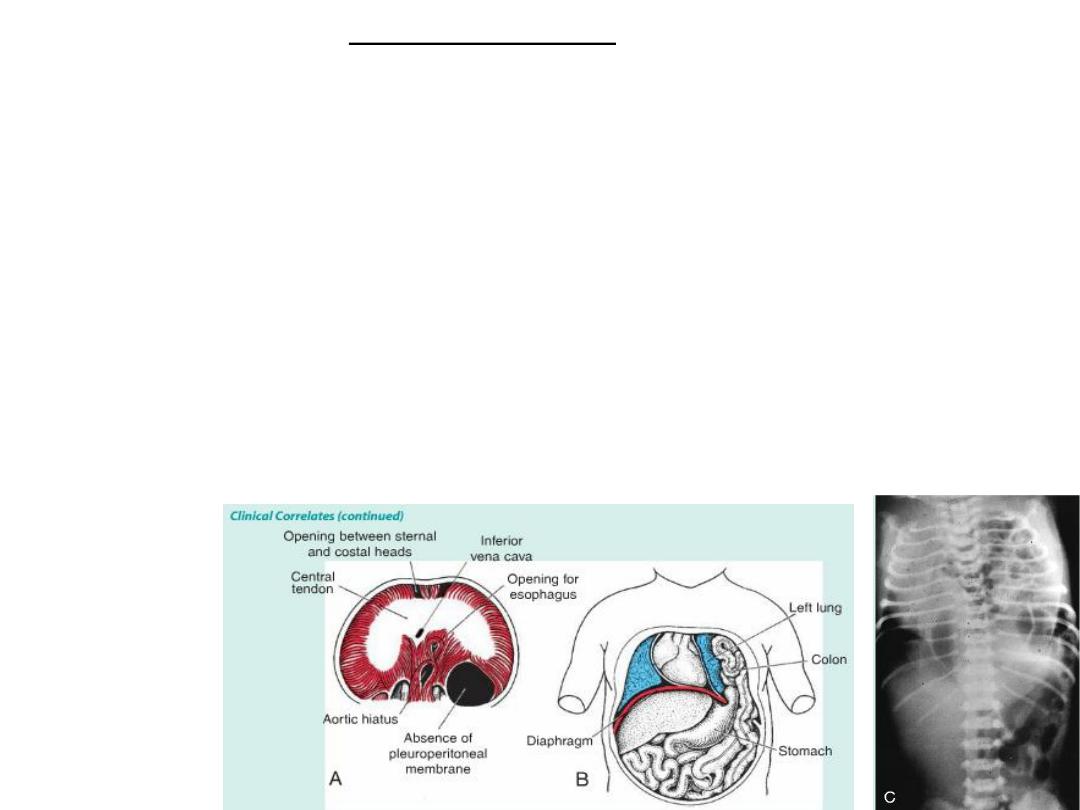
•
Diaphragmatic Hernias
•
A congenital diaphragmatic hernia:
– failure of one or both of the pleuroperitoneal membranes to close the
pericardioperitoneal canals,
– usualy on the left side.
– The abdominal viscera in the chest push the heart anteriorly and compress the lungs,
which are commonly hypoplastic.
– A large defect is associated with a high rate of mortality
•
Parasternal hernia: a small part of the muscular fibers of the diaphragm fails to develop.
•
Esophageal hernia, is due to congenital shortness of the esophagus. Upper portions of the
stomach are retained in the thorax, and the stomach is constricted at the level of the
diaphragm.
Congenital
Diaphragm
atic Hernia