
Chapter 12
Limbs
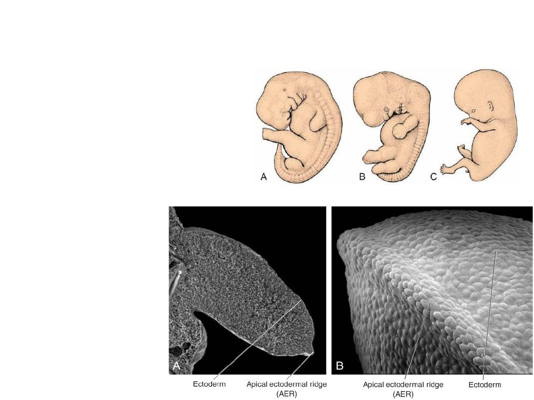
• At the end of the fourth week of
development, limb buds become
visible as outpocketings from the
ventrolateral body wall.
• The forelimb appears first followed
by the hind limb 1 to 2 days later.
•
Initially, the limb buds
consist of a
mesenchymal core
derived from the parietal
(somatic) layer of lateral
plate mesoderm that will
form the bones and
connective tissues of the
limb, covered by a layer
of cuboidal ectoderm.
The appendicular skeleton includes:
1.
Limbs
2.
Shoulder girdle
3.
Pelvic girdle
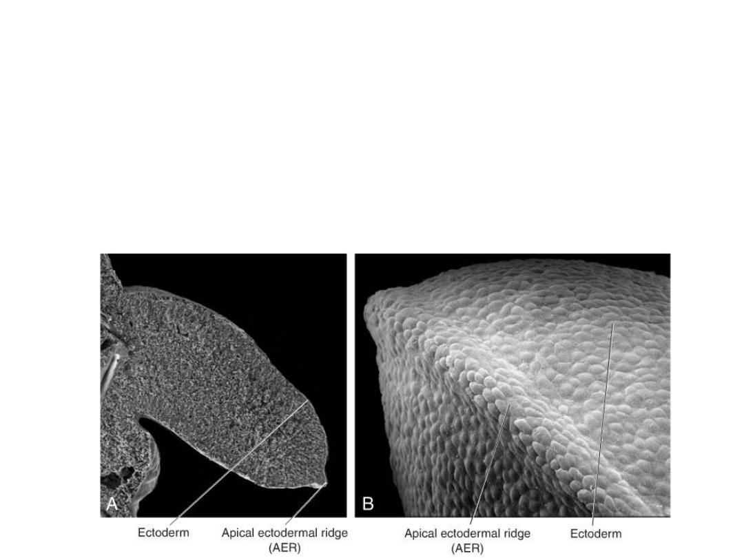
• Ectoderm at the distal border of the limb thickens and forms the apical
ectodermal ridge(AER).
• This ridge exerts an inductive influence on adjacent mesenchyme, causing
it to remain as a population of undifferentiated, rapidly proliferating cells,
the progress zone.
• As the limb grows, cells farther from the influence of the AER begin to
differentiate into cartilage and muscle. In this manner, development of the
limb proceeds proximo-distally.
• In 6-week-old embryos, the terminal
portion of the limb buds becomes
flattened to form the hand and foot
plates and is separated from the
proximal segment by a circular
constriction.
• Later, a second constriction divides
the proximal portion into two
segments, and the main parts of the
extremities can be recognized.
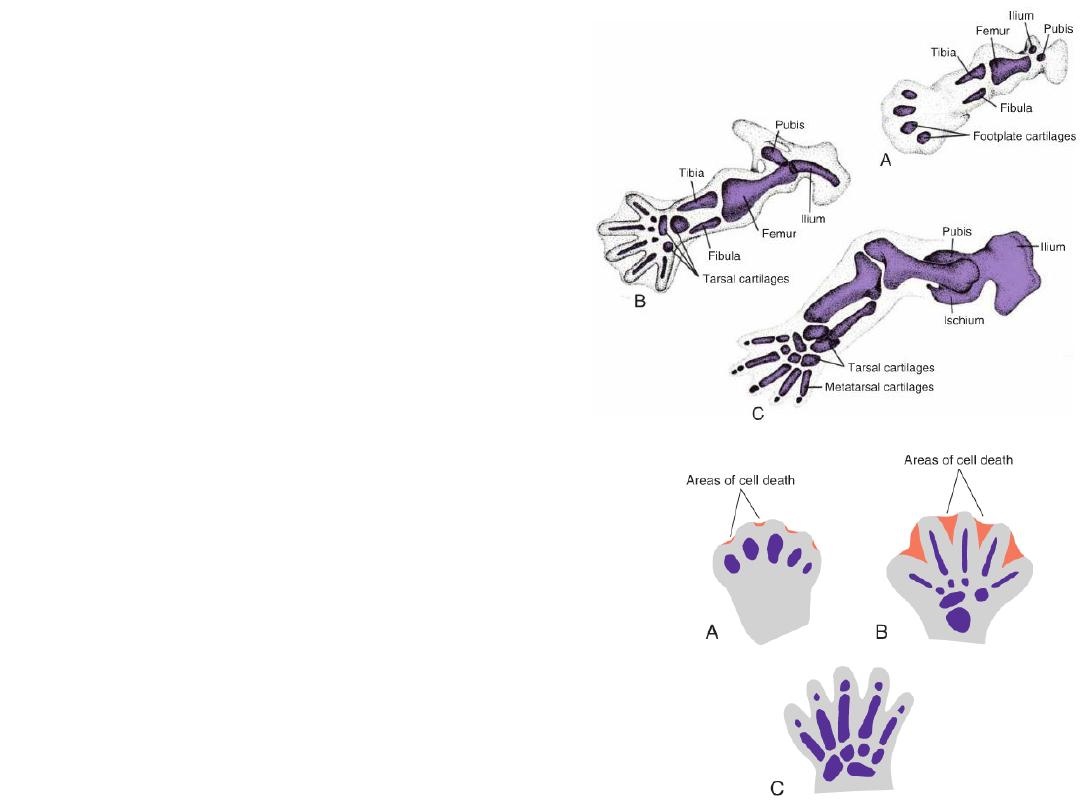
• Fingers and toes are formed when
cell death in the AER separates this
ridge into five parts.
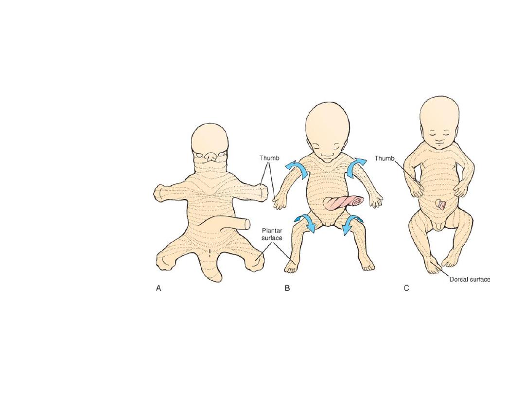
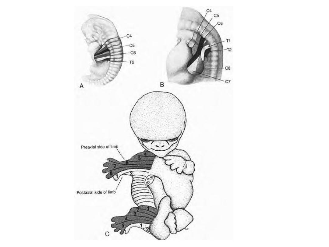
During the seventh week of gestation, the limbs rotate in opposite
directions.
The upper limb rotates 90
°laterally, so that the extensor muscles lie on the
lateral and posterior surface, and the thumbs lie laterally, whereas the lower
limb rotates approximately 90
°medially, placing the extensor muscles on the
anterior surface and the big toe medially.
• While the external shape is being established, mesenchyme in the buds begins to
condense, and these cells differentiate into chondrocytes.
• By the sixth week of development, the first hyaline cartilage models,
foreshadowing the bones of the extremities, are formed by these chondrocytes .
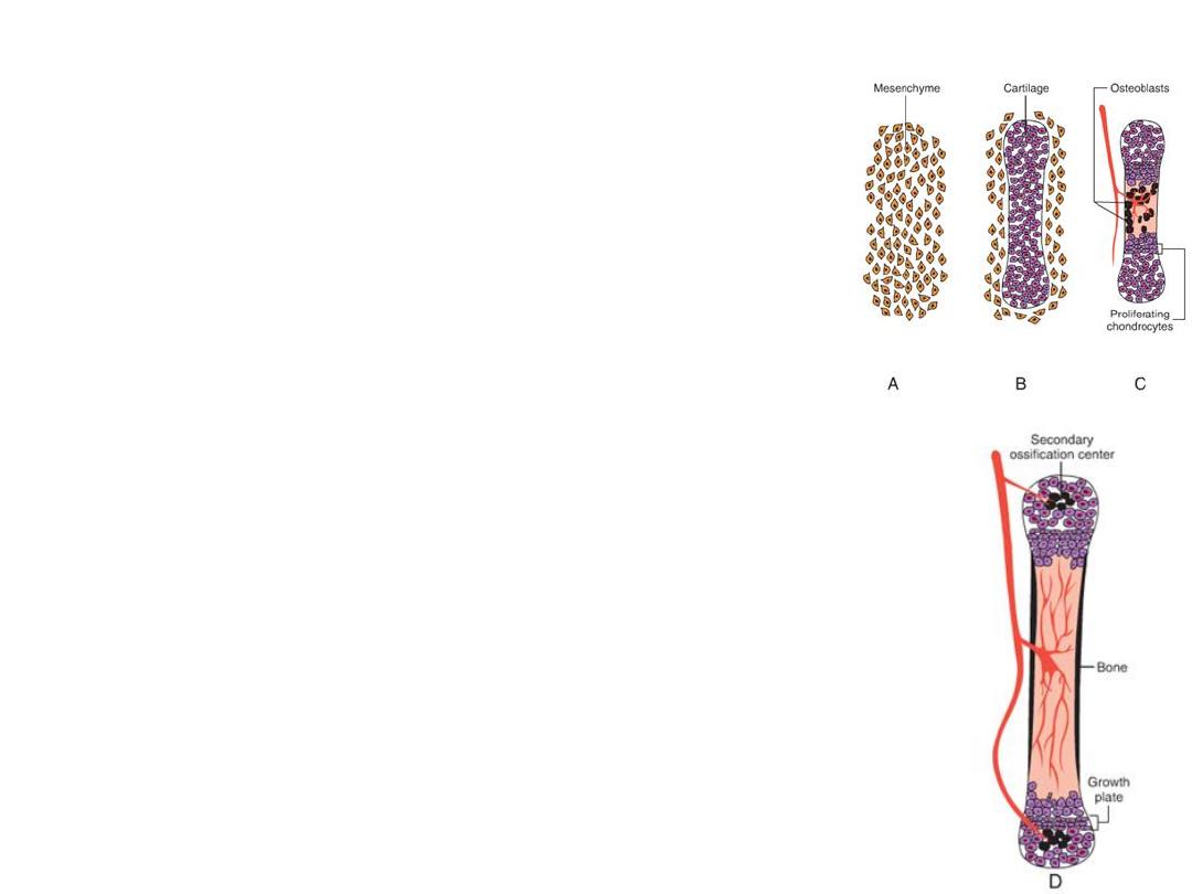
Ossification: Primary ossification centers
Ossification of the bones of the extremities, endochondral
ossification, begins by the end of the embryonic period.
Primary ossification centers are present in all long bones of
the limbs by the 12th week of development.
From the primary center in the shaft or diaphysis of the bone,
endochondral ossification gradually progresses toward the
ends of the cartilaginous model.
Ossification: secondary
ossification centers
At birth, the diaphysis of the bone is usually completely ossified,
but the two ends, the epiphyses, are still cartilaginous.
Shortly thereafter ossification centers arise in the epiphyses
(secondary ossification centers).
Temporarily, a cartilage plate remains between the diaphyseal and
epiphyseal ossification centers. This plate, the epiphyseal plate,
plays an important role in growth in the length of the bones.
Endochondral ossification proceeds on both sides of the plate.
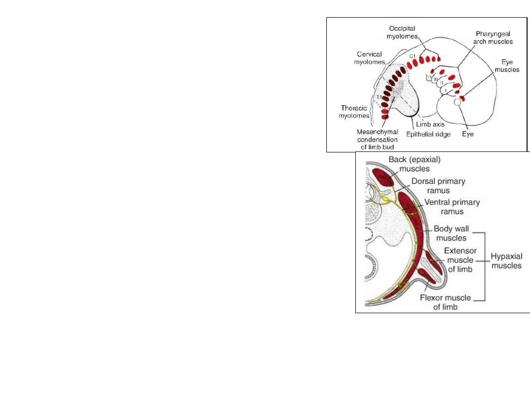
LIMB MUSCULATURE
• Limb musculature is derived from muscle
precursor cells of the somites that migrate
into the limb to form muscles.
• Initially these muscle components are
segmented according to the somite from
which they are derived.
•With elongation of the limb buds, the
muscle tissue splits into flexor and
extensor components
•Additional splitting and fusion occur such
that a single muscle may be formed from
more than one original segment.
•The resultant complex pattern of muscles
is determined by connective tissue derived
from lateral plate mesoderm.
• The upper limb buds lie opposite the lower five cervical and upper two thoracic
segments.
The lower limb buds lie opposite the lower four lumbar and upper two sacral
segments.
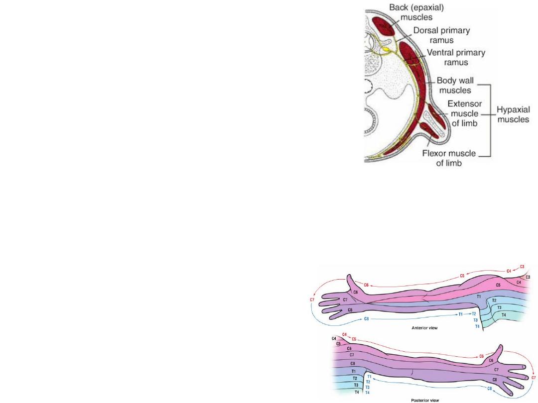
As soon as the buds form, ventral primary rami from the
appropriate spinal nerves and penetrate into the
mesenchyme.
At first, each ventral ramus enters with isolated dorsal
and ventral branches, but soon these branches unite to
form large dorsal and ventral nerves.
Thus, the radial nerve, which supplies the extensor
musculature, is formed by a combination of the dorsal
segmental branches, whereas the ulnar and median
nerves, which supply the flexor musculature, are formed
by a combination of the ventral branches.
Immediately after the nerves have entered the limb buds, they establish an
intimate contact with the differentiating mesodermal
condensations, and the
early contact between the nerve and muscle cells is a prerequisite
for their
complete functional differentiation.
Sensory innervation for dermatomes
Spinal nerves provide sensory innervation for
the dermatomes. Although the original
dermatomal pattern changes with growth of the
extremities, an orderly sequence can still be
recognized in the adult.
Bone Age
• Radiologists use the appearance of various ossification centers to
determine whether a child has reached his or her proper maturation age.
Useful information about bone age is obtained from ossification studies in
the hands and wrists of children.
• Prenatal analysis of fetal bones by ultrasonography provides information
about fetal growth and gestational age.
Limb Defects
• Abnormalities of the limbs vary greatly, and they
may be represented by partial (meromelia) or
complete absence (amelia) of one or more of the
extremities.
• Sometimes the long bones are absent, and
rudimentary hands and feet are attached to the
trunk by small, irregularly shaped bones
(phocomelia, a form of meromelia)
• Sometimes all segments of the extrimities are
present but abnormally short: micromelia
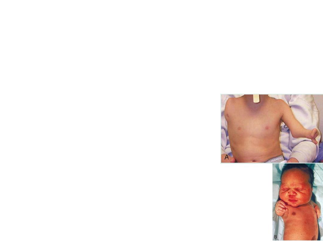
Unilateral amelia
phocomelia
• Thalidomide: a drug widely used as a sleeping pill and antinauseant. It was
subsequently established that thalidomide causes a characteristic syndrome of
malformations consisting of absence or gross deformities of the long bones,
intestinal atresia, and cardiac anomalies.
• Because the drug is now being used to treat AIDS and cancer patients, there is
concern that its return will result in a new wave of limb defects.
• Studies indicate that the most sensitive period for teratogen-induced limb
malformations is the fourth and fifth weeks of development.
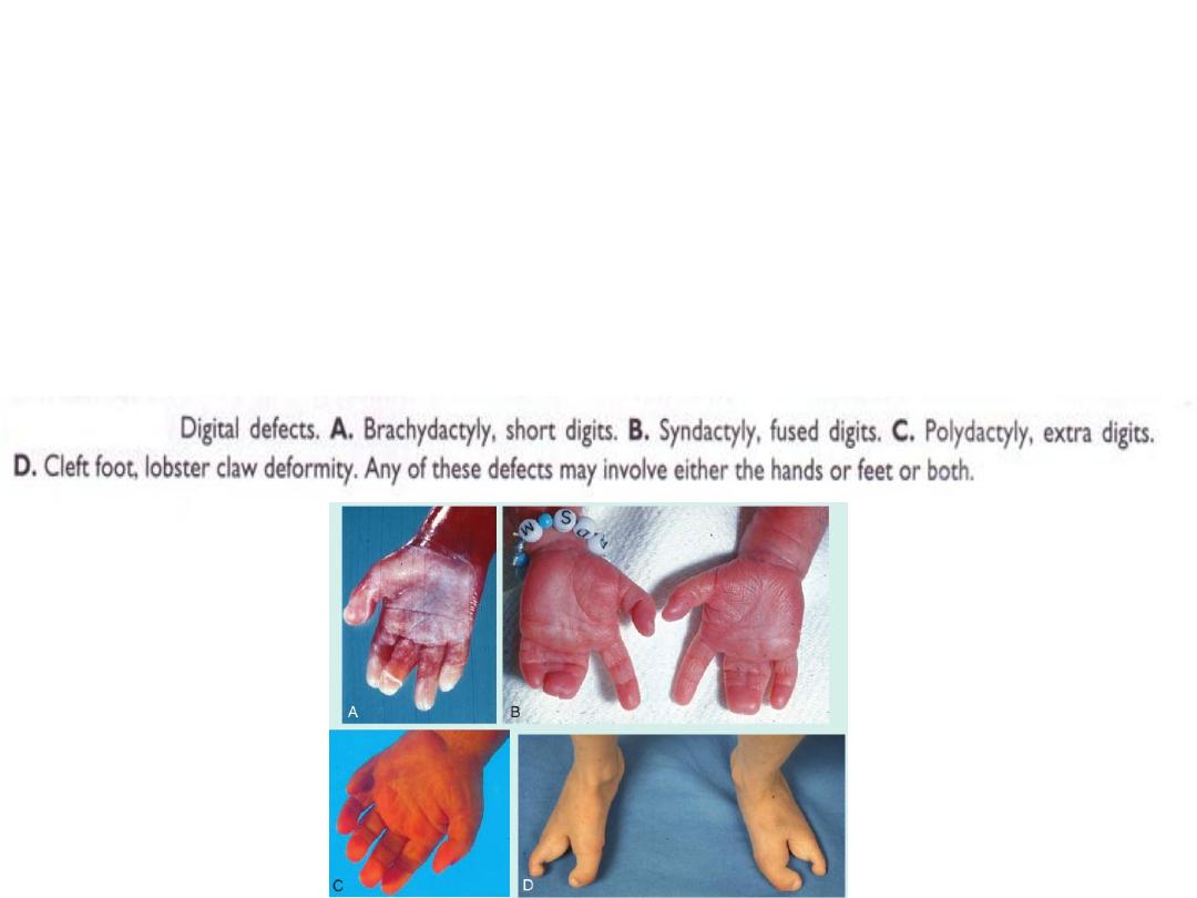
Defects involving the digits
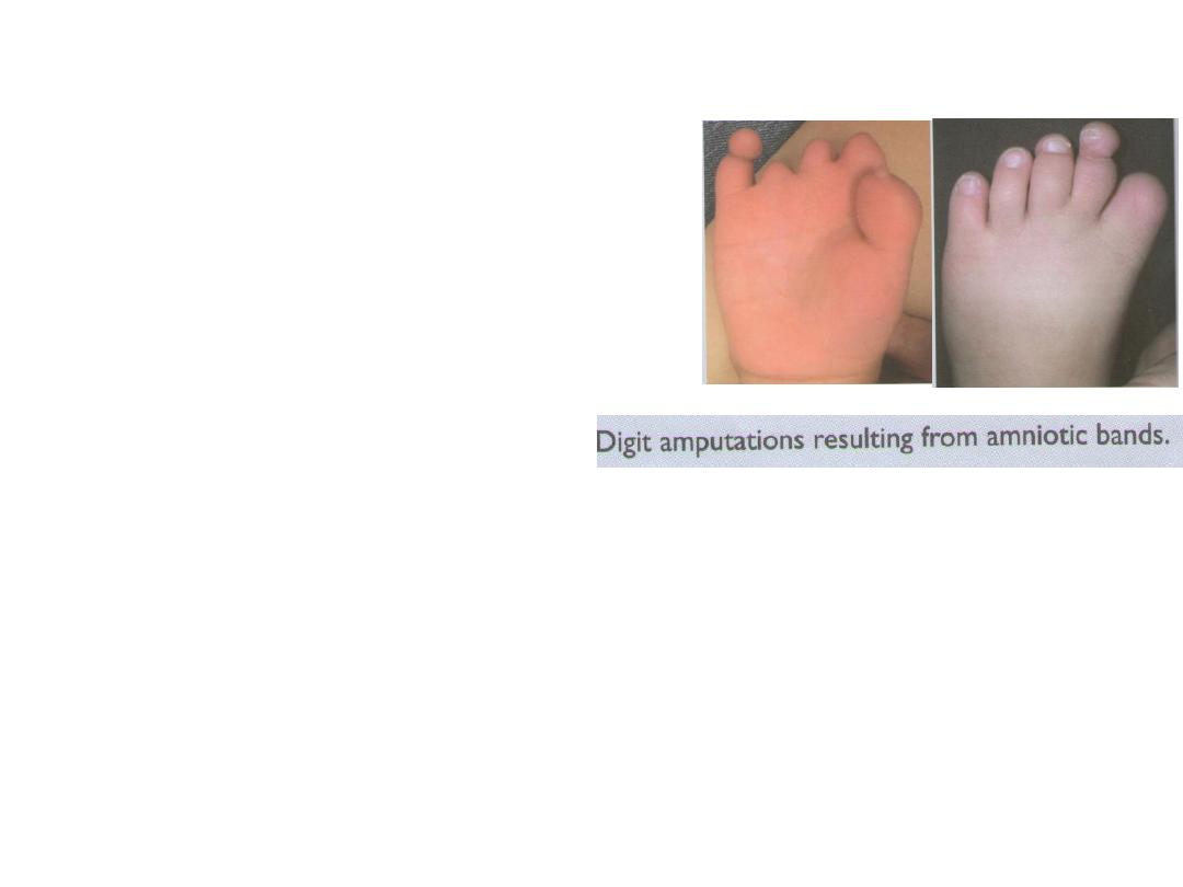
AMNIOTIC BANDS
• Amniotic bands may cause ring constrictions
and amputations of the limbs or digits.
• The origin of the bands is not clear, but they
may represent adhesions between the amnion
and affected structures in the fetus.
• Other investigators believe that bands
originate from tears in the amnion that detach
and surround part of the fetus.
Congenital hip dislocation
• Consists of under development of the acetabulum and head of the femur.
• It is rather common and occurs mostly in female newborns.
• Although dislocation usually occurs after birth, the abnormality of the
bones develops prenatally. Because many babies with congenital hip
dislocation are breech deliveries, it has been thought that breech posture
may interfere with development of the hip joint. It is frequently associated
with laxity of the joint capsule.