
Basic Anatomy
through the joint and emerges beneath the transverse
The tendon of the long head of the biceps muscle passes
lary nerve, and the posterior circumflex humeral vessels
The long head of the triceps muscle, the axil
Inferiorly:
bursa, coracoacromial ligament, and deltoid muscle
The supraspinatus muscle, subacromial
Superiorly:
The infraspinatus and teres minor muscles
Posteriorly:
367
■
■
■
■
■
■
-
ligament.
intercostobrachial nerves
(T1) and the
the arm
medial cutaneous nerve of
of the arm is supplied by the
(C5 and 6). The skin of the armpit and the medial side
a branch of the radial nerve
cutaneous nerve of the arm,
lower lateral
the arm below the deltoid is supplied by the
lary nerve (C5 and 6). The skin over the lateral surface of
a branch of the axil
lateral cutaneous nerve of the arm,
upper
over the lower half of the deltoid is supplied by the
(C3 and 4). The skin
supraclavicular nerves
is from the
point of the shoulder to halfway down the deltoid muscle
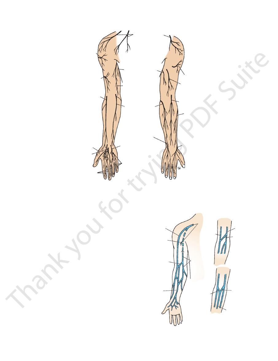
The sensory nerve supply (Fig. 9.38) to the skin over the
Superficial Sensory Nerves
these movements.
shows the direction of pull of the muscles responsible for
summarizes the movements of abduction of the arm and
head is accomplished by rotating the scapula. Figure 9.37
of the acromion. Further elevation of the arm above the
ity of the humerus comes into contact with the lateral edge
At about 120° of abduction of the arm, the greater tuberos
joint and a 1° abduction occurs by rotation of the scapula.
abduction of the arm, a 2° abduction occurs in the shoulder
as well as movement at the shoulder joint. For every 3° of
Abduction of the arm involves rotation of the scapula
clavicular ligament.
of rotation may be considered to pass through the coraco
so that the position of the glenoid fossa is altered, the axis
tone of muscles. When the scapula rotates on the chest wall
cle by the strong coracoclavicular ligament assisted by the
The scapula and upper limb are suspended from the clavi
The Scapular–Humeral Mechanism
-
-
-
The Upper Arm
Skin
-
(T2). The
skin of the back of the arm (Fig. 9.38) is supplied by
the
the radial nerve (C8).
a branch of
posterior cutaneous nerve of the arm,
Stability of the Shoulder Joint
example, diseases of the spinal cord and vertebral column and
Injury to the shoulder joint is followed by pain, limitation of
nerve. The joint is sensitive to pain, pressure, excessive traction,
displacement of the humerus can also stretch and damage the
of skin sensation over the lower half of the deltoid. Downward
nerve, as indicated by paralysis of the deltoid muscle and loss
into the quadrangular space can cause damage to the axillary
muscle. A subglenoid displacement of the head of the humerus
the humerus is no longer bulging laterally beneath the deltoid
shoulder is seen to be lost because the greater tuberosity of
with shoulder dislocation, the rounded appearance of the
violence to the front of the joint. On inspection of the patient
Posterior dislocations are rare and are usually caused by direct
tendons of these muscles are fused to the underlying capsule of
of the short muscles that bind the upper end of the humerus to
ble structure. Its strength almost entirely depends on the tone
The shallowness of the glenoid fossa of the scapula and the lack
of support provided by weak ligaments make this joint an unsta-
the scapula—namely, the subscapularis in front, the supraspi-
natus above, and the infraspinatus and teres minor behind. The
the shoulder joint. Together, these tendons form the rotator cuff.
The least supported part of the joint lies in the inferior loca-
tion, where it is unprotected by muscles.
Dislocations of the Shoulder Joint
The shoulder joint is the most commonly dislocated large joint.
Anterior Inferior Dislocation
Sudden violence applied to the humerus with the joint fully
abducted tilts the humeral head downward onto the inferior
weak part of the capsule, which tears, and the humeral head
comes to lie inferior to the glenoid fossa. During this move-
ment, the acromion has acted as a fulcrum. The strong flexors
and adductors of the shoulder joint now usually pull the humeral
head forward and upward into the subcoracoid position.
Posterior Dislocations
radial nerve.
Shoulder Pain
The synovial membrane, capsule, and ligaments of the shoulder
joint are innervated by the axillary nerve and the suprascapular
and distention. The muscles surrounding the joint undergo reflex
spasm in response to pain originating in the joint, which in turn
serves to immobilize the joint and thus reduce the pain.
movement, and muscle atrophy owing to disuse. It is important
to appreciate that pain in the shoulder region can be caused by
disease elsewhere and that the shoulder joint may be normal; for
the pressure of a cervical rib (see page XXX) can cause shoul-
der pain. Irritation of the diaphragmatic pleura or peritoneum
can produce referred pain via the phrenic and supraclavicular
nerves.
C L I N I C A L N O T E S
Basic Anatomy
369
supraclavicular
nerves
intercostobrachial
nerve
medial cutaneous
nerve of arm
medial cutaneous
nerve of forearm
posterior cutaneous branch
of ulnar nerve
palmar cutaneous branch
of ulnar nerve
ulnar nerve
anterior surface
median nerve
palmar cutaneous
branch of
median nerve
superficial branch
of radial nerve
lateral
cutaneous nerve
of forearm
lower lateral
cutaneous nerve
of arm
upper lateral
cutaneous nerve
of arm
posterior surface
upper lateral cutaneous
nerve of arm
posterior cutaneous
nerve of arm
posterior cutaneous
nerve of forearm
superficial branch of
radial nerve
lateral cutaneous
nerve of forearm
FIGURE 9.38
Cutaneous innervation of the upper limb.
Superficial Lymph Vessels
similar to those of the arteries.
that provide vasomotor tone. The origin of these fibers is
is innervated by sympathetic postganglionic nerve fibers
Like the arteries, the smooth muscle in the wall of the veins
Nerve Supply of the Veins
form the axillary vein.
major joins the venae comitantes of the brachial artery to
pierces the deep fascia and at the lower border of the teres
medial side of the biceps (Fig. 9.39). Halfway up the arm, it
ascends in the superficial fascia on the
basilic vein
The
lar fossa, drains into the axillary vein.
lateral side of the biceps and, on reaching the infraclavicu
ascends in the superficial fascia on the
cephalic vein
The
superficial fascia.
The superficial veins of the arm (Fig. 9.39) lie in the
in pairs, and the axillary vein.
comitantes, which accompany all the large arteries, usually
superficial and deep. The deep veins comprise the venae
The veins of the upper limb can be divided into two groups:
Superficial Veins
-
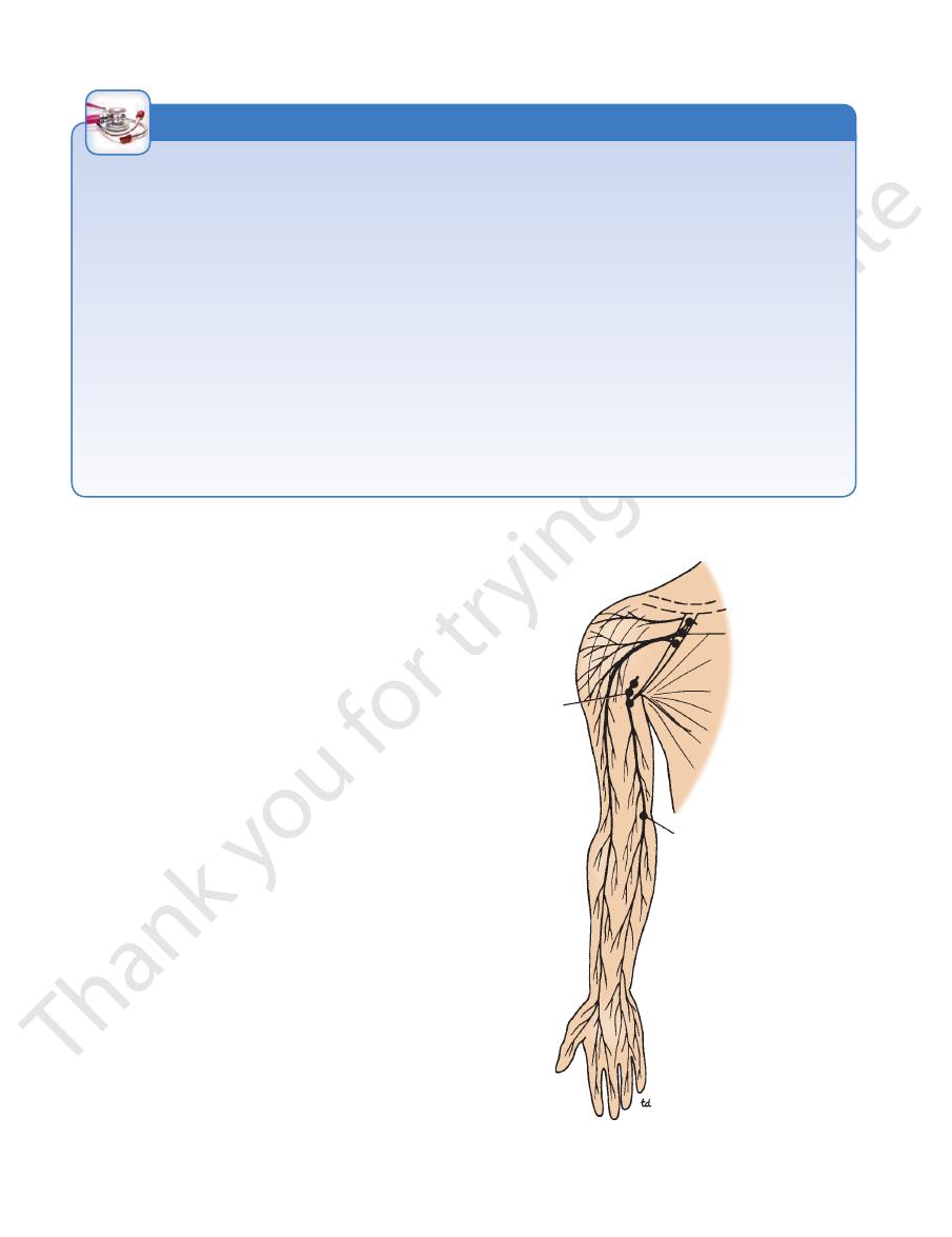
The superficial lymph vessels draining the superficial tissues
of the upper arm pass upward to the axilla (Fig. 9.40).
cephalic vein
venae
comitantes
of brachial
artery
median
cubital
vein
anterior
median vein
of forearm
axillary
vein
basilic vein
cephalic vein
median
cephalic
vein
basilic vein
median
cubital
vein
median basilic
vein
anterior median
vein of forearm
FIGURE 9.39
Superficial veins of the upper limb. Note the
common variations seen in the region of the elbow.
370
CHAPTER 9
weak flexor of the shoulder joint.
The biceps also is a powerful flexor of the elbow joint and a
the cork or driving the screw into wood with a screwdriver.
this action is made use of in twisting the corkscrew into
Note that the biceps brachii is a powerful supinator, and
in Figures 9.43 and 9.44 and are described in Table 9.5.
The muscles of the anterior fascial compartment are shown
Muscles of the Anterior Fascial Compartment
part of the compartment.
and basilic vein. The radial nerve is present in the lower
locutaneous, median, and ulnar nerves; brachial artery
Muscu
Structures passing through the compartment:
Musculocutaneous nerve
Nerve supply to the muscles:
Brachial artery (Fig. 9.42)
Blood supply:
Biceps brachii, coracobrachialis, and brachialis
Muscles:
of the Upper Arm
Contents of the Anterior Fascial Compartment
ment, each having its muscles, nerves, and arteries.
divided into an anterior and a posterior fascial compart
the humerus, respectively. By this means, the upper arm is
attached to the medial and lateral supracondylar ridges of
one on the lateral side, extend from this sheath and are
(Fig. 9.41). Two fascial septa, one on the medial side and
The upper arm is enclosed in a sheath of deep fascia
axillary nodes.
deep structures of the arm drain into the lateral group of
draining the muscles and
deep lymphatic vessels
The
axillary nodes.
medial side follow the basilic vein to the lateral group of
vein to the infraclavicular group of nodes; those from the
Those from the lateral side of the arm follow the cephalic
The Upper Limb
Fascial Compartments of the Upper Arm
-
■
■
■
■
■
■
■
■
-
Venipuncture and Blood Transfusion
termination, the cephalic vein joins the axillary vein at a right angle.
the clavicle and join the external jugular vein. In its usual method of
topectoral triangle. One or more of these branches may ascend over
The cephalic vein does not increase in size as it ascends the arm,
valves in the axillary vein may be troublesome, but abduction of the
diameter and is in direct line with the axillary vein (Fig. 9.39). The
basilic vein reaches the axillary vein, the basilic vein increases in
tral venous catheterization, because from the cubital fossa until the
crosses in front of the clavicle. Fracture of the clavicle can result
municates with the external jugular vein by a small vein that
obtain blood from the arm. When a patient is in a state of shock,
The superficial veins are clinically important and are used for
venipuncture, transfusion, and cardiac catheterization. Every
clinical professional, in an emergency, should know where to
the superficial veins are not always visible. The cephalic vein
lies fairly constantly in the superficial fascia, immediately pos-
terior to the styloid process of the radius. In the cubital fossa,
the median cubital vein is separated from the underlying brachial
artery by the bicipital aponeurosis. This is important because it
protects the artery from the mistaken introduction into its lumen
of irritating drugs that should have been injected into the vein.
The cephalic vein, in the deltopectoral triangle, frequently com-
in rupture of this communicating vein, with the formation of a
large hematoma.
Intravenous Transfusion and Hypovolemic Shock
In extreme hypovolemic shock, excessive venous tone may
inhibit venous blood flow and thus delay the introduction of intra-
venous blood into the vascular system.
Anatomy of Basilic and Cephalic Vein Catheterization
The median basilic or basilic veins are the veins of choice for cen-
shoulder joint may permit the catheter to move past the obstruction.
and it frequently divides into small branches as it lies within the del-
It may be difficult to maneuver the catheter around this angle.
C L I N I C A L N O T E S
lateral group
of axillary
nodes
infraclavicular group
of nodes
supratrochlear lymph node
FIGURE 9.40
Superficial lymphatics of the upper limb. Note
the positions of the lymph nodes.
Basic Anatomy
371
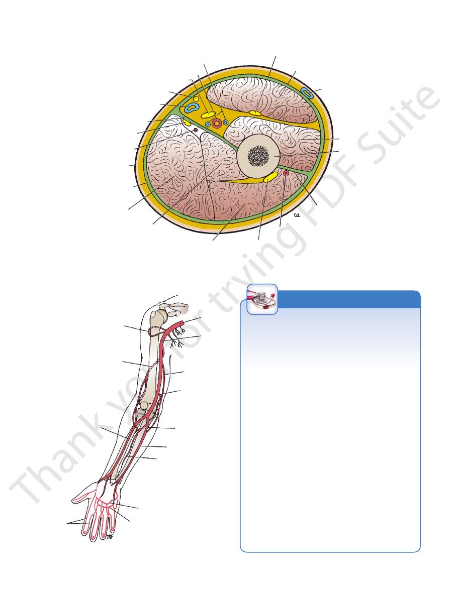
venae comitantes
brachial artery
median nerve
medial cutaneous nerve
of forearm
basilic vein
medial
intermuscular septum
ulnar nerve
superior ulnar
collateral artery
skin
deep fascia
long head of triceps
medial head of triceps
lateral head of triceps
radial nerve
profunda artery
lateral intermuscular septum
humerus
brachialis
cephalic vein
biceps brachii
musculocutaneous nerve
FIGURE 9.41
Cross section of the upper arm just below the level of insertion of the deltoid muscle. Note the division of the
arm by the humerus and the medial and lateral intermuscular septa into anterior and posterior compartments.
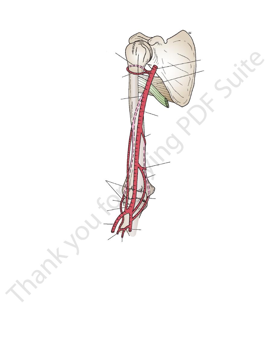
anterior and posterior
cicumflex humeral arteries
profunda artery
radial artery
axillary
artery
brachial
artery
superior ulnar
collateral artery
inferior ulnar
collateral artery
common
interosseous artery
ulnar artery
anterior interosseous
artery
deep palmar arch
superficial palmar arch
digital
arteries
FIGURE 9.42
The main arteries of the upper limb.
Lymphangitis
glenoid tubercle within the shoulder joint. Advanced osteo
lary nodes; those from the middle, ring, and little fingers and
are characteristic of the condition. The lymph vessels from
Infection of the lymph vessels (lymphangitis) of the arm is
common. Red streaks along the course of the lymph vessels
the thumb and index finger and the lateral part of the hand
follow the cephalic vein to the infraclavicular group of axil-
from the medial part of the hand follow the basilic vein to the
supratrochlear node, which lies in the superficial fascia just
above the medial epicondyle of the humerus, and thence to
the lateral group of axillary nodes.
Lymphadenitis
Once the infection reaches the lymph nodes, they become
enlarged and tender, a condition known as lymphadenitis.
Most of the lymph vessels from the fingers and palm pass to
the dorsum of the hand before passing up into the forearm.
This explains the frequency of inflammatory edema, or even
abscess formation, which may occur on the dorsum of the
hand after infection of the fingers or palm.
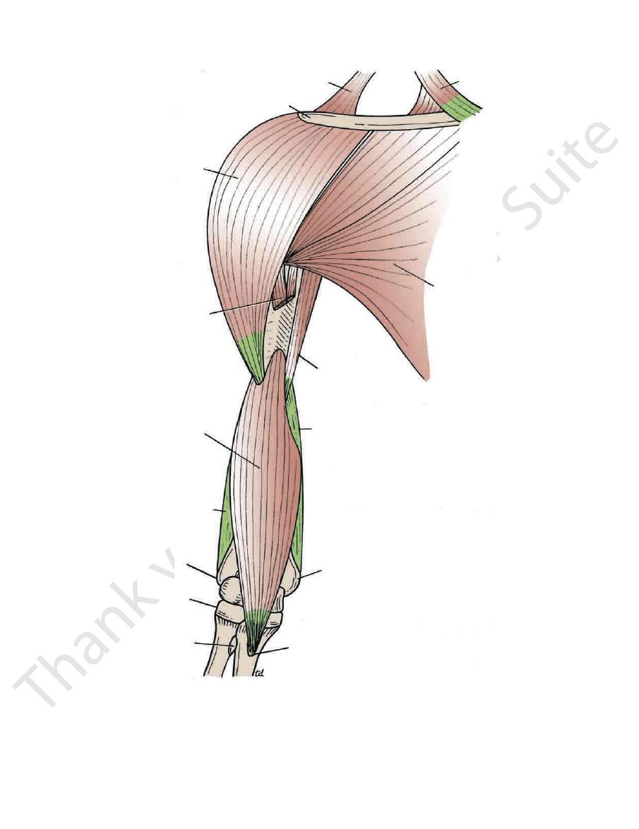
Biceps Brachii and Osteoarthritis of the Shoulder Joint
The tendon of the long head of biceps is attached to the supra-
-
arthritic changes in the joint can lead to erosion and fraying
of the tendon by osteophytic outgrowths, and rupture of the
tendon can occur.
C L I N I C A L N O T E S
372
CHAPTER 9
The Upper Limb
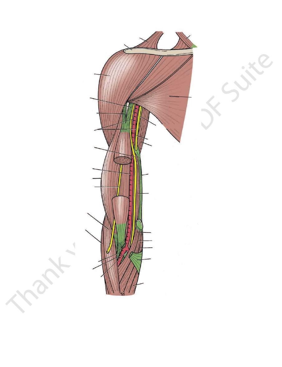
trapezius
clavicle
deltoid
short head of biceps
long head of biceps
coracobrachialis
median nerve
brachial artery
brachialis
musculocutaneous nerve
brachioradialis
extensor carpi radialis longus
biceps tendon
radial artery
ulnar artery
flexor carpi ulnaris
bicipital aponeurosis
palmaris longus
flexor carpi radialis
pronator teres
brachialis
medial intermuscular septum
triceps
ulnar nerve
pectoralis major
sternocleidomastoid
radial
nerve
FIGURE 9.43
hii has been removed to show the muscu
Anterior view of the upper arm. The middle portion of the biceps brac
locutaneous nerve lying in front of the brachialis.
-
Basic Anatomy
373
trapezius
clavicle
sternocleidomastoid
pectoralis major
deltoid
biceps
coracobrachialis
medial intermuscular septum
brachialis
lateral intermuscular
septum
lateral epicondyle
head of radius
bicipital tuberosity
coronoid process of ulna
medial epicondyle
FIGURE 9.44
Anterior view of the upper arm showing the insertion of the deltoid and the origin and insertion of the brachialis.

374
CHAPTER 9
(Fig. 9.38).
of the forearm as the lateral cutaneous nerve of the forearm
fascia just above the elbow. It runs down the lateral aspect
lateral margin of the biceps tendon and pierces the deep
biceps and brachialis muscles (Fig. 9.43). It appears at the
cle (Fig. 9.15), and then passes downward between the
downward and laterally, pierces the coracobrachialis mus
(C5, 6, and 7) in the axilla is described on page 352. It runs
taneous nerve from the lateral cord of the brachial plexus
The origin of the musculocu
Musculocutaneous Nerve
around the elbow joint (Fig. 9.45).
mination of the artery and takes part in the anastomosis
arises near the ter
inferior ulnar collateral artery
The
(Fig. 9.45).
middle of the upper arm and follows the ulnar nerve
arises near the
superior ulnar collateral artery
The
ral groove of the humerus (Fig. 9.45).
brachial artery and follows the radial nerve into the spi
arises near the beginning of the
profunda artery
The
to the humerus
nutrient artery
The
upper arm
to the anterior compartment of the
Muscular branches
Branches
(Fig. 9.43).
lies lateral to the artery in the lower part of its course
lis and biceps muscles above; the tendon of the biceps
The median nerve and the coracobrachia
Laterally:
median nerve lies on its medial side (Fig. 9.43).
upper part of the arm; in the lower part of the arm, the
The ulnar nerve and the basilic vein in the
Medially:
brachialis insertion, and the brachialis (Fig. 9.43).
The artery lies on the triceps, the coraco
Posteriorly:
(Fig. 9.43).
part; and the bicipital aponeurosis crosses its lower part
of the upper part; the median nerve crosses its middle
The medial cutaneous nerve of the forearm lies in front
from the lateral side by the coracobrachialis and biceps.
The vessel is superficial and is overlapped
Anteriorly:
Relations
of the radius by dividing into the radial and ulnar arteries.
supply to the arm (Fig. 9.42). It terminates opposite the neck
tinuation of the axillary artery. It provides the main arterial
begins at the lower border of the teres major muscle as a con
The brachial artery (Figs. 9.42 and 9.43)
Brachial Artery
Compartment
Structures Passing through the Anterior Fascial
The Upper Limb
-
■
■
■
■
-
■
■
■
■
-
■
■
■
■
■
■
-
■
■
■
■
-
-
-
Muscles of the Arm
T A B L E 9 . 5
Triceps
Tuberosity of radius
Muscle
Origin
Insertion
Nerve Supply
Nerve Roots
a
Action
Anterior Compartment
Biceps brachii
Long head
Supraglenoid
tubercle of scapula
and bicipital
aponeurosis into
deep fascia of
forearm
Musculocutaneous
nerve
C5, 6
Supinator of forearm
and flexor of elbow
joint; weak flexor
of shoulder joint
Short head
Coracoid process of
scapula
Coracobrachialis
Coracoid process of
scapula
Medial aspect of shaft
of humerus
Musculocutaneous
nerve
C5, 6, 7
Flexes arm and also
weak adductor
Brachialis
Front of lower half of
humerus
Coronoid process of
ulna
Musculocutaneous
nerve
C5, 6
Flexor of elbow joint
Posterior Compartment
Long head
Infraglenoid tubercle
of scapula
Lateral head
Upper half of
posterior surface
of shaft of humerus
Olecranon process of
ulna
Radial nerve
C6, 7, 8
Extensor of elbow
joint
Medial head
Lower half of
posterior surface
of shaft of humerus
a
The predominant nerve root supply is indicated by boldface type.
Basic Anatomy
Ulnar Nerve
chial artery.
(Fig. 9.22), except for a small vasomotor nerve to the bra
The median nerve has no branches in the upper arm
further course of this nerve is described on page XXX.
at the elbow, it is crossed by the bicipital aponeurosis. The
The nerve, like the artery, is therefore superficial, but
downward on its medial side.
upper arm, it crosses the brachial artery and continues
side of the brachial artery (Fig. 9.43). Halfway down the
is described on page 352. It runs downward on the lateral
medial and lateral cords of the brachial plexus in the axilla
The origin of the median nerve from the
Median Nerve
to the elbow joint
Articular branches
of the forearm down as far as the root of the thumb.
supplies the skin of the front and lateral aspects
forearm
lateral cutaneous nerve of the
Cutaneous branches;
brachialis (Fig. 9.22)
to the biceps, coracobrachialis, and
Muscular branches
Branches
375
■
■
■
■
the
■
■
-
The origin of the ulnar nerve from the
pierces the medial fascial septum, accompanied by the
Here, at the insertion of the coracobrachialis, the nerve
brachial artery as far as the middle of the arm (Fig. 9.43).
on page 353. It runs downward on the medial side of the
medial cord of the brachial plexus in the axilla is described
superior ulnar collateral artery, and enters the
ior
poster
axillary artery
anterior circumflex
posterior circumflex
humeral artery
humeral artery
profunda artery
interosseous recurrent artery
radial recurrent artery
neck of radius
radial artery
anterior interosseous artery
common interosseous artery
ulnar artery
posterior ulnar recurrent artery
anterior ulnar recurrent artery
inferior ulnar collateral artery
superior ulnar collateral artery
brachial artery
teres major
posterior interosseous
artery
FIGURE 9.45
Main arteries of the upper arm. Note the arterial anastomosis around the elbow joint.
376
CHAPTER 9
Radial Nerve
partment of the upper arm (Fig. 9.23).
The ulnar nerve has no branches in the anterior com
medial epicondyle of the humerus.
compartment of the arm; the nerve passes behind the
The Upper Limb
-
On leaving the axilla, the radial nerve immedi
rior cord of the brachial plexus in the axilla is described on
The origin of the radial nerve from the poste
Radial Nerve
Compartment
Structures Passing through the Posterior Fascial
in Table 9.5.
The triceps muscle is seen in Figure 9.46 and is described
Muscle of the Posterior Fascial Compartment
nerve and ulnar nerve
Radial
Structures passing through the compartment:
arteries
Profunda brachii and ulnar collateral
Blood supply:
Radial nerve
Nerve supply to the muscle:
The three heads of the triceps muscle
Muscle:
of the Upper Arm
Contents of the Posterior Fascial Compartment
the anterior compartment just above the lateral epicondyle.
ately enters the posterior compartment of the arm and enters
-
■
■
■
■
■
■
■
■
-
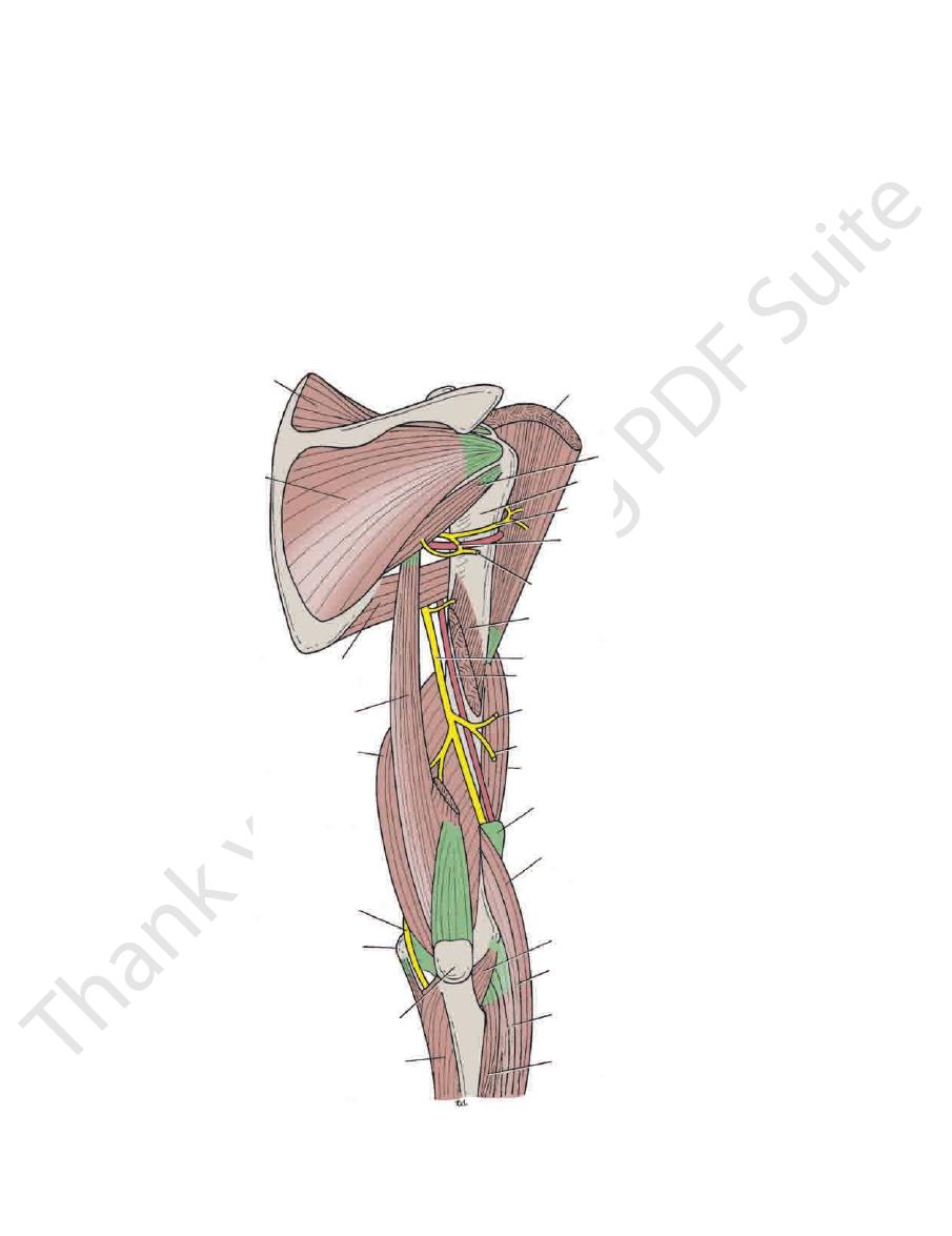
supraspinatus
infraspinatus
teres major
long head of triceps
medial head of triceps
ulnar nerve
medial epicondyle
olecranon process of ulna
flexor carpi ulnaris
extensor carpi radialis longus
extensor carpi radialis brevis
anconeus
brachioradialis
lateral intermuscular septum
brachialis
posterior cutaneous nerve of forearm
lower lateral cutaneous nerve of arm
profunda artery
radial nerve
lateral head of triceps
upper lateral cutaneous nerve of arm
posterior division of
axillary nerve
anterior division of
axillary nerve
surgical neck of humerus
teres minor
deltoid
extensor carpi ulnaris
FIGURE 9.46
Posterior view of the upper arm. The lateral head of the triceps has been divided to display the radial nerve and
the profunda artery in the spiral groove of the humerus.
Basic Anatomy
medial epicondyle of the humerus (Fig. 9.46) on the medial
ulnar collateral vessels. At the elbow, it lies behind the
of the triceps. The nerve is accompanied by the superior
behind the septum, covered posteriorly by the medial head
halfway down the upper arm, the ulnar nerve descends
Having pierced the medial fascial septum
Ulnar Nerve
to the elbow joint.
branches
articular
radialis longus muscles (Fig. 9.47). It also gives
the brachialis, the brachioradialis, and the extensor carpi
has pierced the lateral fascial septum, it gives branches to
after the nerve
anterior compartment of the arm,
In the
of the forearm as far as the wrist.
runs down the middle of the back
nerve of the forearm
posterior cutaneous
of the lower part of the arm. The
supplies the skin over the lateral and anterior aspects
lower lateral cutaneous nerve of the arm
anconeus. The
the lateral and medial heads of the triceps and to the
(Fig. 9.46), branches are given to
spiral groove
In the
is given off.
neous nerve of the arm
posterior cuta
and medial heads of the triceps, and the
branches (Fig. 9.25) are given to the long
axilla,
In the
Branches
directly in contact with the shaft of the humerus (Fig. 9.46).
the nerve is accompanied by the profunda vessels, and it lies
and brachioradialis muscles (Fig. 9.47). In the spiral groove,
cubital fossa in front of the elbow, between the brachialis
septum above the elbow and continues downward into the
heads of the triceps (Fig. 9.46). It pierces the lateral fascial
the spiral groove on the back of the humerus between the
page 353. The nerve winds around the back of the arm in
377
■
■
-
■
■
■
■
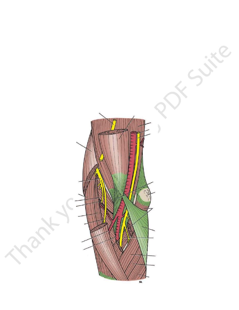
musculocutaneous nerve
brachioradialis
radial nerve
extensor carpi
radialis longus
supinator
deep branch
of radial nerve
extensor carpi
radialis brevis
superficial branch of
radial nerve
radial artery
ulnar artery
flexor carpi ulnaris
palmaris longus
ulnar head of pronator teres
bicipital aponeurosis
humeral head of pronator teres
medial epicondyle
biceps tendon
median nerve
brachial artery
brachialis
biceps brachii
flexor carpi radialis
FIGURE 9.47
Right cubital fossa.
378
CHAPTER 9
The Upper Limb
ligament of the elbow joint. It continues downward to enter
of the fossa is formed by the supinator muscle
floor
The
drawn between the two epicondyles of the humerus.
of the triangle is formed by an imaginary line
The
The pronator teres muscle
Medially:
The brachioradialis muscle
Laterally:
of the elbow (Figs. 9.47 and 9.48).
The cubital fossa is a triangular depression that lies in front
elbow joint.
brachial artery and take part in the anastomosis around the
superior and inferior ulnar collateral arteries arise from the
The
Superior and Inferior Ulnar Collateral Arteries
sis around the elbow joint.
supplies the triceps muscle, and takes part in the anastomo
It accompanies the radial nerve through the spiral groove,
arises from the brachial artery near its origin (Fig. 9.45).
The profunda brachii artery
Profunda Brachii Artery
(Fig. 9.23).
The ulnar nerve has an articular branch to the elbow joint
Branches
carpi ulnaris (see page 390).
the forearm between the two heads of origin of the flexor
-
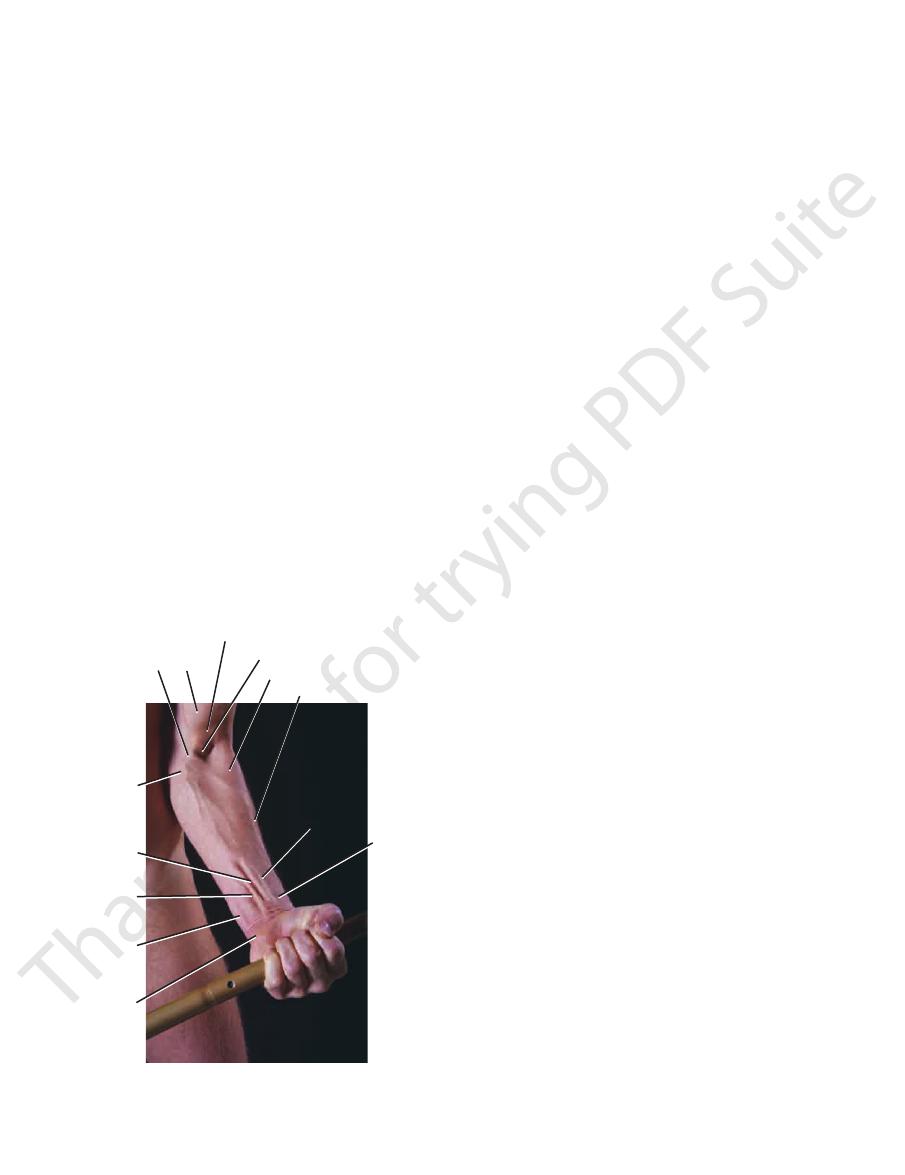
The Cubital Fossa
Boundaries
■
■
■
■
base
bicipital
aponeurosis
basilic vein
palmaris
longus
flexor
digitorum
superficialis
flexor carpi
ulnaris
pisiform
bone
biceps
brachii
biceps brachii tendon
cubital fossa
brachioradialis
cephalic vein
flexor carpi
radialis
site fo
palpati
of radi
artery
FIGURE 9.48
The cubital fossa and anterior surface of the
forearm in a 27-year-old man.
laterally and the brachialis muscle medially. The
joint and with the head of the radius at the proximal
proximal end articulates with the humerus at the elbow
The ulna is the medial bone of the forearm (Fig. 9.49). Its
radius are shown in Figure 9.49.
The important muscles and ligaments attached to the
of the extensor pollicis longus (Fig. 9.49).
which is grooved on its medial side by the tendon
tubercle,
dorsal
terior aspect of the distal end is a small tubercle, the
articulates with the scaphoid and lunate bones. On the pos
the round head of the ulna. The inferior articular surface
which articulates with
ulnar notch,
medial surface is the
projects distally from its lateral margin (Fig. 9.49). On the
styloid process;
At the distal end of the radius is the
its lateral side.
tion of the pronator teres muscle, lies halfway down on
for the inser
pronator tubercle,
and ulna together. The
ment of the interosseous membrane that binds the radius
medially for the attach
interosseous border
has a sharp
of the ulna, is wider below than above (Fig. 9.49). It
The shaft of the radius, in contradistinction to that
for the insertion of the biceps muscle.
bicipital tuberosity
Below the neck is the
neck.
to form the
notch of the ulna. Below the head, the bone is constricted
The circumference of the head articulates with the radial
and articulates with the convex capitulum of the humerus.
(Fig. 9.49). The upper surface of the head is concave
head
At the proximal end of the radius is the small circular
radioulnar joint.
of the hand at the wrist joint and with the ulna at the distal
distal end articulates with the scaphoid and lunate bones
joint and with the ulna at the proximal radioulnar joint. Its
Its proximal end articulates with the humerus at the elbow
The radius is the lateral bone of the forearm (Fig. 9.49).
The forearm contains two bones: the radius and the ulna.
nodes (Fig. 9.40).
pass up to the axilla and enter the lateral axillary group of
the medial side of the forearm. The efferent lymph vessels
fourth, and fifth fingers; the medial part of the hand; and
(Fig. 9.40). It receives afferent lymph vessels from the third,
fascia over the upper part of the fossa, above the trochlea
lies in the superficial
supratrochlear lymph node
The
and the radial nerve and its deep branch.
ulnar and radial arteries, the tendon of the biceps muscle,
median nerve, the bifurcation of the brachial artery into the
tures, enumerated from the medial to the lateral side: the
The cubital fossa (Fig. 9.47) contains the following struc
aponeurosis.
formed by skin and fascia and is reinforced by the bicipital
is
roof
Contents
-
Bones of the Forearm
Radius
-
-
this
-
Ulna