450
CHAPTER 10
The Lower Limb
sciatic nerve
L4 L5 S1 S2 S3
pelvis
sacral plexus
gluteal region
sciatic nerve
hip joint
tibial nerve
common peroneal nerve
back of thigh
semitendinosus
biceps femoris
(long head)
semimembranosus
adductor magnus
(hamstring part)
lower leg
knee joint
gastrocnemius
soleus
plantaris
popliteus
tibialis posterior
flexor digitorum longus
flexor hallucis longus
skin of ankle
ankle joint
sural nerve
sole of foot
medial plantar nerve
adductor hallucis
flexor digitorum brevis
flexor hallucis brevis
first lumbrical
joints
of foot
skin of sole
of foot
lateral plantar nerve
skin of sole
of foot
flexor digitorum accessorius
abductor digiti minimi
flexor digiti minimi brevis
second, third, fourth lumbricals
abductor hallucis
all interossei
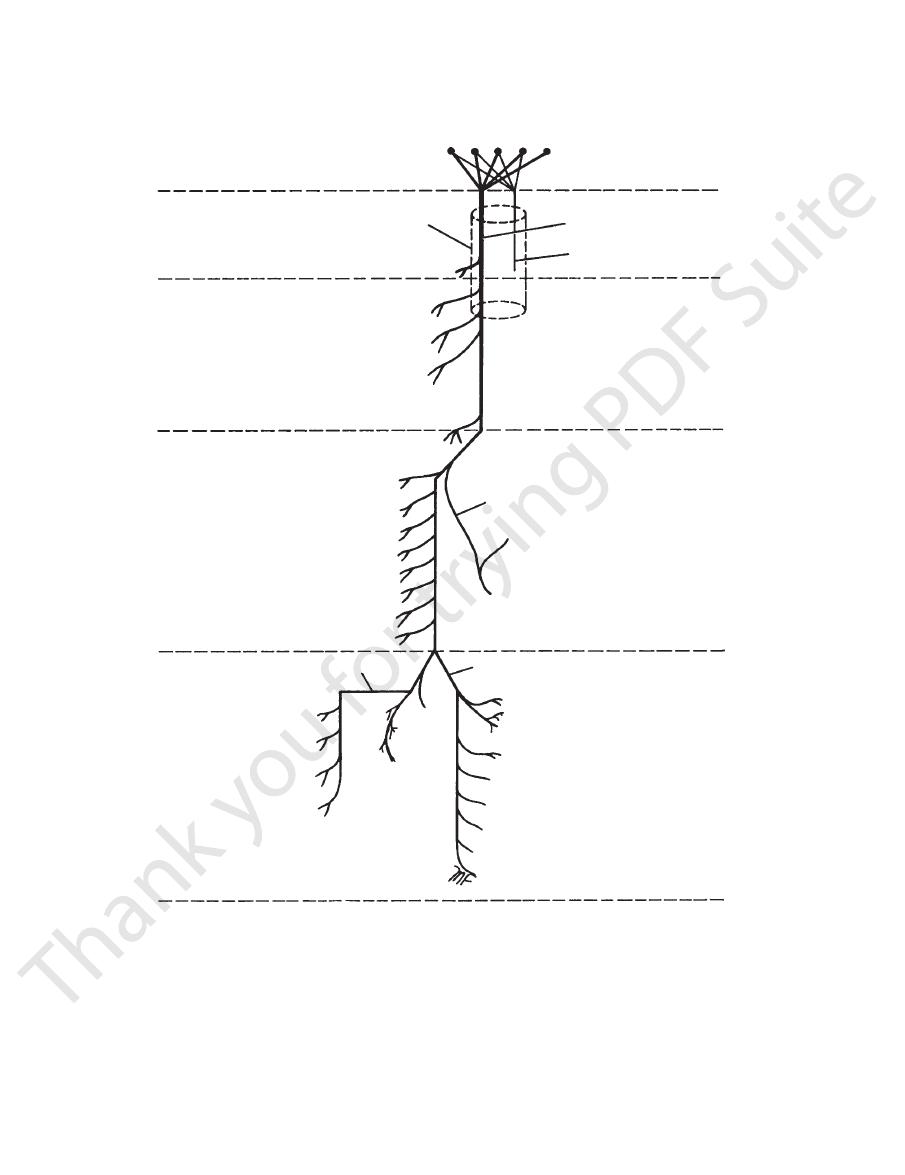
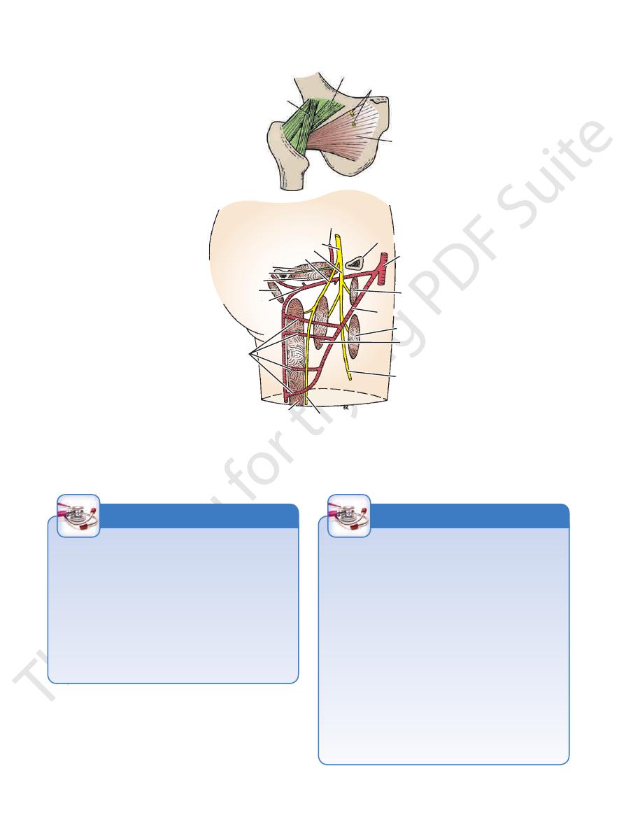
FIGURE 10.17
Summary of the origin of the sciatic nerve and the main branches of the tibial nerve.
a branch of the
lateral cutaneous nerve of the thigh,
The
Cutaneous Nerves
The Front and Medial Aspects
of the Thigh
Skin of the Thigh
lumbar plexus (L2 and 3), enters the thigh behind the lateral
the cremaster muscle (see page 222).
small area of skin (Fig. 10.2). The genital branch supplies
behind the middle of the inguinal ligament and supplies a
branch of the lumbar plexus (L1 and 2), enters the thigh
femoral branch of the genitofemoral nerve,
The
skin of the lower lateral quadrant of the buttock (Fig. 10.1).
the lateral aspect of the thigh and knee. It also supplies the
into anterior and posterior branches, it supplies the skin of
end of the inguinal ligament (Fig. 10.2). Having divided
a

Basic Anatomy
451
artery
arterial supply
femoral arteries
from circumflex
arterial supply
synovial sheath
small branch of obturator artery
obturator artery
transverse acetabular ligament
acetabular labrum
articular surface
synovial membrane
synovial sheath
epiphyseal line
acetabular
acetabulum
synovial membrane
acetabular labrum
capsule
head of femur
fossa
pad of fat
ligament
of head
of femur
ligament of head of femur
from obturator
ligament of head of femur
A
B
FIGURE 10.18
Coronal section of the right hip joint
opening in the deep fascia and joins the femoral vein about
thigh. It passes through the lower part of the saphenous
the knee and curves forward around the medial side of the
cia over the medial side of the leg. The vein passes behind
in company with the saphenous nerve in the superficial fas
the medial malleolus (Fig. 10.19). It then ascends
in front of
directly
dorsal venous arch of the foot and passes upward
drains the medial end of the
great saphenous vein
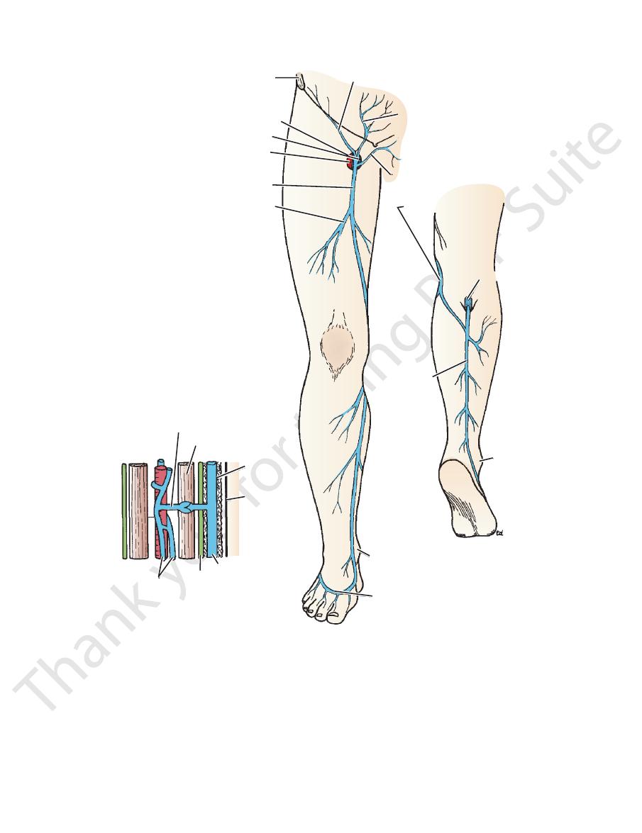
The
of great clinical importance.
saphenous veins and their tributaries (Fig. 10.19). They are
The superficial veins of the leg are the great and small
Superficial Veins
infrapatellar branch of the saphenous nerve (Fig. 10.2).
mediate, and medial cutaneous nerves of the thigh and the
formed from the terminal branches of the lateral, inter
lies in front of the knee and is
patellar plexus
The
the thigh (Fig. 10.2).
supply a variable area of skin on the medial aspect of
nerve
obturator
Branches from the anterior division of the
plexus (Fig. 10.2).
supply the anterior aspect of the thigh and joins the patellar
branch of the femoral nerve, divides into two branches that
intermediate cutaneous nerve of the thigh,
The
and joins the patellar plexus (Fig. 10.2).
the femoral nerve, supplies the medial aspect of the thigh
a branch of
medial cutaneous nerve of the thigh,
The
nal ligament.
and to a small skin area below the medial part of the ingui
ris and adjacent part of the labium majus in the female)
penis and adjacent part of the scrotum (or root of the clito
(Fig. 10.2). It is distributed to the skin of the root of the
(L1), enters the thigh through the superficial inguinal ring
a branch of the lumbar plexus
ilioinguinal nerve,
The
head of the femur
and articular surfaces of the right hip joint and arterial supply of the
(A)
(B).
-
-
a
-
-
452
CHAPTER 10
The Lower Limb
perforating vein
saphenous opening
anterior superior
iliac spine
femoral vein
femoral artery
great saphenous vein
accessory vein
dorsal venous arch
medial malleolus
small
saphenous
vein
superficial external pudendal vein
pubic tubercle
superficial epigastric vein
superficial circumflex iliac vein
popliteal vein
lateral
malleolus
muscle
superficial
fascia
saphenous vein
deep fascia
"venous pump"
venae comitantes
skin
great saphenous vein
FIGURE 10.19
ance of the valved perforating veins in the “venous pump.”
Superficial veins of the right lower limb. Note the import
is described on page 487.
small saphenous vein
The
up at the saphenous opening.
joins the main vein about the middle of the thigh or higher
usually
accessory vein,
An additional vein, known as the
artery found in this region.
veins correspond with the three branches of the femoral
These
superficial external pudendal vein.
vein,
superficial epigastric
superficial circumflex iliac vein,
variable in size and arrangement (Figs. 10.3 and 10.19): the
saphenous vein usually receives three tributaries that are
At the saphenous opening in the deep fascia, the great
along the medial side of the calf (Fig. 10.19).
connect the great saphenous vein with the deep veins
veins
perforating
branches that pass behind the knee. Several
and is connected to the small saphenous vein by one or two
The great saphenous vein possesses numerous valves
1.5 in. (4 cm) below and lateral to the pubic tubercle (Figs.
10.3 and 10.19).
the
and the
454
CHAPTER 10
The Lower Limb
medial malleolus of tibia
edge of saphenous
opening in deep
fascia
femoral artery
femoral vein
great saphenous
vein
A
B
C
saphenous nerve
great saphenous vein
saphenous nerve
great saphenous vein
pubic
tubercle
anterior superior
iliac spine
D
FIGURE 10.20
Great saphenous vein cutdown.
of the group receive superficial lymph vessels from the anterior
inguinal ligament (Figs. 10.3 and 10.4). The medial members
lies just below and parallel to the
horizontal group
The
a vertical group (Figs. 10.3 and 10.4).
inguinal ligament and can be divided into a horizontal and
The superficial nodes lie in the superficial fascia below the
Superficial Inguinal Lymph Nodes
deep groups.
The inguinal lymph nodes are divided into superficial and
Inguinal Lymph Nodes
below and lateral to the pubic tubercle.
At the groin. The great saphenous vein drains into the femoral vein two fingerbreadths
At the ankle. The great saphenous vein is constantly found in front of the
A, B.
medial malleolus of the tibia. C, D.
abdominal wall below the level of the umbilicus and from the
perineum (Fig. 10.4). The lymph vessels from the urethra,
the external genitalia of both sexes (but not the testes), and
is attached to the deep fascia (fascia lata) about a
anterior abdominal wall extends into the thigh and
of the
membranous layer of the superficial fascia
The
the external iliac artery (see Fig. 5.76).
passing through the femoral canal to lymph nodes along
the efferent vessels from these nodes enter the abdomen by
lie along the medial side of the femoral vein (Fig. 10.21);
The deep nodes are located beneath the deep fascia and
Deep Inguinal Lymph Nodes
cia and join the deep inguinal nodes.
nodes pass through the saphenous opening in the deep fas
The efferent lymph vessels from the superficial inguinal
lymph vessels of the lower limb (Figs. 10.3 and 10.4).
great saphenous vein and receives most of the superficial
lies along the terminal part of the
vertical group
The
from the back below the level of the iliac crests (Fig. 10.4).
lateral members of the group receive superficial lymph vessels
the lower half of the anal canal are drained by this route. The
-
Superficial Fascia of the Thigh
Lymphatics of the Lower Limb
node caused by lymphatic spread of pathogenic organisms
The superficial and deep inguinal lymph nodes not only drain
all the lymph from the lower limb, but also drain lymph from
the skin and superficial fascia of the anterior and posterior
abdominal walls below the level of the umbilicus; lymph from
the external genitalia and the mucous membrane of the lower
half of the anal canal also drains into these nodes. Remember
the large distances the lymph has had to travel in some
instances before it reaches the inguinal nodes. For example,
a patient may present with an enlarged, painful inguinal lymph
that entered the body through a small scratch on the under-
surface of the big toe.
C L I N I C A L N O T E S
Basic Anatomy
455
inguinal ligament
femoral nerve
iliopsoas
femoral artery
femoral sheath femoral vein
femoral canal
pubic tubercle
lymphatic vessel
pectineus
external iliac artery and vein
femoral sheath
femoral artery
lymphatic vessel
femoral ring
femoral canal
internal oblique
external oblique
inguinal ligament
membranous layer
fatty layer
superficial fascia
deep fascia
lymph node
femoral canal
pubis
fascia
iliaca
peritoneum
extraperitoneal fat
fascia transversalis
transversus
FIGURE 10.21
Right femoral sheath and its contents.
fingerbreadth below the inguinal ligament (Figs. 10.3 and
The deep fascia encloses the thigh like a trouser leg
over the lower limb without interruption (Fig. 10.21).
abdominal wall extends into the thigh and continues down
on the anterior
fatty layer of the superficial fascia
The
described in Chapter 4.
extravasation of urine after a rupture of the urethra is fully
10.21). The importance of this fact in connection with
Deep Fascia of the Thigh (Fascia Lata)
(Fig.
r end is attached to the pelvis and
10.22) and at its uppe
are described in Table 10.2.
The muscles are seen in Figures 10.6, 10.23, and 10.24 and
of the Thigh
Muscles of the Anterior Fascial Compartment
Femoral nerve
Nerve supply:
Femoral artery
Blood supply:
ceps femoris
Sartorius, iliacus, psoas, pectineus, and quadri
Muscles:
compartments are anterior, medial, and posterior in position.
partments, each having muscles, nerves, and arteries. The
10.22). By this means, the thigh is divided into three com
cial sheath of the thigh to the linea aspera of the femur (Fig.
Three fascial septa pass from the inner aspect of the deep fas
cribriform fascia.
tissue called the
The saphenous opening is filled with loose connective
the superior ramus of the pubis.
the femoral vessels, to be attached to the pectineal line of
then curves upward and medially, and then laterally behind
to the femoral vessels (Fig. 10.3). The border of the opening
the lower lateral border of the opening, which lies anterior
is
falciform margin
and lateral to the pubic tubercle. The
saphenous opening is situated about 1.5 in. (4 cm) below
of the femoral artery, and lymph vessels (Fig. 10.3). The
transmits the great saphenous vein, some small branches
the front of the thigh just below the inguinal ligament. It
is a gap in the deep fascia in
saphenous opening
The
the tensor fasciae latae and the gluteus maximus muscles.
gluteal region, the deep fascia forms sheaths, which enclose
gluteus maximus muscle (see Figs. 10.5 and 10.6). In the
tion of the tensor fasciae latae and the greater part of the
condyle of the tibia. The iliotibial tract receives the inser
attached above to the iliac tubercle and below to the lateral
(Figs. 10.6 and 10.22), which is
iliotibial tract
to form the
the inguinal ligament. On its lateral aspect, it is thickened
-
Fascial Compartments of the Thigh
-
-
Contents of the Anterior Fascial
Compartment of the Thigh
■
■
-
■
■
■
■
456
CHAPTER 10
The Lower Limb
rectus femoris
deep fascia
vastus medialis
nerve to vastus medialis
saphenous nerve
femoral vein
femoral artery
sartorius
great saphenous
vein
adductor longus
gracilis
adductor magnus
semimembranosus
posterior cutaneous nerve of thigh
semitendinosus
biceps femoris
sciatic nerve
iliotibial tract
profunda femoris
artery
vastus lateralis
vastus intermedius
medial
lateral
FIGURE 10.22
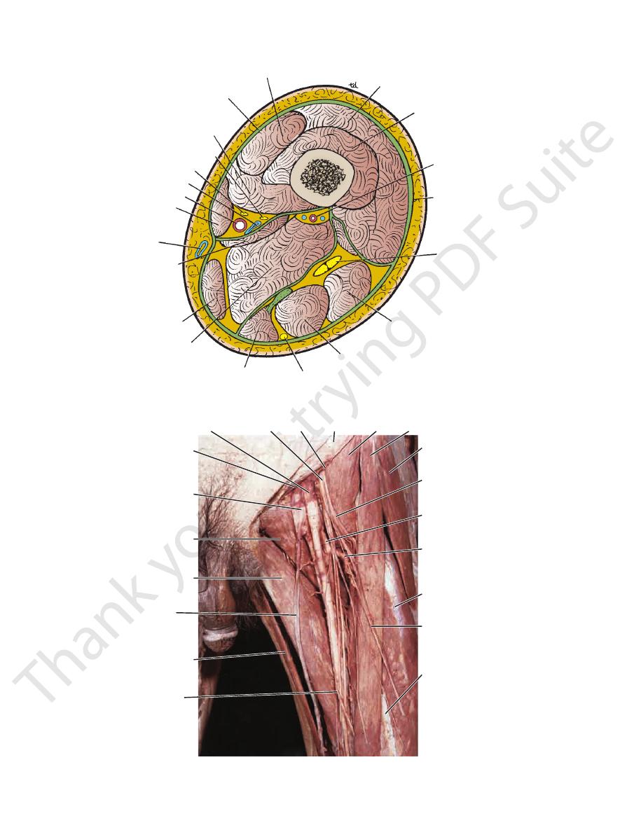
Transverse section through the middle of the right thigh as seen from above.
femoral artery femoral nerve psoas inguinal ligament iliacus sartorius
tensor
fasciae
latae
muscular
branches
of femoral
nerve
profunda
femoris
artery
lateral
femoral
circumflex
artery
vastus
lateralis
intermediate
cutaneous
nerve of
the thigh
medial
cutaneous
nerve of
the thigh
rectus
femoris
gracilis
great
saphenous
vein
adductor
longus
pectineus
femoral vein
site of
femoral
canal
FIGURE 10.23
Dissection of the femoral triangle in the left lower limb.
Basic Anatomy
457
iliacus
femoral sheath
obturator externus
adductor brevis
profunda femoris artery
perforating arteries
femoral vein
descending
genicular artery
femoral artery
adductor magnus
posterior division
obturator nerve
anterior division
pectineus
lacunar ligament
femoral ring
femoral vessels
psoas
femoral nerve
adductor longus
FIGURE 10.24
Relationship between the obturator nerve and the adductor muscles in the right lower limb.
Adductor (Subsartorial) Canal
tributaries, and the deep inguinal lymph nodes.
femoral artery and its branches, the femoral vein and its
femoral nerve and its branches, the femoral sheath, the
The femoral triangle contains the terminal part of the
is formed by the skin and fasciae of the thigh.
roof
by the iliopsoas, the pectineus, and the adductor longus. Its
is gutter shaped and formed from lateral to medial
floor
Its
The adductor longus muscle
Medially:
The sartorius muscle
Laterally:
The inguinal ligament
Superiorly:
as follows:
below the inguinal ligament (Fig. 10.6). Its boundaries are
ated in the upper part of the medial aspect of the thigh just
The femoral triangle is a triangular depressed area situ
Femoral Triangle
The rectus femoris muscle also flexes the hip joint.
the quadriceps muscle greatly strengthens the knee joint.
during contraction of the quadriceps muscle. The tone of
zontal and prevent the patella from being pulled laterally
lowest muscle fibers of the vastus medialis are almost hori
join the capsule of the knee joint and strengthen it. The
which
retinacula,
eralis and vastus medialis form bands, or
knee joint. Some of the tendinous fibers of the vastus lat
10.25). Together, they provide a powerful extensor of the
mentum patellae, is attached to the tibial tuberosity (Fig.
vastus medialis, is inserted into the patella and, via the liga
femoris, the vastus intermedius, the vastus lateralis, and the
The quadriceps femoris muscle, consisting of the rectus
(Quadriceps Mechanism)
Action of Quadriceps Femoris Muscle
Note the following:
-
-
-
-
■
■
■
■
■
■
The adductor canal is an intermuscular cleft situated on the
terminal part of the obturator nerve.
saphenous nerve, the nerve to the vastus medialis, and the
oral artery, the femoral vein, the deep lymph vessels, the
The adductor canal contains the terminal part of the fem
is formed by the vastus medialis.
lateral wall
The
and magnus.
is formed by the adductor longus
posterior wall
The
cle and fascia.
is formed by the sartorius mus
anteromedial wall
The
and a lateral wall.
is triangular, having an anteromedial wall, a posterior wall,
at the opening in the adductor magnus. In cross section, it
above at the apex of the femoral triangle and ends below
sartorius muscle (Figs. 10.6 and 10.22). It commences
medial aspect of the middle third of the thigh beneath the
■
■
-
■
■
■
■
-

458
CHAPTER 10
The Lower Limb
Vastus
Vastus medialis
Vastus lateralis
bodies, and intervertebral
Transverse processes,
Muscle
Origin
Insertion
Nerve
Supply
Nerve Root
a
Action
Sartorius
Anterior superior iliac
spine
Upper medial
surface of
shaft of tibia
Femoral
nerve
L2, 3
Flexes, abducts,
laterally rotates
thigh at hip
joint; flexes and
medially rotates
leg at knee joint
Iliacus
Iliac fossa of hip bone
With psoas into
lesser trochanter
of femur
Femoral
nerve
L2, 3
Flexes thigh on
trunk; if thigh is
fixed, it flexes the
trunk on the thigh
as in sitting up
from lying down
Psoas
discs of the 12th
thoracic and five lumbar
vertebrae
With iliacus into
lesser trochanter
of femur
Lumbar
plexus
L1, 2, 3
Flexes thigh on
trunk; if thigh is
fixed, it flexes the
trunk on thigh as
in sitting up from
lying down
Pectineus
Superior ramus of pubis
Upper end of linea
aspera of shaft of
femur
Femoral
nerve
L2, 3
Flexes and adducts
thigh at hip joint
Quadriceps
femoris
Rectus femoris
Straight head: anterior
inferior iliac spine
Reflected head: ilium
above acetabulum
Quadriceps tendon
into patella, then
via ligamentum
patellae into
tubercle of tibia
Femoral
nerve
L2, 3, 4
Extension of leg at
knee joint; flexes
thigh at hip joint
Upper end and shaft of
femur
Quadriceps tendon
into patella, then
via ligamentum
patellae into
tubercle of tibia
Femoral
nerve
L2, 3, 4
Extension of leg at
knee joint
Upper end and shaft of
femur
Quadriceps tendon
into patella, then
via ligamentum
patellae into
tubercle of tibia
Femoral
nerve
L2, 3, 4
Extension of leg
at knee joint;
stabilizes patella
intermedius
Anterior and lateral
surfaces of shaft of
femur
Quadriceps tendon
into patella, then
via ligamentum
patellae into
tubercle of tibia
Femoral
nerve
L2, 3, 4
Extension of leg
at knee joint;
articularis genus
retracts synovial
membrane
a
The predominant nerve root supply is indicated by boldface type.
Muscles of the Anterior Fascial Compartment of the Thigh
T A B L E 1 0 . 2
Basic Anatomy
459
rectus femoris
retinaculum
muscular fibers of
vastusmedialis
vastus lateralis
large lateral
femoral condyle
retinaculum
ligamentum patellae
fibula
tuberosity of tibia
semitendinosus
gracilis
sartorius
patella
vastus medialis
FIGURE 10.25
The quadriceps femoris mechanism. The lateral and upward pull of the powerful rectus femoris and the vastus
lateral condyle of the femur, which projects forward.
lateralis muscles on the patella is counteracted by the lowest horizontal muscular fibers of the vastus medialis and the large
Quadriceps Femoris as a Knee Joint Stabilizer
knee joint when the quadriceps femoris muscle is actively
This can occur when a sudden flexing force is applied to the
proximally, leaving a gap that may be palpable on the anterior
can be tested by measuring the circumference of each thigh a
The quadriceps femoris is a most important extensor muscle for
the knee joint. Its tone greatly strengthens the joint; therefore,
this muscle mass Vmust be carefully examined when disease of
the knee joint is suspected. Both thighs should be examined, and
the size, consistency, and strength of the quadriceps muscles
should be tested. Reduction in size caused by muscle atrophy
fixed distance above the superior border of the patella.
The vastus medialis muscle extends farther distally than the
vastus lateralis. Remember that the vastus medialis is the first
part of the quadriceps muscle to atrophy in knee joint disease
and the last to recover.
Rupture of the Rectus Femoris
The rectus femoris muscle can rupture in sudden violent exten-
sion movements of the knee joint. The muscle belly retracts
surface of the thigh. In complete rupture of the muscle, surgical
repair is indicated.
Rupture of the Ligamentum Patellae
contracting
C L I N I C A L N O T E S

460
CHAPTER 10
sheath surrounds the femoral vessels and lymphatics for
versalis, and its posterior wall with the fascia iliaca. The
anterior wall is continuous above with the fascia trans
envelope lining the abdominal walls (see page XXX). Its
is a downward protrusion into the thigh of the fascial
The femoral sheath (Figs. 10.3, 10.6, 10.21, and 10.24)
Femoral Sheath
The Lower Limb
-
about 1 in. (2.5 cm) below the inguinal ligament. The
partment
medial com
by a fibrous septum and occupy the most
vessels, as they leave the thigh, are separated from the vein
The lymph
intermediate compartment.
and occupies the
medial side and is separated from it by a fibrous septum
as it leaves the thigh, lies on its
femoral vein,
sheath. The
of the
lateral compartment
nal ligament, occupies the
as it enters the thigh beneath the ingui
femoral artery,
-
-
(Fig. 10.21).
femoral septum,
The
femoral ring.
long, and its upper opening is called the
the lymph vessels (Fig. 10.21). It is about 0.5 in. (1.3 cm)
is the small medial compartment for
femoral canal
The
which is a condensation of
eritoneal
extrap
tissue, closes the ring. The femoral canal contains fatty
connective tissue, all the efferent lymph vessels from the
described below.
and is
femoral hernia
it. Such a condition is known as a
the femoral canal, pushing the femoral septum before
men. A protrusion of peritoneum could be forced down
this site that forms a potentially weak area in the abdo
not adherent to the walls of the small lymph vessels; it is
sheath that forms the medially located femoral canal is
adventitia of these vessels. The part of the femoral
blood vessels and inferiorly blends with the tunica
The femoral sheath is adherent to the walls of the
lymph nodes.
deep inguinal lymph nodes, and one of the deep inguinal
-
The femoral ring (Fig. 10.21) has the following important
relations: anteriorly, the inguinal ligament;
iorly, the
poster
The lower end of the canal is normally closed by
ment; and laterally, the femoral vein.
superior ramus of the pubis; medially, the lacunar liga-
the adherence of its medial wall to the tunica adventitia of
the deep fascia of the thigh (Fig. 10.3).
the femoral vein. It lies close to the saphenous opening in
Femoral Sheath and Femoral Hernia
ral vessels and lymphatic vessels for about 1 in. (2.5 cm) below
thigh of the fascial lining of the abdomen. It surrounds the femo
The hernial sac descends through the femoral canal within the
femoral sheath.
The femoral sheath is a prolongation downward into the
-
the inguinal ligament (see Fig. 10.21). The femoral artery, as it
enters the thigh below the inguinal ligament, occupies the lateral
compartment of the sheath. The femoral vein, which lies on its
medial side and is separated from it by a fibrous septum, occu-
pies the intermediate compartment. The lymphatics, which are
separated from the vein by a fibrous septum, occupy the most
medial compartment.
The femoral canal, the compartment for the lymphatic ves-
sels, occupies the medial part of the sheath. It is about 0.5 in.
(1.3 cm) long, and its upper opening is referred to as the
other anatomic structures close to the inguinal ligament. For
hernia, it is important to consider diseases that may involve
When considering the differential diagnosis of a femoral
A femoral hernia is a dangerous condition and should always be
(strangulated hernia).
ring, seriously impairing its blood supply
neck, and its blood vessels may be compressed by the femoral
strains or coughs, a piece of bowel may be forced through the
Furthermore, after the patient
abdominal viscus has passed through the neck into the body of
structures, the neck of the sac is unable to expand. Once an
and laterally to the femoral vein. Because of these anatomic
pubis, medially to the sharp free edge of the lacunar ligament,
teriorly to the pectineal ligament and the superior ramus of the
ring. The ring is related anteriorly to the inguinal ligament, pos
tubercle. The neck of the sac is narrow and lies at the femoral
an inguinal hernia, which lies above and medial to the pubic
(see page 145). This serves to distinguish it from
below and lateral to the
the thigh deep to the deep fascia (see page 143). With further
femoral canal, it expands to form a swelling in the upper part of
femoral
ring. The femoral septum, which is a condensation of extraperi-
toneal tissue, plugs the opening of the femoral ring.
A femoral hernia is more common in women than in men
(possibly because of their wider pelvis and femoral canal). The
hernial sac passes down the femoral canal, pushing the femo-
ral septum before it. On escaping through the lower end of the
expansion, the hernial sac may turn upward to cross the anterior
surface of the inguinal ligament.
The neck of the sac always lies
pubic tubercle
-
the sac, it may be difficult to push it up and return it to the abdom-
inal cavity (irreducible hernia).
treated surgically.
example:
■
■
Inguinal canal: The swelling of an inguinal hernia lies above
the medial end of the inguinal ligament. Should the hernial
sac emerge through the superficial inguinal ring to start its
descent into the scrotum, the swelling will lie above and me-
dial to the pubic tubercle. The sac of a femoral hernia lies
below and lateral to the pubic tubercle.
■
■
Superficial inguinal lymph nodes: Usually, more than one
lymph node is enlarged. In patients with inflammation of the
nodes (lymphadenitis), carefully examine the entire area of
the body that drains its lymph into these nodes. A small, un-
noticed skin abrasion may be found. Never forget the mucous
membrane of the lower half of the anal canal—it may have an
undiscovered carcinoma.
C L I N I C A L N O T E S
(continued)
Basic Anatomy
461
■
lateral femoral
medial
its origin, it gives off the
At
fourth perforating artery.
It ends by becoming the
partment of the thigh (Figs. 10.23, 10.24, and 10.27).
the femoral vessels and enters the medial fascial com
(Figs. 10.6, 10.23, and 10.26). It passes medially behind
artery about 1.5 in. (4 cm) below the inguinal ligament
branch that arises from the lateral side of the femoral
is a large and important
profunda femoris artery
The
majus).
medially and supplies the skin of the scrotum (or labium
(Fig. 10.6) runs
deep external pudendal artery
The
scrotum (or labium majus).
small branch that runs medially to supply the skin of the
(Fig. 10.3) is a
superficial external pudendal artery
The
the umbilicus (Fig. 10.3).
crosses the inguinal ligament and runs to the region of
is a small branch that
superficial epigastric artery
The
spine (Fig. 10.3).
that runs up to the region of the anterior superior iliac
is a small branch
superficial circumflex iliac artery
The
Branches
(Fig. 10.6).
The femoral nerve and its branches
Laterally:
part of its course (Figs. 10.6 and 10.23).
It is related to the femoral vein in the upper
Medially:
the artery and the adductor longus.
longus (Fig. 10.6). The femoral vein intervenes between
it from the hip joint, the pectineus, and the adductor
The artery lies on the psoas, which separates
Posteriorly:
course, it passes behind the sartorius muscle (Fig. 10.6).
and is covered by skin and fascia. In the lower part of its
In the upper part of its course, it is superficial
Anteriorly:
Relations
popliteal artery (Fig. 10.24).
tor magnus muscle by entering the popliteal space as the
tubercle of the femur and ends at the opening in the adduc
limb. It descends almost vertically toward the adductor
The femoral artery is the main arterial supply to the lower
the anterior superior iliac spine and the symphysis pubis.
(Figs. 10.6, 10.23, and 10.26). Here, it lies midway between
nal ligament, as a continuation of the external iliac artery
The femoral artery enters the thigh from behind the ingui
Femoral Artery
Compartment of the Thigh
Blood Supply of the Anterior Fascial
crease in size when the patient is asked to cough. (Elevated
part of the great saphenous vein, a
■
Great saphenous vein: A localized dilatation of the terminal
saphenous varix, can
cause confusion, especially because a hernia and a varix in-
intra-abdominal pressure drives the blood downward.) The
presence of varicose veins elsewhere in the leg should help
in the diagnosis.
■
■
Psoas sheath: Tuberculous infection of a lumbar vertebra can
result in the extravasation of pus down the psoas sheath into
the thigh. The presence of a swelling above and below the
inguinal ligament, together with clinical signs and symptoms
referred to the vertebral column, should make the diagnosis
obvious.
■
■
Femoral artery: An expansile swelling lying along the course
of the femoral artery that fluctuates in time with the pulse
rate should make the diagnosis of aneurysm of the femoral
artery certain.
-
-
■
■
■
■
■
■
■
■
■
■
■
■
■
■
■
■
■
■
-
and
circumflex arteries,
that arises from the femoral artery near its termination
is a small branch
descending genicular artery
The
(Fig. 10.27).
three perforating arteries
and during its course it gives off
■
■
(Fig. 10.24). It assists in supplying the knee joint.
external iliac artery
moral artery
circumflex artery
medial femoral
moral artery
moris artery
perforating
popliteal arte
posterior tibial artery
peroneal arte
arcuate artery
artery
artery
anterior tibial
circumflex artery
profunda artery
inguinal ligament
lateral femoral
dorsalis pedis
ry
ry
branches
of profunda
fe
fe
fe
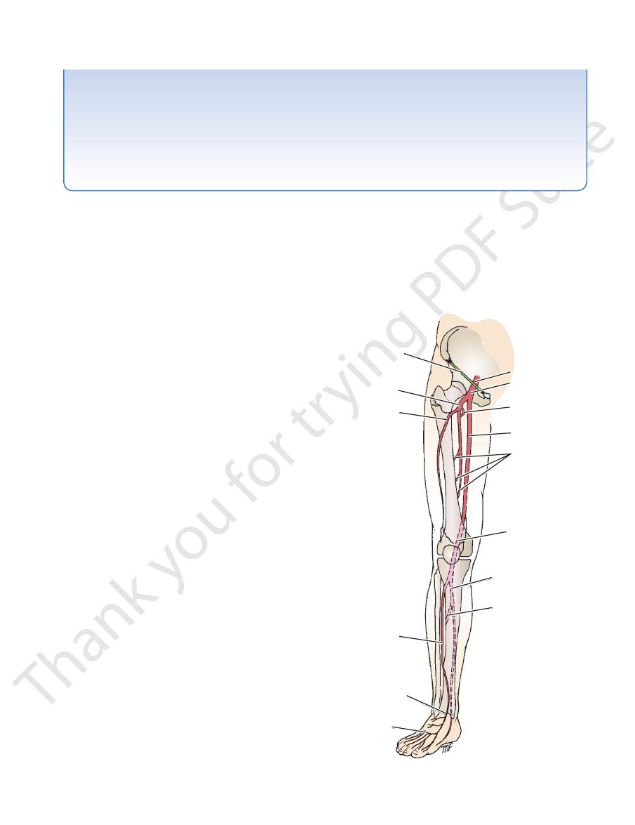
FIGURE 10.26
Major arteries of the lower limb.
462
CHAPTER 10
The Lower Limb
obturator artery
obturator nerve
anterior division
posterior division
obturator internus
obturator membrane
obturator externus
quadratus femoris
medial femoral circumflex artery
perforating arteries
posterior
adductor magnus
articular branch to knee joint
anterior
cutaneous branch
adductor brevis
adductor longus
profunda femoris artery
pectineus
femoral artery
superior ramus of pubis
pubofemoral ligament
divisions of obturator nerve
obturator externus
iliofemoral ligament
A
B
FIGURE 10.27
Obturator externus muscle
tomosis between the perforating arteries and the medial femoral circumflex artery.
courses taken by the obturator nerve and its divisions and the profunda femoris artery and its branches. Note also the anas
and vertical section of the medial compartment of the thigh
(A)
(B). Note the
-
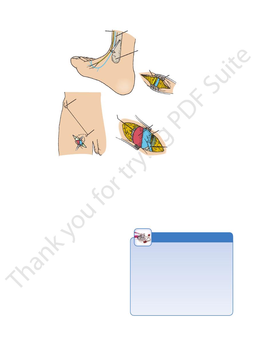
Femoral Artery Catheterization
passed into the inferior mesenteric, superior mesenteric,
A long, fine catheter can be inserted into the femoral artery
as it descends through the femoral triangle. The catheter is
guided under fluoroscopic view along the external and com-
mon iliac arteries into the aorta. The catheter can then be
celiac, or renal arteries. Contrast medium can then be injected
into the artery under examination and a permanent record
obtained by taking a radiograph. Pressure records can also be
obtained by guiding the catheter through the aortic valve into
the left ventricle.
C L I N I C A L N O T E S
Femoral Vein
the thigh, lying at first on the lateral side of the artery, then
popliteal vein (Figs. 10.23 and 10.24). It ascends through
opening in the adductor magnus as a continuation of the
The femoral vein enters the thigh by passing through the
Femoral Vein Catheterization
tionship to the medial side of the femoral artery just below the
large vein is needed. The femoral vein has a constant rela
Femoral vein catheterization is used when rapid access to a
-
inguinal ligament and is easily cannulated. However, because
of the high incidence of thrombosis with the possibility of fatal
pulmonary embolism, the catheter should be removed once
the patient is stabilized.
Anatomy of the Procedure
1.
The skin of the thigh below the inguinal ligament is sup-
plied by the genitofemoral nerve; this nerve is blocked with
a local anesthetic.
2.
The femoral pulse is palpated midway between the ante-
rior superior iliac spine and the symphysis pubis, and the
femoral vein lies immediately medial to it.
3.
At a site about two fingerbreadths below the inguinal liga-
ment, the needle is inserted into the femoral vein.
C L I N I C A L N O T E S
Basic Anatomy
lateral side of the femoral artery in the femoral triangle,
The profunda femoris is a large artery that arises from the
Profunda Femoris Artery
Compartment of the Thigh
Blood Supply of the Medial Fascial
downward into the popliteal space.
mits the femoral vessels to pass from the adductor canal
the attachment of this muscle to the femur, which per
is a gap in
adductor hiatus
hamstring portions. The
a large, triangular muscle consisting of adductor and
The adductor magnus (Figs. 10.24, 10.27, and 10.29) is
Note the following:
Table 10.3.
Figures 10.22, 10.23, 10.24, and 10.27 and are described in
The muscles of the medial fascial compartment are seen in
of the Thigh
Muscles of the Medial Fascial Compartment
Obturator nerve
Nerve supply:
artery
Profunda femoris artery and obturator
Blood supply:
adductor magnus, and obturator externus
Gracilis, adductor longus, adductor brevis,
Muscles:
supply the knee joint.
the hip joint; the branches to the three vasti muscles also
of the rectus femoris also supplies
muscular branch
The
the region of the ball of the big toe.
along the medial border of the foot, where it terminates in
nous vein. It passes in front of the medial malleolus and
the medial side of the leg in company with the great saphe
dons of sartorius and gracilis (Fig. 10.2). It then runs down
emerges on the medial side of the knee between the ten
ral artery from its lateral to its medial side (Fig. 10.6). It
runs downward and medially and crosses the femo
nerve
saphenous
cular branches to the quadriceps muscle. The
off one cutaneous branch, the saphenous nerve, and mus
The posterior division (Fig. 10.28) gives
Posterior Division
supply the sartorius and the pectineus.
respectively (Figs. 10.2 and 10.6). The muscular branches
the skin of the medial and anterior surfaces of the thigh,
that supply
intermediate cutaneous nerves
thigh
medial cutaneous nerve of the
neous branches are the
off two cutaneous and two muscular branches. The cuta
The anterior division (Fig. 10.28) gives
Anterior Division
Branches
not enter the thigh within the femoral sheath.
Note that the femoral nerve does
of the thigh (Fig. 10.6).
nerve supplies all the muscles of the anterior compartment
dividing into anterior and posterior divisions. The femoral
1.5 in. (4 cm) below the inguinal ligament, it terminates by
the inguinal ligament (Figs. 10.6, 10.21, and 10.23). About
lateral to the femoral artery and the femoral sheath, behind
iliacus. It lies behind the fascia iliaca and enters the thigh
passes downward in the interval between the psoas and
the psoas muscle within the abdomen (see page 222) and
plexus (L2, 3, and 4). It emerges from the lateral border of
The femoral nerve is the largest branch of the lumbar
Femoral Nerve
Compartment of the Thigh
Nerve Supply of the Anterior Fascial
the femoral canal and drain into the external iliac nodes.
inguinal nodes ascend into the abdominal cavity through
the popliteal nodes. The efferent lymph vessels from the deep
vessels alongside the arteries, some having passed through
structures of the lower limb that have ascended in lymph
saphenous opening. They also receive lymph from the deep
lymph vessels that pass through the cribriform fascia of the
receive all the lymph from the superficial inguinal nodes via
rior is usually located in the femoral canal (Fig. 10.21). They
the terminal part of the femoral vein, and the most supe
there are commonly three. They lie along the medial side of
are variable in number, but
deep inguinal lymph nodes
The
Compartment of the Thigh
Lymph Nodes of the Anterior Fascial
external pudendal veins drain into the great saphenous vein.
circumflex iliac vein, the superficial epigastric vein, and the
branches of the femoral artery (Fig. 10.3). The superficial
and veins that correspond to the
great saphenous vein
The tributaries of the femoral vein are the
Tributaries
become the external iliac vein.
femoral sheath and passes behind the inguinal ligament to
It leaves the thigh in the intermediate compartment of the
posterior to it, and finally on its medial side (Fig. 10.6).
463
-
-
and the
-
-
-
-
Contents of the Medial Fascial
Compartment of the Thigh
■
■
■
■
■
■
■
■
-
L2 L3 L4
femoral nerve
abdomen
lumbar plexus
iliacus
pectineus
hip joint
intermediate cutaneous
nerve of thigh
medial cutaneous
nerve of thigh
sartorius
quadriceps
femoris
knee joint
femoral artery
saphenous nerve
branch to subsartorial
plexus
infrapatellar
branch to skin
to skin of medial
side of leg
skin on medial
side of foot
as far as ball
of big toe
front
of
thigh
lower
leg
foot
FIGURE 10.28
Summary of the main branches of the femoral
nerve.
464
CHAPTER 10
The Lower Limb
Muscles of the Medial Fascial Compartment of the Thigh
T A B L E 1 0 . 3
Hamstring portion:
Adductor portion:
of shaft of femur,
Muscle
Origin
Insertion
Nerve Supply
Nerve Root
a
Action
Gracilis
Inferior ramus of pubis,
ramus of ischium
Upper part of shaft
of tibia on medial
surface
Obturator
nerve
L2, 3
Adducts thigh at hip
joint; flexes leg at
knee joint
Adductor longus
Body of pubis, medial to
pubic tubercle
Posterior surface
of shaft of femur
(linea aspera)
Obturator
nerve
L2, 3, 4
Adducts thigh at hip
joint and assists in
lateral rotation
Adductor brevis
Inferior ramus of pubis
Posterior surface
of shaft of femur
(linea aspera)
Obturator
nerve
L2, 3, 4
Adducts thigh at hip
joint and assists in
lateral rotation
Adductor magnus
Inferior ramus of pubis,
ramus of ischium,
ischial tuberosity
Posterior surface
adductor tubercle
of femur
obturator nerve
sciatic nerve
L2, 3, 4
Adducts thigh at hip
joint and assists
in lateral rotation;
hamstring portion
extends thigh at hip
joint
Obturator externus
Outer surface of obturator
membrane and pubic
and ischial rami
Medial surface of
greater trochanter
Obturator nerve
L3, 4
Laterally rotates thigh
at hip joint
a
The predominant nerve root supply is indicated by boldface type.
gluteus
maximus
semimembranosus
semitendinosus
genicular artery
adductor tubercle
semimembranosus
tibial nerve
popliteus
head of fibula
plantaris
common peroneal
nerve
short head
long head
biceps
femoris
sciatic nerve
linea aspera
biceps femoris
gluteus maximus
adductor magnus
gemellus superior
piriformis
gluteus minimus
gluteus medius
popliteal vein
popliteal artery
opening in
adductor magnus
quadratus femoris
greater trochanter
gemellus inferior
obturator internus
nerve to hamstrings
adductor magnus
(hamstring part)
FIGURE 10.29
Deep structures in the posterior aspect of the
(see page 256). It passes forward on the lateral wall of
The obturator artery is a branch of the internal iliac artery
Obturator Artery
to the branches of the artery. It drains into the femoral vein.
The profunda femoris vein receives tributaries that correspond
Profunda Femoris Vein
branches of the popliteal artery below.
the circumflex femoral arteries above and the muscular
one another and with the inferior gluteal artery and
supply the muscles and terminate by anastomosing with
ward, piercing the various muscle layers as they go. They
artery (Fig. 10.27). The perforating arteries run back
perforating artery is the terminal part of the profunda
branches of the profunda femoris artery; the fourth
Three of these arise as
Four perforating arteries:
the cruciate anastomosis.
muscles of the region and takes part in the formation of
(Fig. 10.6). It breaks up into branches that supply the
ally between the terminal branches of the femoral nerve
This passes later
Lateral femoral circumflex artery:
takes part in the formation of the cruciate anastomosis.
medial fascial compartment of the thigh (Fig. 10.27). It
femoral triangle and gives off muscular branches in the
ward between the muscles that form the floor of the
This passes back
Medial femoral circumflex artery:
Branches
artery (Fig. 10.27).
adductor magnus, where it ends as the fourth perforating
adductor longus and adductor brevis and then lies on the
10.24, and 10.26). It descends in the interval between the
about 1.5 in. (4 cm) below the inguinal ligament (Figs. 10.6,
right thigh.
■
■
-
■
■
-
■
■
-
Basic Anatomy
and passes downward behind the adductor brevis and in
pierces the obturator externus
posterior division
The
the thigh.
rial plexus and supplies the skin on the medial side of
artery. It contributes a variable branch to the subsarto
terminates as a small nerve that supplies the femoral
pectineus. It gives articular branches to the hip joint and
tor brevis, and adductor longus, and occasionally to the
10.30). It gives muscular branches to the gracilis, adduc
the pectineus and adductor longus (Figs. 10.27 and
obturator externus and the adductor brevis and behind
passes downward in front of the
anterior division
The
Branches
anterior and posterior divisions (Fig. 10.27).
obturator foramen (see Fig. 6.12), where it divides into
the lateral wall of the pelvis to reach the upper part of the
cle within the abdomen (see page 222). It runs forward on
and 4) and emerges on the medial border of the psoas mus
The obturator nerve arises from the lumbar plexus (L2, 3,
Compartment of the Thigh
Nerve Supply of the Medial Fascial
vein.
the branches of the artery. It drains into the internal iliac
The obturator vein receives tributaries that correspond to
Obturator Vein
branches and an articular branch to the hip joint.
surface of the obturator membrane. It gives off muscular
eral branches, which pass around the margin of the outer
compartment of the thigh, it divides into medial and lat
tor foramen) (Fig. 10.27). On entering the medial fascial
the obturator canal (i.e., the upper part of the obtura
the pelvis and accompanies the obturator nerve through
465
-
-
Obturator Nerve
-
■
■
-
-
■
■
front of the adductor magnus (Fig. 10.27). It terminates
by descending through the opening in the
or
adduct
tor brevis.
of the adductor magnus, and occasionally to the adduc
branches to the obturator externus, to the adductor part
magnus to supply the knee joint. It gives muscular
-
group of muscles and permits slow recovery of the muscles
crushed. This operation overcomes the spasm of the adductor
severe cases, the posterior division of the obturator nerve is
the anterior division of the obturator nerve. In addition, in some
form a tenotomy of the adductor longus tendon and to divide
In patients with cerebral palsy who have marked spasticity of
Adductor Muscles and Cerebral Palsy
the adductor group of muscles, it is common practice to per-
supplied by the posterior division of the obturator nerve.
C L I N I C A L N O T E S
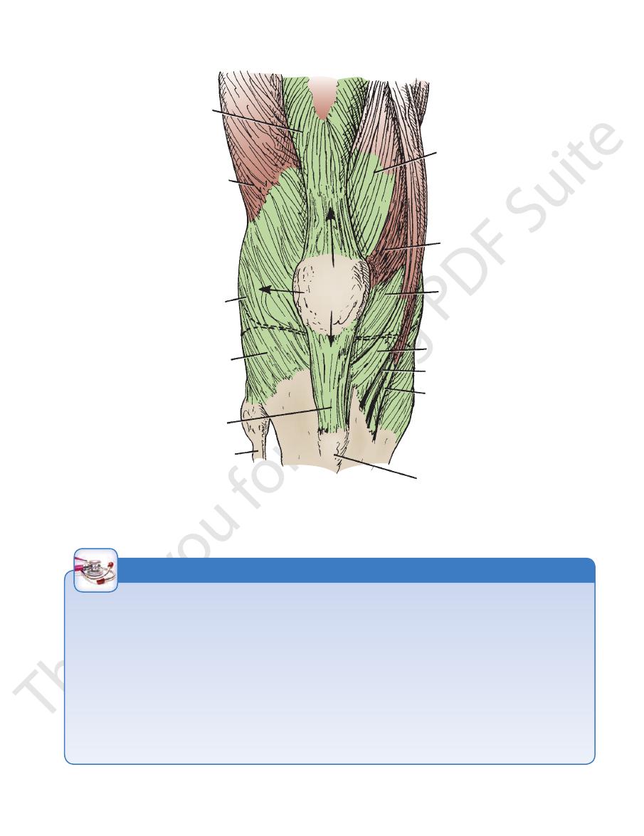
The Back of the Thigh
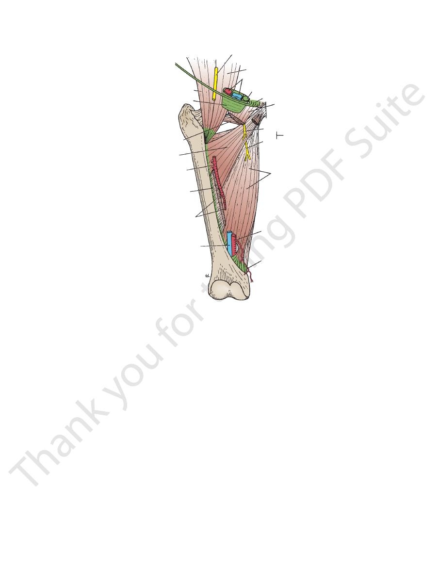
in Figure 10.31 and are described in Table 10.4.
The muscles of the posterior fascial compartment are seen
Sciatic nerve
Nerve supply:
Branches of the profunda femoris artery
Blood supply:
string muscles)
nosus, and a small part of the adductor magnus (ham
Biceps femoris, semitendinosus, semimembra
Muscles:
group of superficial inguinal lymph nodes (Fig. 10.4).
of the thigh drains upward and forward into the vertical
Lymph from the skin and superficial fascia on the back
Lymph Vessels
vein in the popliteal fossa.
lower part of the back of the thigh join the small saphenous
saphenous vein (Fig. 10.19). Superficial veins from the
aspects of the thigh and ultimately drain into the great
Many small veins curve around the medial and lateral
Superficial Veins
(Fig. 10.1).
skin on the back of the thigh and the upper part of the leg
supplies the skin. It gives off numerous branches to the
and in the popliteal fossa it pierces the deep fascia and
muscle (Fig. 10.1). It descends on the back of the thigh,
from beneath the lower border of the gluteus maximus
the sacral plexus, leaves the gluteal region by emerging
a branch of
posterior cutaneous nerve of the thigh,
The
Cutaneous Nerves
Skin
Contents of the Posterior Fascial
Compartment of the Thigh
■
■
-
-
■
■
■
■
L2 L3 L4
obturator nerve
abdomen
lumbar plexus
pelvis
peritoneum on lateral
wall of pelvis
anterior division
posterior division
adductor
region
of thigh
hip joint
pectineus ?
adductor longus
adductor brevis
gracilis
adductor magnus
(adductor portion)
adductor brevis
knee joint
popliteal artery
femoral artery
subsartorial plexus
with medial cutaneous
nerve of thigh and branch
of saphenous nerve
FIGURE 10.30
Summary of the main branches of the obtura
tor nerve.
-