
Basic Anatomy
the knee joint is a profuse anastomosis of small branches
which occurs during extreme flexion of the knee, around
To compensate for the narrowing of the popliteal artery,
479
Arterial Anastomosis Around
the Knee Joint
of the femoral artery with muscular and articular branches
ris muscle, which arises high up in the popliteal fossa
branch to the short head of the biceps femo
Muscular
the skin on the lateral side of the back of the leg (Figs.
supplies
lateral cutaneous nerve of the calf
nerve. The
10.16 and 10.41) runs downward and joins the sural
(Figs.
sural communicating branch
The
Cutaneous:
Branches
is subcutaneous and can easily be rolled against the bone.
nerve lies on the lateral aspect of the neck of the fibula, it
neal nerve and the deep peroneal nerve (Fig. 10.44). As the
divides into two terminal branches: the superficial pero
neck of the bone, pierces the peroneus longus muscle, and
behind the head of the fibula, winds laterally around the
lateral head of the gastrocnemius muscle. It then passes
(Fig. 10.42). It leaves the fossa by crossing superficially the
closely following the medial border of the biceps muscle
of the thigh. It runs downward through the popliteal fossa,
467), the common peroneal nerve arises in the lower third
The smaller terminal branch of the sciatic nerve (see page
branches supply the knee joint.
Articular
mius and the plantaris, soleus, and popliteus (Figs. 10.41
branches supply both heads of the gastrocne
Muscular
border of the foot and the lateral side of the little toe.
malleolus and is distributed to the skin along the lateral
panies the small saphenous vein behind the lateral
the calf and the back of the leg. The sural nerve accom
branches arise from the sural nerve to supply the skin of
peroneal nerve (Figs. 10.41 and 10.17). Numerous small
branch of the common
sural communicating
by the
heads of the gastrocnemius muscle and is usually joined
descends between the two
sural nerve
The
Cutaneous:
Branches
course is described on page 489.
of the leg by passing beneath the soleus muscle. Its further
out its course. The nerve enters the posterior compartment
liteal vein lies between the nerve and the artery through
and finally medial to it (Figs. 10.41 and 10.42). The pop
the lateral side of the popliteal artery, then posterior to it,
It runs downward through the popliteal fossa, lying first on
467), the tibial nerve arises in the lower third of the thigh.
The larger terminal branch of the sciatic nerve (see page
Tibial Nerve
and posterior tibial arteries.
and from deep lymph vessels accompanying the anterior
popliteal fossa. They also receive lymph from the knee joint
and leg; these accompany the small saphenous vein into the
superficial lymph vessels from the lateral side of the foot
tive tissue of the popliteal fossa (Fig. 10.4). They receive
About six lymph nodes are embedded in the fatty connec
Popliteal Lymph Nodes
and posterior tibial arteries.
of the popliteal artery and with branches of the anterior
-
-
-
■
■
-
■
■
-
and 10.42).
■
■
Common Peroneal Nerve
-
■
■
10.1 and 10.41).
■
■
-
(Fig. 10.42).
branches to the knee joint.
Articular
■
■
is exposed to direct trauma or is involved in fractures of the
Common Peroneal Nerve Injury
The common peroneal nerve is extremely vulnerable to
injury as it winds around the neck of the fibula. At this site, it
upper part of the fibula. Injury to the common peroneal nerve
causes footdrop.
C L I N I C A L N O T E S
Posterior Cutaneous Nerve of the Thigh
as pulleys.
the long tendons around the ankle joint in position and act
The retinacula are thickenings of the deep fascia that keep
(see Figs. 10.44 and 10.45).
together and provides attachment for neighboring muscles
The interosseous membrane binds the tibia and fibula
having its own muscles, blood supply, and nerve supply.
three compartments—anterior, lateral, and posterior—each
together with the interosseous membrane, divide the leg into
pass from its deep aspect to be attached to the fibula. These,
borders of the tibia (Fig. 10.45). Two intermuscular septa
it is attached to the periosteum on the anterior and medial
with the deep fascia of the thigh. Below the tibial condyles,
The deep fascia surrounds the leg and is continuous above
10.42). The nerve terminates by supplying the knee joint.
passing through the opening in the adductor magnus (Fig.
465. It leaves the subsartorial canal with the femoral artery by
in the medial compartment of the thigh is described on page
The course of the posterior division of the obturator nerve
over the popliteal fossa (Fig. 10.1).
described on page 465. It terminates by supplying the skin
through the gluteal region and the back of the thigh is
The course of the posterior cutaneous nerve of the thigh
Obturator Nerve
Fascial Compartments of the Leg
Interosseous Membrane
Retinacula of the Ankle
480
CHAPTER 10
The Lower Limb
ligamentum patellae
tibialis anterior
peroneus longus
extensor digitorum longus
anterior tibial artery
deep peroneal nerve
extensor hallucis longus
superficial peroneal nerve
peroneus brevis
peroneus longus
superior extensor retinaculum
inferior extensor retinaculum
extensor digitorum brevis
peroneus tertius
extensor digitorum longus
deep peroneal nerve
extensor hallucis longus
dorsalis pedis artery
tibialis anterior
medial malleolus
soleus
interosseous membrane
gastrocnemius
saphenous nerve
great saphenous vein
sartorius
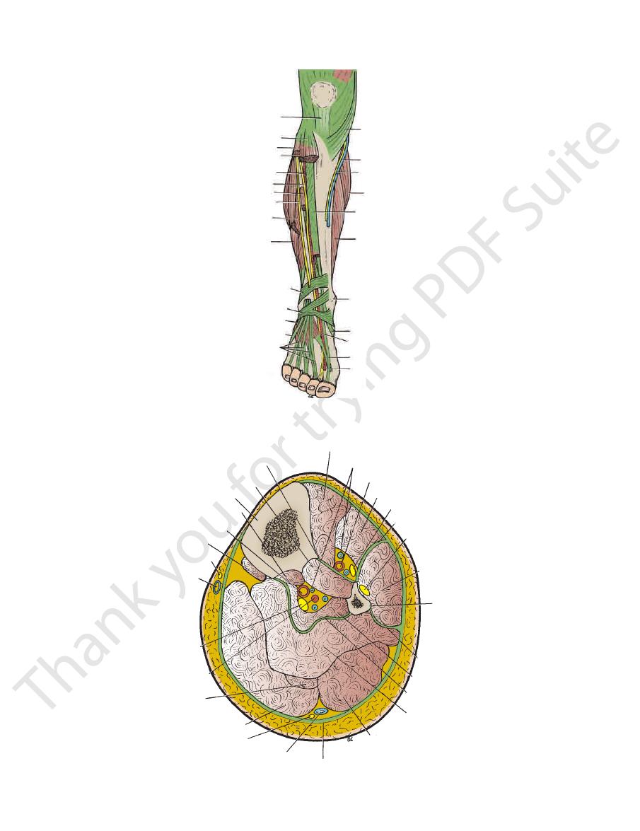
FIGURE 10.44
Deep structures in the anterior and lateral aspects of the right leg and the dorsum of the foot.
interosseous membrane
tibialis posterior
tibia
flexor digitorum longus
popliteus
saphenous nerve
great saphenous vein
tibial nerve
peroneal artery
plantaris
gastrocnemius (medial head)
sural nerve
small saphenous vein
deep fascia
gastrocnemius (lateral head)
soleus
deep transverse fascia
flexor hallucis longus
posterior fascial septum
fibula
peroneus longus
superficial peroneal nerve
anterior fascial septum
deep peroneal nerve
extensor digitorum longus
anterior tibial artery
venae comitantes
tibialis anterior
posterior tibial artery
medial
lateral
FIGURE 10.45
Transverse section through the middle of the right leg as seen from above.
Basic Anatomy
tal ends of the anterior borders of the fibula and the tibia
The superior extensor retinaculum is attached to the dis
Superior Extensor Retinaculum
481
-
(Figs. 10.46, 10.47, and 10.50).
The inferior extensor retinaculum is a Y-shaped band
Inferior Extensor Retinaculum
located in front of the ankle joint (Figs. 10.44, 10.46, and
The superior peroneal retinaculum connects the lateral
Superior Peroneal Retinaculum
lined by a synovial sheath.
tendons lie in compartments (Fig. 10.48), each of which is
medial malleolus as they pass forward to enter the sole. The
the deep muscles of the back of the leg to the back of the
face of the calcaneum (Fig. 10.49). It binds the tendons of
downward and backward to be attached to the medial sur
The flexor retinaculum extends from the medial malleolus
Flexor Retinaculum
synovial sheath.
ments (Figs. 10.48 and 10.50), each of which is lined by a
10.47). Fibrous bands separate the tendons into compart
-
-
malleolus to the lateral surface of the calcaneum (Fig. 10.49).
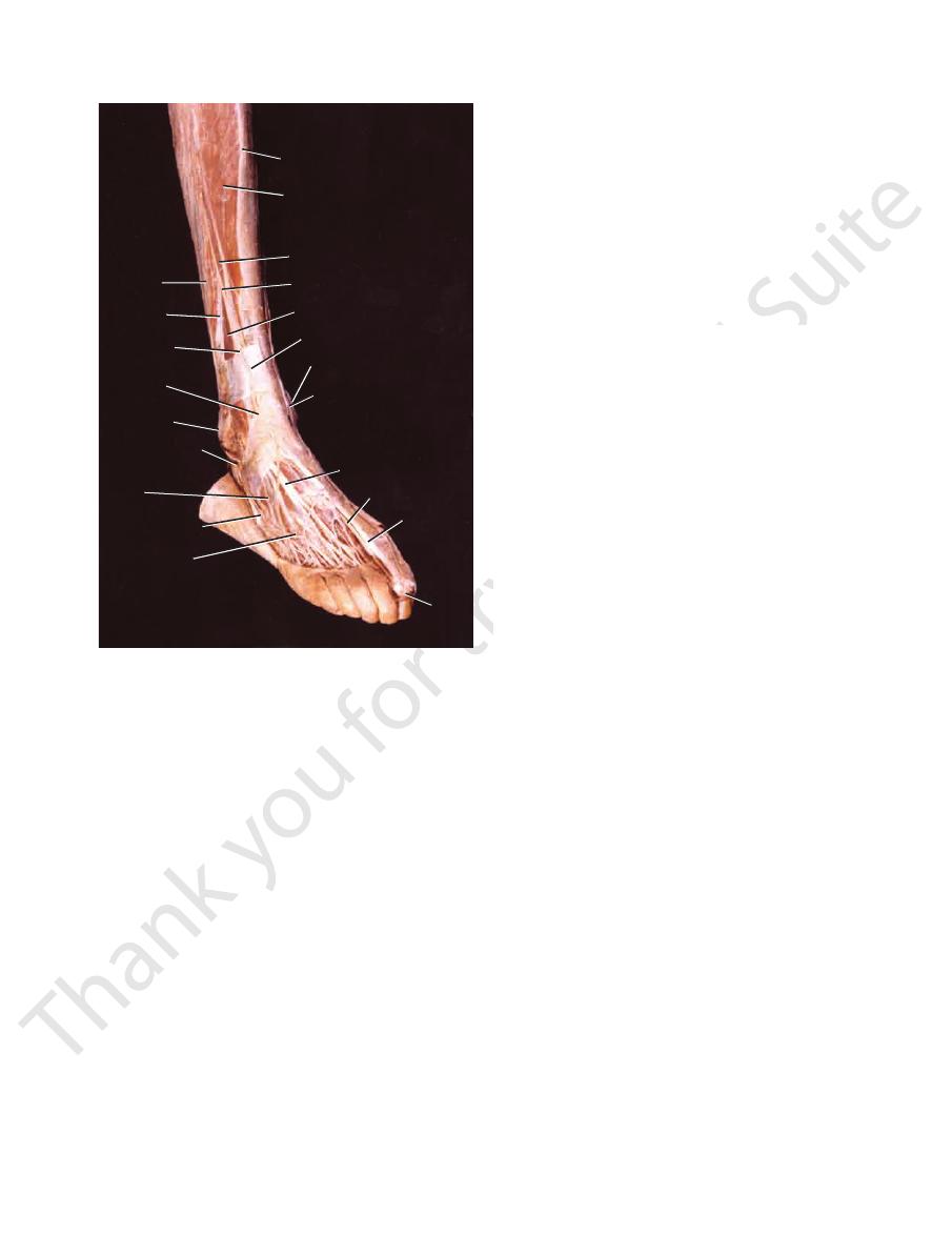
big
toe
peroneus
longus
anterior border
of tibia
tibialis anterior
extensor hallucis longus
superficial peroneal nerve
extensor digitorum longus
superior extensor
retinaculum
medial malleolus
great saphenous vein
extensor digitorum longus
dorsalis pedis artery
peroneus
brevis
superficial
peroneal nerve
inferior extensor
retinaculum
lateral
malleolus
peroneus longus
and brevis tendons
extensor digitorum
brevis
peroneus
tertius
dorsal
venous
network
extensor
hallucis
longus
FIGURE 10.46
Dissection of the front of the right leg and
mon peroneal nerve (see page 479), supplies the skin of the
a branch of the com
superficial peroneal nerve,
The
on the upper part of the lateral surface of the leg (Fig. 10.1).
common peroneal nerve (see page 479), supplies the skin
a branch of the
lateral cutaneous nerve of the calf,
The
Cutaneous Nerves
retinacula is described on pages 490.
The arrangement of the tendons beneath the different
mon sheath.
synovial sheath, which is continuous above with the com
of the calcaneum (Fig. 10.49). The tendons each possess a
the peroneus longus and brevis muscles to the lateral side
The inferior peroneal retinaculum binds the tendons of
Inferior Peroneal Retinaculum
with a common synovial sheath.
the back of the lateral malleolus. The tendons are provided
It binds the tendons of the peroneus longus and brevis to
dorsum of the foot.
-
The Front of the Leg
Skin
-
lower part of the anterolateral surface of the leg (Fig. 10.2).
joint along with the other muscles in this compartment
The peroneus tertius muscle extends the foot at the ankle
of the foot away from the ground.
Extension, or dorsiflexion of the ankle, is the movement
Note the following:
10.48, and 10.49 and are described in Table 10.5.
The muscles are seen in Figures 10.44, 10.45, 10.46, 10.47,
Muscles of the Anterior Fascial Compartment
Deep peroneal nerve
Nerve supply:
Anterior tibial artery
Blood supply:
gus, peroneus tertius, and extensor hallucis longus
The tibialis anterior, extensor digitorum lon
Muscles:
(Fig. 10.4).
the small saphenous vein and drain into the popliteal nodes
of the front of the leg may pass via vessels that accompany
10.4). A small amount of lymph from the upper lateral part
the vertical group of superficial inguinal lymph nodes (Fig.
in vessels that follow the great saphenous vein, to end in
fascia on the front of the leg drains upward and medially
The greater part of the lymph from the skin and superficial
Lymph Vessels
(Fig. 10.51).
the leg and ultimately drain into the great saphenous vein
Numerous small veins curve around the medial aspect of
Superficial Veins
face of the leg (Fig. 10.2).
(see page 463), supplies the skin on the anteromedial sur
a branch of the femoral nerve
saphenous nerve,
The
-
Contents of the Anterior Fascial
Compartment of the Leg
■
■
-
■
■
■
■
of the Leg
■
■
■
■