Surface Anatomy
the lower part of the anterior abdominal wall.
bodies of the pubic bones can be felt on palpation through
and the
upper margin of the symphysis pubis
10.80). The
in the midline between the bodies of the pubic bones (Fig.
is a cartilaginous joint that lies
symphysis pubis
The
pubic tubercle (Figs. 10.83 and 10.84).
ally to the anterior superior iliac spine and medially to the
groin and can be felt along its length. It is attached later
lies beneath the skin fold in the
inguinal ligament
The
and the ischial tuberosity (Figs. 10.82 and 10.83).
down, midway between the tip of the greater trochanter
superior iliac spine and the ischial tuberosity and, lower
ward, it is situated at first midway between the posterior
gluteus maximus muscle. As it curves laterally and down
in the buttock lies under cover of the
sciatic nerve
The
the lower border of the gluteus maximus muscle.
standing position; its lower border does not correspond to
is most prominent in the
fold of the buttocks
The
be palpated with a gloved finger in the anal canal.
the anus (Fig. 10.83). The anterior surface of the coccyx can
the cleft between the buttocks about 1 in. (2.5 cm) behind
can be palpated beneath the skin in
coccyx
The tip of the
cleft between the buttocks.
crest can be felt beneath the skin in the upper part of the
The
median sacral crest.
fused with each other to form the
of the sacrum (Fig. 10.79) are
spinous processes
The
ischial tuberosity (Fig. 10.83).
on a line connecting the anterior superior iliac spine to the
hip joint, the upper border of the greater trochanter lies
and extended. It is important to verify that, in the normal
moves beneath the examining finger as the hip joint is flexed
lateral surface of the thigh (Figs. 10.82 and 10.83) and
of the femur can be felt on the
greater trochanter
The
a pad of fat.
tuberosity is separated from the skin by only a bursa and
and supports the weight of the body; in this position, the
from beneath the lower border of the gluteus maximus
mus. In the sitting position, the ischial tuberosity emerges
ing position, the tuberosity is covered by the gluteus maxi
part of the buttock (Figs. 10.82 and 10.83). In the stand
can be palpated in the lower
ischial tuberosity
The
rior iliac spine (Fig. 10.83).
iliac crest about 2 in. (5 cm) posterior to the anterior supe
is a prominence felt on the outer surface of the
tubercle
vertebra and the middle of the sacroiliac joint. The
lies beneath a skin dimple at the level of the second sacral
(Fig. 10.82); the latter
posterior superior iliac spine
(Figs. 10.79 and 10.80) and behind
rior superior iliac spine
ante
(Figs. 10.82 and 10.83). Each crest ends in front at the
are easily palpable along their entire length
iliac crests
The
513
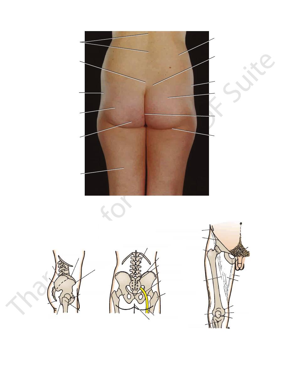
Gluteal Region
-
at the
iliac
-
-
-
-
Inguinal Region
-
FIGURE 10.69
y.)
Ectromelia. (Courtesy of G. Aver
FIGURE 10.70
Radiograph of bilateral congenital dislocation
shallow acetabular fossae. (Courtesy of J. Adams.)
of the hip showing that the femoral heads are not within the
FIGURE 10.71
Talipes equinovarus. (Courtesy of J. Adams.)
Surface Anatomy
519
spinous processes
of lumbar vertebrae
fused spinous
processes
of sacrum
greater trochanter
of femur
position
of sciatic nerve
site of ischial
tuberosity
hamstring
group of muscles
iliac crest
posterior superior
iliac spine
gluteus medius
gluteus maximus
natal cleft
fold of buttock
FIGURE 10.82
The gluteal region and the posterior aspect of the thigh of a 25-year-old woman.
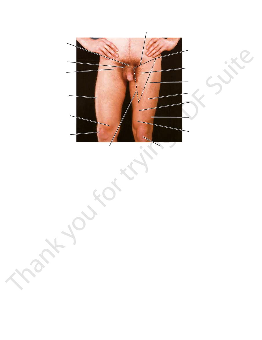
iliac tubercle
anterior superior
iliac spine
pubic tubercle
greater
trochanter
ischial tuberosity
coccyx
posterior
superioriliac
spine
iliac crest
posterior superior iliac spine
iliac crest
sacral spines
iliac tubercle
sciatic nerve
greater trochanter
fold of buttock
natal cleft
anterior superior iliac spine
inguinal ligament
greater
trochanter
sartorius
lateral
condyle
tibial
tuberosity
patella
medial condyle
adductor
tubercle
femoral
triangle
adductor
longus
pubic
tubercle
iliac tubercle
e
e
e
anterior superior
iliac spine
pubic tubercle
greater
trochanter
chial tuberosity
c
crest
posterior superior iliac spine
iliac crest
sacral spines
iliac tubercle
sciatic nerve
greater trochanter
fold of buttock
natal cleft
anterior superior iliac spine
inguinal ligament
greater
trochanter
sartorius
lateral
condyle
tibial
tuberosity
patella
medial co
adductor
tubercle
femora
triangle
adduc
longus
p
t
FIGURE 10.83
Surface markings in the gluteal region and the front of the thigh.
520
CHAPTER 10
The Lower Limb
of femoral artery
pubic tubercle
symphysis pubis
site for palpation
rectus femoris
vastus medialis
patella
adductor longus
patella
vastus medialis
vastus lateralis
subsartorial
(adductor canal)
rectus femoris
sartorius
femoral triangle
anterior superior
iliac spine
inguinal ligament
FIGURE 10.84
The broken lines indicate the boundaries of the femoral
Anterior aspect of the thigh of a 27-year-old man.
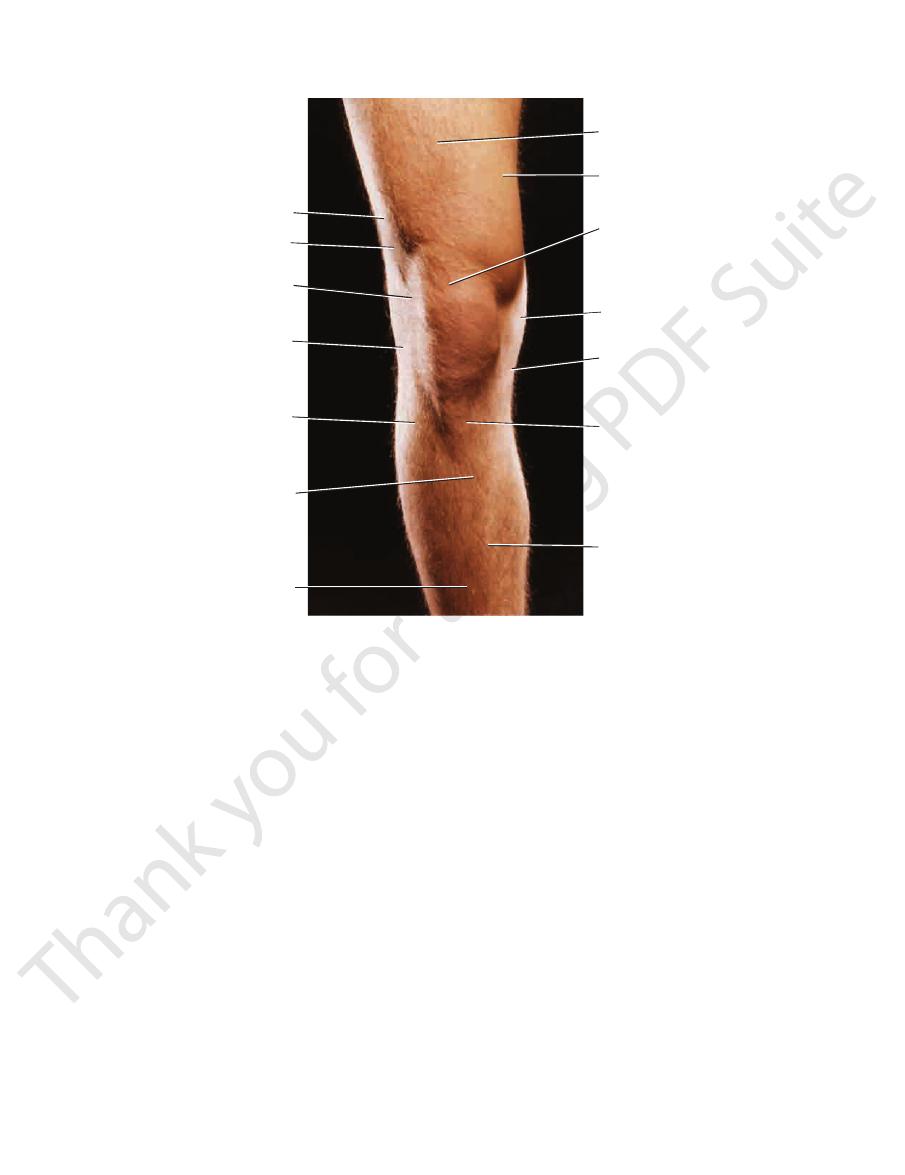
to their bony attachments. Because the ligaments cover the
sides of the joint line; they can be followed above and below
can be palpated on the
lateral collateral ligament
rounded
medial collateral ligament
The bandlike
between them (Fig. 10.85).
on the sides of the knee, and the joint line can be identified
can be recognized
condyles of the femur and tibia
The
tuberosity of the tibia.
tum patellae can be traced downward to its attachment to
can be easily palpated (Fig. 10.85). The ligamen
patellae
ligamentum
patella
In front of the knee joint, the
and the saphenous nerve.
longus and magnus muscles. It contains the femoral vessels
the vastus medialis muscle and posteriorly by the adductor
beneath the sartorius muscle and is bounded laterally by
the femoral triangle. It is an intermuscular cleft situated
of the thigh (Fig. 10.84), immediately distal to the apex of
lies in the middle third
adductor (subsartorial) canal
The
tubercle (Figs. 10.3 and 10.19).
femoral vein 1.5 in. (4 cm) below and lateral to the pubic
ing in the deep fascia (fascia lata) of the thigh and joins the
pierces the saphenous open
great saphenous vein
The
femoral artery (Fig. 10.6).
of the inguinal ligament—that is, lateral to the pulsating
enters the thigh behind the midpoint
femoral nerve
The
lateral to the pubic tubercle (Figs. 10.3 and 10.6).
lies below and
femoral canal
The lower opening of the
(Fig. 10.6).
inguinal ligament medial to the pulsating femoral artery
leaves the thigh by passing behind the
femoral vein
The
sations are easily felt (Fig. 10.84).
symphysis pubis to the anterior superior iliac spine; its pul
ligament (Fig. 10.6) at the midpoint of a line joining the
enters the thigh behind the inguinal
femoral artery
The
and parallel to the inguinal ligament (Fig. 10.3).
can be palpated in the superficial fascia just below
nodes
superficial inguinal lymph
The horizontal group of
by the adductor longus muscle.
medial border
by the sartorius muscle,
lateral border
inguinal ligament, the
of the triangle is formed by the
and laterally rotated. The
triangle can be identified when the thigh is flexed, abducted,
and 10.84). In a thin, muscular subject, the boundaries of the
fold of the groin in the upper part of the thigh (Figs. 10.83
The femoral triangle can be seen as a depression below the
Femoral Triangle
of the body of the pubis, medial to the pubic tubercle (Figs.
is the ridge of bone on the upper surface
pubic crest
The
the lateral margin of the labium majus.
examining finger. In the female, it can be palpated through
palpated in the male by invaginating the scrotum with the
medial end of the inguinal ligament. The tubercle is easily
of the pubis (Figs. 10.83 and 10.84). Attached to it is the
can be felt on the upper border
pubic tubercle
The
triangle. The right leg is laterally rotated at the hip joint.
10.7 and 10.8).
base
and the
-
-
Adductor Canal
Knee Region
and the
-
the
and the
Surface Anatomy
521
subcutaneous
surface of tibia
ligamentum
patellae (attached
to tuberosity of tibia)
medial condyle
of tibia
medial condyle
of femur
patella (upper margin)
vastus medialis
rectus femoris
lateral
medial
vastus lateralis
lateral condyle
of femur
iliotibial tract
tibialis anterior
anterior border
of tibia
position of
joint line
fibula
FIGURE 10.85
Anterior aspect of the right knee of a 27-year-old man.
the examining finger just below the head of the fibula
can be rolled beneath
common peroneal nerve
The
(Fig. 10.85).
head of the fibula
on the lateral aspect of the knee and can be traced down to
can be felt as a rounded structure
tendon of biceps
The
and the medial and lateral collateral ligaments, respectively.
palpated on the joint line between the ligamentum patellae
the outer edges of the medial and lateral menisci can be
femoral and tibial condyles. Although not recognizable,
are located in the interval between the
menisci
The
collateral ligaments (Fig. 10.61).
joint line, the joint line cannot be palpated at the sites of the
the
(Fig. 10.86); here, it passes forward around the lateral side
posterior
tendon of flexor digitorum longus,
rior,
tendon of tibialis poste
structures, in the order named: the
and the medial surface of the calcaneum lie the following
In the interval behind the medial malleolus (Fig. 10.86)
the tip of the lateral malleolus (Figs. 10.86 and 10.87).
the tibia lies about 0.5 in. (1.3 cm) proximal to the level of
of
medial malleolus
(Figs. 10.86 and 10.87). The tip of the
lateral malleolus
can be followed downward to form the
In the region of the ankle, the fibula is subcutaneous and
(Fig. 10.85).
subcutaneous and can be felt throughout their length
are
tibia
The medial surface and anterior border of the
Tibia
cia is fully relaxed by passively flexing the knee joint.
the depths of the popliteal fossa, provided that the deep fas
can be felt by gentle palpation in
popliteal artery
The
beneath the finger.
With the knee joint partially flexed, the nerve can be rolled
as the latter passes to its insertion on the head of the fibula.
medial side of the tendon of the biceps femoris (Fig. 10.86),
can be palpated on the
common peroneal nerve
The
each side by one of the heads of the gastrocnemius muscle.
and semitendinosus muscles. Its lower part is bounded on
by the tendons of the semimembranosus
medially
cle and
by the tendon of the biceps femoris mus
laterally
bounded
and the boundaries are easily defined. Its upper part is
flexed, the deep fascia, which roofs over the fossa, is relaxed
(Fig. 10.86). When the knee is
popliteal fossa
sion called the
Behind the knee joint is a diamond-shaped skin depres
it (Fig. 10.86).
can be felt passing to
adductor magnus
string part of the
aspect of the femur just above the medial condyle; the ham
can be palpated on the medial
adductor tubercle
The
of the bone.
-
-
-
-
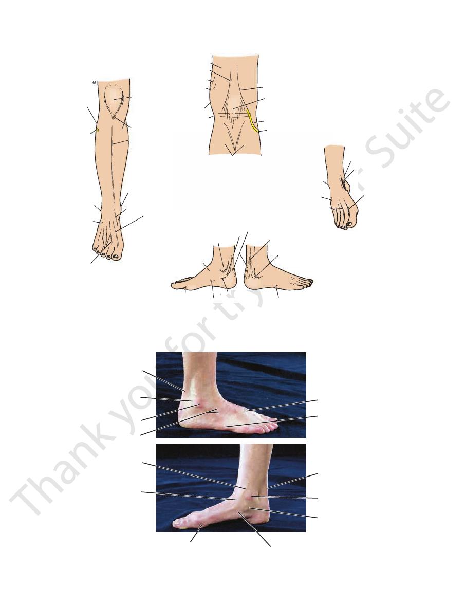
Ankle Region and Foot
-
the
the
522
CHAPTER 10
The Lower Limb
medial
semimembranosus
semitendinosus
adductor magnus tendon
adductor tubercle
patella
condyle of femur
joint line
ligamentum
patellae
anterior border
of tibia
head of fibula
common peroneal
nerve
lateral malleolus
extensor digitorum
brevis
extensor digitorum
longus tendons
head of talus
tuberosity of navicular bone
sustentaculum tali
tuberosity of fifth metatarsal
lateral malleolus
peroneus longus and brevis tendons
tendo calcaneus
tibialis posterio tendon
medial malleolus
extensor hallucis longus tendon
tibialis anterior
tendon
medial malleolus
gastrocnemius
(medial and lateral heads)
lateral malleolus
talus
extensor digitorum
longus tendons
extensor hallucis
longus tendon
tibialis anterior tendon
medial malleolus
common peroneal nerve
head of fibula
popliteal fossa
biceps femoris
lateral
medial aspect of foot
lateral aspect of foot
metatarsophalangeal joint of big toe
FIGURE 10.86
Surface markings in the popliteal fossa, the front of the leg, and the foot.
dorsal venous
tuberosity of navicular
great saphenous vein
digitorum brevis
extensor
longus and brevis
tendo calcaneus
tendons of peroneus
lateral malleolus
head of talus
head of first metatarsal
sustentaculum tali
medial malleolus
tendo calcaneus
tuberosity of fifth
metatarsal
arch
A
B
FIGURE 10.87
Lateral aspect
of the right ankle of a 29-year-old woman.
(A) and medial aspect (B)
Surface Anatomy
of the calcaneum can be palpated about 1 in. (2.5 cm) below
peroneal tubercle
On the lateral aspect of the foot, the
lateral malleolus (Fig. 10.19).
behind
the lateral part of the plexus and passes up
drains
small saphenous vein
malleolus (Fig. 10.87). The
of the medial
in front
part of the plexus and passes upward
leaves the medial
great saphenous vein
and 10.87). The
dorsal surface of the foot proximal to the toes (Figs. 10.19
can be seen on the
or
dorsal venous arch
The
toes (Fig. 10.86).
can be made prominent by dorsiflexing the
lucis longus
extensor hal
tendons of extensor digitorum longus
be palpated just in front of the malleoli (Fig. 10.87). The
head of the talus
On the dorsum of the foot, the
(Achilles tendon) (Fig. 10.88).
tendo calcaneus
heel is the
Above the
calcaneum.
nence of the heel is formed by the
On the posterior surface of the ankle joint, the promi
longus, midway between the two malleoli on the front of
tendons of extensor hallucis longus and extensor digitorum
can be felt between the
dorsalis pedis artery
sations of the
The pul
peroneus tertius.
extensor digitorum longus
tendons of
Lateral to the extensor hallucis longus lie the
stand out by extending the big toe (Figs. 10.86 and 10.88).
lies lateral to it and can be made to
sor hallucis longus
tendon of exten
and inverted (Figs. 10.86 and 10.88). The
can be seen when the foot is dorsiflexed
tibialis anterior
tendon of
On the anterior surface of the ankle joint, the
(Figs. 10.87
longus
tendons of peroneus brevis
are the
lus and the heel (Fig. 10.88). Behind the lateral malleolus
can be felt halfway between the medial malleo
tibial artery
posterior
The pulsations of the
of flexor hallucis longus.
tendon
posterior tibial nerve,
tibial vessels,
523
the
and the
-
and
and 10.88).
-
and
-
the ankle.
-
can
and
-
plexus
the
extensor
digitorum longus
lateral malleolus
dorsal venous arch
medial malleolus
site for palpation
of posterior tibial artery
tendons of peroneus
longus and brevis
lateral malleolus
tendo calcaneus
tendon of extensor
hallucis longus
tendon of tibialis
anterior
medial malleolus
A
B
FIGURE 10.88
Anterior aspect
of the right foot and ankle of a 29-year-old woman.
(A) and posterior aspect (B)
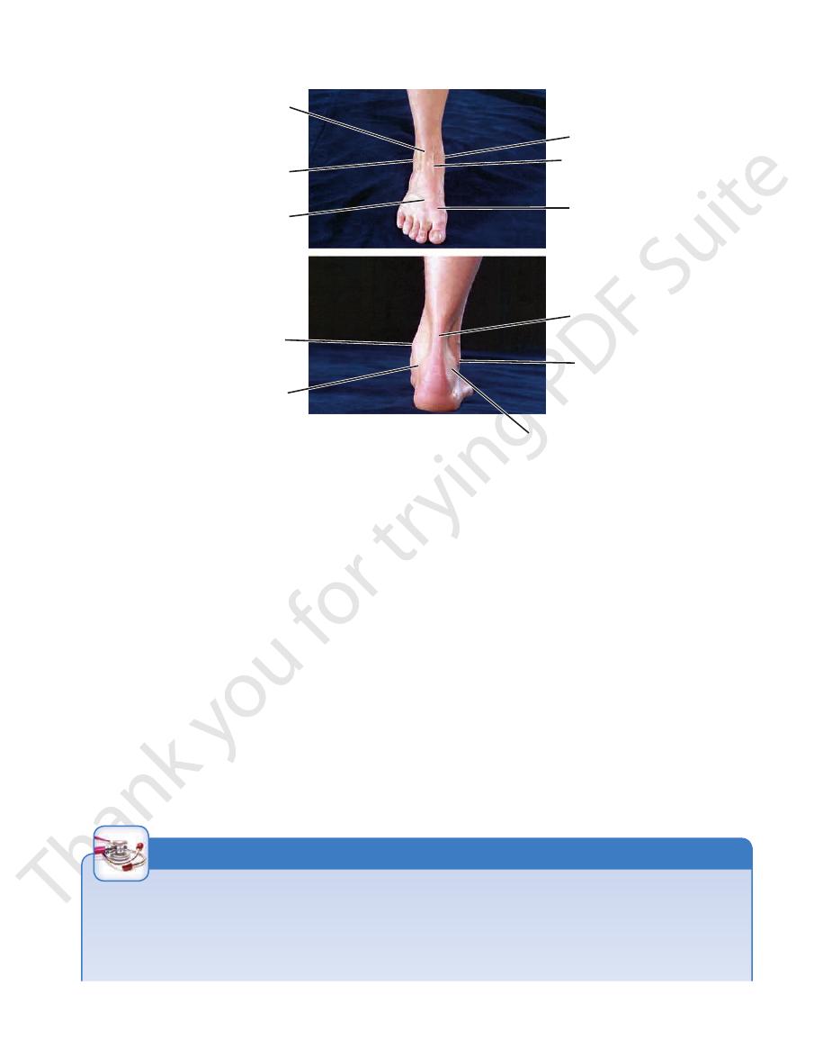
Arterial Palpation
and the symphysis pubis (Fig. 10.84). The artery is easily palpated
ment at a point midway between the anterosuperior iliac spine
Every health professional should know the precise position of the
main arteries within the lower limb, for he or she may be called
on to arrest a severe hemorrhage or palpate different parts of
the arterial tree in patients with arterial occlusion.
The femoral artery enters the thigh behind the inguinal liga-
here because it can be pressed backward against the pectineus
and the superior ramus of the pubis.
C L I N I C A L N O T E S O N T H E A R T E R I E S O F T H E L O W E R L I M B
(continued)
526
CHAPTER 10
alis posterior muscle.
receives the main part of the tendon of insertion of the tibi
can be seen and palpated (Fig. 10.87). It
navicular bone
tuberosity of the
In front of the sustentaculum tali, the
lower surface.
and the tendon of flexor hallucis longus winds around its
don of flexor digitorum longus crosses its medial surface;
rior lies immediately above the sustentaculum tali; the ten
medial malleolus (Fig. 10.87). The tendon of tibialis poste
can be palpated about 1 in. (2.5 cm) below the tip of the
sustentaculum tali
On the medial aspect of the foot, the
enter the groove on the under aspect of the cuboid bone.
passes forward to
tendon of peroneus longus
tubercle, the
(Fig. 10.87). Below the
5th metatarsal bone
base of the
forward to its insertion on the prominent tuberosity on the
tendon of peroneus brevis
Above the tubercle, the
and in front of the tip of the lateral malleolus (Fig. 10.86).
The Lower Limb
passes
-
-
-
Clinical Cases
www.thePoint.lww.com/Snell9e.
and
Review Questions
are available online at
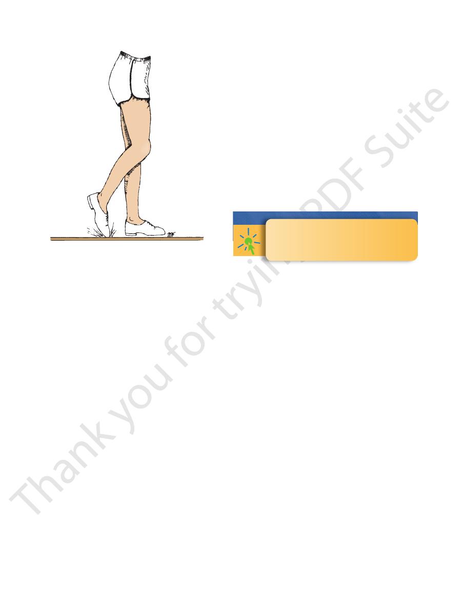
FIGURE 10.89
Footdrop. With this condition, the individual
catches his or her toes on the ground when walking.