
Basic Anatomy
the ground.
ning by using the foot as a lever and raising the heel off
the main forward propulsive force in walking and run
powerful plantar flexors of the ankle joint. They provide
Together, the soleus, gastrocnemius, and plantaris act as
Note the following:
described in Table 10.7.
The muscles are seen in Figures 10.45 and 10.53 and are
of the Leg: Superficial Group
Muscles of the Posterior Fascial Compartment
Tibial nerve
Nerve supply:
Posterior tibial artery
Blood supply:
gus, flexor hallucis longus, and tibialis posterior
Popliteus, flexor digitorum lon
Deep group of muscles:
and soleus
Gastrocnemius, plantaris,
Superficial group of muscles:
superficial and deep groups (see Fig. 10.45).
divides the muscles of the posterior compartment into
of the leg is a septum that
deep transverse fascia
The
liteal nodes (Fig. 10.4).
group of superficial inguinal nodes or drain into the pop
around the medial side of the leg to end in the vertical
the back of the leg drain upward and either pass forward
Lymph vessels from the skin and superficial fascia on
Lymph Vessels
great saphenous vein.
division joining the popliteal and the other joining the
join the great saphenous vein; or it may split in two, one
subject to variation: It may join the popliteal vein; it may
The mode of termination of the small saphenous vein is
medially and join the great saphenous vein (Fig. 10.19)
that run upward and
anastomotic branches
Important
with the deep veins of the foot
Communicating veins
from the back of the leg
small veins
Numerous
Tributaries
small saphenous vein has numerous valves along its course.
and 10.40); it ends in the popliteal vein (see page 478). The
muscle in the lower part of the popliteal fossa (Figs. 10.19
fascia and passes between the two heads of the gastrocnemius
up the middle of the back of the leg. The vein pierces the deep
lows the lateral border of the tendo calcaneus and then runs
the lateral malleolus in company with the sural nerve. It fol
behind
dorsal venous arch of the foot (Fig. 10.19). It ascends
arises from the lateral part of the
small saphenous vein
The
Superficial Veins
the posteromedial surface of the leg (Fig. 10.1).
(see page 463), gives off branches that supply the skin on
a branch of the femoral nerve
saphenous nerve,
The
eral surface of the leg (Fig. 10.1).
479), supplies the skin on the lower part of the posterolat
a branch of the tibial nerve (see page
sural nerve,
The
(Fig. 10.1).
on the upper part of the posterolateral surface of the leg
common peroneal nerve (see page 479), supplies the skin
a branch of the
lateral cutaneous nerve of the calf,
The
of the back of the leg (Fig. 10.1).
supplies the skin over the popliteal fossa and the upper part
the back of the thigh (see page 465). In the popliteal fossa, it
of the thigh descends on
posterior cutaneous nerve
The
Cutaneous Nerves
of the little toe (see page 498).
cent sides of the first and second toes and the lateral side
dorsal surfaces of the skin of all the toes, except the adja
the dorsum of the foot. In addition, branches supply the
to the skin on the lower part of the front of the leg and
Medial and lateral branches are distributed
Cutaneous:
(Fig. 10.44)
branches to the peroneus longus and brevis
Muscular
Branches
cutaneous (Figs. 10.47 and 10.50).
brevis muscles, and in the lower part of the leg it becomes
and 10.50). It descends between the peroneus longus and
the lateral side of the neck of the fibula (Figs. 10.44, 10.46,
487
■
■
■
■
-
The Back of the Leg
Skin
-
-
■
■
■
■
■
■
-
Contents of the Posterior Fascial
Compartment of the Leg
■
■
■
■
-
■
■
■
■
■
■
-
fibers of the soleus or partial tearing of the tendo calcaneus is
gastrocnemius and soleus muscles retract proximally, leaving
tion. A sudden, sharp pain is felt, with immediate disability. The
Tearing of the gastrocnemius or soleus muscles will produce
Gastrocnemius and Soleus Muscle Tears
severe localized pain over the damaged muscle. Swelling may
be present.
Ruptured Tendo Calcaneus
Rupture of the tendo calcaneus is common in middle-aged
men and frequently occurs in tennis players. The rupture
occurs at its narrowest part, about 2 in. (5 cm) above its inser-
a palpable gap in the tendon. It is impossible for the patient to
actively plantar flex the foot. The tendon should be sutured as
soon as possible and the leg immobilized with the ankle joint
plantar flexed and the knee joint flexed.
Rupture of the Plantaris Tendon
Rupture of the plantaris tendon is rare, although tearing of the
frequently diagnosed as such a rupture.
Plantaris Tendon and Autografts
The plantaris muscle, which is often missing, can be used for
tendon autografts in repairing severed flexor tendons to the
fingers; the tendon of the palmaris longus muscle can also be
used for this purpose.
C L I N I C A L N O T E S
488
CHAPTER 10
The Lower Limb
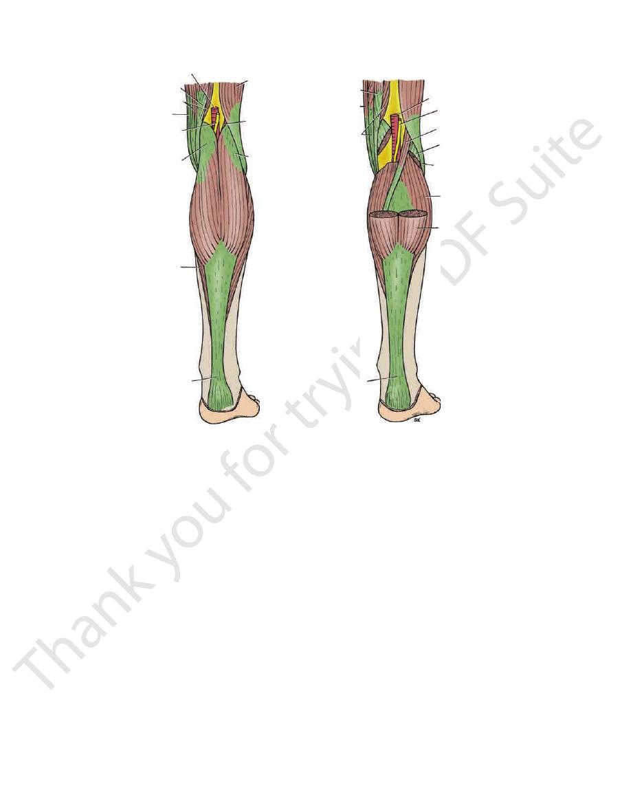
popliteal artery
semitendinosus
semimembranosus
gracilis
tibial nerve
gastrocnemius
soleus
tendo calcaneus
tendo calcaneus
gastrocnemius
soleus
biceps femoris
lateral head of
gastrocnemius
plantaris
tibial nerve
popliteal artery
semitendinosus
gracilis
semimembranosus
gastrocnemius
plantaris
biceps femoris
A
B
FIGURE 10.53
Structures in the posterior aspect of the right leg. In
The posterior tibial artery is one of the terminal branches
Posterior Tibial Artery
Artery of the Posterior Fascial Compartment
knee joint.
The popliteus muscle is responsible for “unlocking” the
of the femur and the tibia.
is freer to move and adapt to the surfaces of the condyle
so that the meniscus is not tethered to the ligament and
eral ligament of the knee joint from the lateral meniscus
terior surface of the tibia. The tendon separates the lat
knee joint and is inserted into the upper part of the pos
The popliteus muscle arises inside the capsule of the
Note the following:
10.49 and are described in Table 10.7.
The muscles are seen in Figures 10.43, 10.45, 10.48, and
of the Leg: Deep Group
Muscles of the Posterior Fascial Compartment
part of the gastrocnemius has been removed.
B,
■
■
-
-
■
■
of the Leg
of the popliteal artery (see page 477). It begins at the level
form the popliteal vein.
those of the anterior tibial artery in the popliteal fossa to
of the posterior tibial artery join
Venae comitantes
(see page 495).
Medial and lateral plantar arteries
around the ankle joint.
which join other arteries
Anastomotic branches,
Nutrient artery to the tibia.
posterior compartment of the leg.
are distributed to muscles in the
Muscular branches
of the leg.
seous membrane to reach the lower part of the front
pierces the interos
perforating branch
ankle joint. A
and ends by taking part in the anastomosis around the
nutrient artery to the fibula
muscular branches
posterior to it. The peroneal artery gives off numerous
the substance of the flexor hallucis longus muscle or
10.44). It descends behind the fibula, either within
close to the origin of the posterior tibial artery (Fig.
which is a large artery that arises
Peroneal artery,
Branches
dividing into medial and lateral plantar arteries (Fig. 10.49).
malleolus deep to the flexor retinaculum and terminates by
by skin and fascia. The artery passes behind the medial
below. In the lower part of the leg, the artery is covered only
rior muscle above and on the posterior surface of the tibia
10.44). It lies on the posterior surface of the tibialis poste
deep transverse fascia of the leg (Figs. 10.41, 10.42, and
downward deep to the gastrocnemius and soleus and the
of the lower border of the popliteus muscle and passes
-
■
■
and a
-
■
■
■
■
■
■
■
■

Basic Anatomy
489
Muscles of the Posterior Fascial Compartment of the Leg
T A B L E 1 0 . 7
Tibial nerve
Tuberosity of
Tibialis posterior
supports medial longitudinal
plantar flexes foot at ankle joint;
Flexes distal phalanx of big toe;
Tibial nerve
Tibial nerve
Tibial nerve
Together with gastrocnemius and
Tibial nerve
Via tendo calcaneus
Tibial nerve
Tibial nerve
Via tendo
Muscle
Origin
Insertion
Nerve Supply
Nerve Root
a
Action
Superficial Group
Gastrocnemius
Lateral head from
lateral condyle of
femur and medial
head from above
medial condyle
calcaneus into
posterior surface
of calcaneum
S1, 2
Plantar flexes foot at ankle joint;
flexes knee joint
Plantaris
Lateral
supracondylar
ridge of femur
Posterior surface of
calcaneum
S1, 2
Plantar flexes foot at ankle joint;
flexes knee joint
Soleus
Shafts of tibia and
fibula
into posterior
surface of
calcaneum
S1, 2
plantaris is powerful plantar
flexor of ankle joint; provides
main propulsive force in
walking and running
Deep Group
Popliteus
Lateral surface of
lateral condyle of
femur
Posterior surface
of shaft of tibia
above soleal line
L4, 5; S1
Flexes leg at knee joint; unlocks
knee joint by lateral rotation
of femur on tibia and slackens
ligaments of joint
Flexor digitorum
longus
Posterior surface of
shaft of tibia
Bases of distal
phalanges of
lateral four toes
S2, 3
Flexes distal phalanges of lateral
four toes; plantar flexes foot
at ankle joint; supports medial
and lateral longitudinal arches
of foot
Flexor hallucis
longus
Posterior surface of
shaft of fibula
Base of distal
phalanx of big toe
S2, 3
arch of foot
Posterior surface
of shafts of tibia
and fibula and
interosseous
membrane
navicular bone
and other
neighboring
bones
L4, 5
Plantar flexes foot at ankle
joint; inverts foot at subtalar
and transverse tarsal joints;
supports medial longitudinal
arch of foot
a
The predominant nerve root supply is indicated by boldface type.
lum and divides into the medial and lateral plantar nerves.
longus (Fig. 10.50). It is covered here by the flexor retinacu
tendons of the flexor digitorum longus and the flexor hallucis
the artery, passes behind the medial malleolus, between the
terior to it, and finally lies on its lateral side. The nerve, with
tibial artery and lies at first on its medial side, then crosses pos
of the tibia (Fig. 10.43). The nerve accompanies the posterior
posterior and, lower down the leg, on the posterior surface
10.43 and 10.53). It lies on the posterior surface of the tibialis
passes deep to the gastrocnemius and soleus muscles (Figs.
(see page 467). It descends through the popliteal fossa and
nerve (Fig. 10.17) in the lower third of the back of the thigh
The tibial nerve is the larger terminal branch of the sciatic
Tibial Nerve
Nerve of the Posterior Fascial Compartment
of the Leg
-
-
Deep Vein Thrombosis and Long-Distance Air
often fatal. Preventative measures include stretching of the
the heart and lungs, causing pulmonary embolism, which is
Should the thrombus become dislodged, it passes rapidly to
vein thrombosis can also occur with no signs or symptoms.
ness in the calf and calf muscle tenderness. However, deep
of the veins of the soleus muscle gives rise to mild pain or tight
are very prone to deep vein thrombosis in the legs. Thrombosis
Passengers who sit immobile for hours on long-distance flights
Travel
-
legs every hour to improve the venous circulation.
C L I N I C A L N O T E S

490
CHAPTER 10
to absorb shocks, such as in jumping.
shape to uneven surfaces. It also serves as a resilient spring
structed in the form of arches, which enable it to adapt its
age for walking and running. It is unique in that it is con
The foot supports the body weight and provides lever
(see Fig. 10.48).
The fat and the large tendo calcaneus lie behind the ankle
Structures That Lie Directly behind the Ankle
rate sheaths.
beneath the inferior peroneal retinaculum, they have sepa
10.59) share a common synovial sheath. Lower down,
The peroneus longus and brevis tendons (Figs. 10.48 and
Malleolus beneath the Superior Peroneal
Structures That Pass behind the Lateral
Small saphenous vein (see Fig. 10.48)
The sural nerve
Malleolus Superficial to the Superior Peroneal
Structures That Pass behind the Lateral
lum, it is surrounded by a synovial sheath.
As each of these tendons passes beneath the flexor retinacu
Flexor hallucis longus (Figs. 10.48 and 10.49)
Tibial nerve
Posterior tibial artery with venae comitantes
Flexor digitorum longus
Tibialis posterior tendon
From Medial to Lateral
Malleolus beneath the Flexor Retinaculum
Structures That Pass behind the Medial
Saphenous nerve (Figs. 10.48 and 10.51)
Great saphenous vein
Structures That Pass in Front of the Medial
oneus tertius share a common synovial sheath.
The tendons of extensor digitorum longus and the per
extensor retinacula, it is surrounded by a synovial sheath.
As each of the above tendons passes beneath or through the
Peroneus tertius (Fig. 10.48)
Extensor digitorum longus tendons
Deep peroneal nerve
Anterior tibial artery with venae comitantes
Extensor hallucis longus tendon
Tibialis anterior tendon
Extensor Retinacula from Medial to Lateral
Structures That Pass Beneath or Through the
(Fig. 10.48)
Superficial peroneal nerve (medial and lateral branches)
the medial malleolus)
(in front of
Saphenous nerve and great saphenous vein
Retinacula from Medial to Lateral
Structures That Pass Anterior to the Extensor
identify as many of the structures as possible.
to lateral. At the same time, examine your own ankle and
in Figure 10.48; on it, identify the structures from medial
A transverse section through the ankle joint is shown
site for fractures, sprains, and dislocations.
joint. From the clinical standpoint, the ankle is a common
the tendons, arteries, and nerves in the region of the ankle
a student have a sound knowledge of the arrangement of
Before learning the anatomy of the foot, it is essential that
See pages 496 and
Medial and lateral plantar nerves:
to the ankle joint.
Articular branch
skin over the medial surface of the heel (Fig. 10.49).
supplies the
medial calcaneal branch
The
Cutaneous:
gus, flexor hallucis longus, and tibialis posterior.
to the soleus, flexor digitorum lon
Muscular branches
Branches in the Leg (Below the Popliteal Fossa)
The Lower Limb
■
■
-
■
■
■
■
■
■
497.
The Region of the Ankle
Anterior Aspect of the Ankle
■
■
■
■
■
■
■
■
■
■
■
■
■
■
■
■
-
Malleolus
■
■
■
■
Posterior Aspect of the Ankle
■
■
■
■
■
■
■
■
■
■
-
Retinaculum
■
■
■
■
Retinaculum
-
The Foot
-
-
plantar aponeurosis
The
Deep Fascia
the sole (Figs. 10.1 and 10.54).
which innervate the lateral third of
lateral plantar nerve,
the medial two thirds of the sole; and branches from the
which innervate
medial plantar nerve,
branches from the
tibial nerve, which innervates the medial side of the heel;
of the
medial calcaneal branch
foot is derived from the
to the skin of the sole of the
sensory nerve supply
The
large numbers.
the sites of skin movement. Sweat glands are present in
ous fibrous bands. The skin shows a few flexure creases at
firmly bound down to the underlying deep fascia by numer
The skin of the sole of the foot is thick and hairless. It is
The Sole of the Foot
Skin
-
is a triangular thickening of the
into the toes.
base of the aponeurosis divides into five slips that pass
the medial and lateral tubercles of the calcaneum. The
vessels, and muscles (Fig. 10.54). Its apex is attached to
deep fascia that protects the underlying nerves, blood