
The Thigh (Front)
Lab Session 4
Dr. Hayder Jalil Al-Assam
MBChB (Iraq), MRes Anatomy (UK)
: dr_hayder_anatomy@yahoo.com
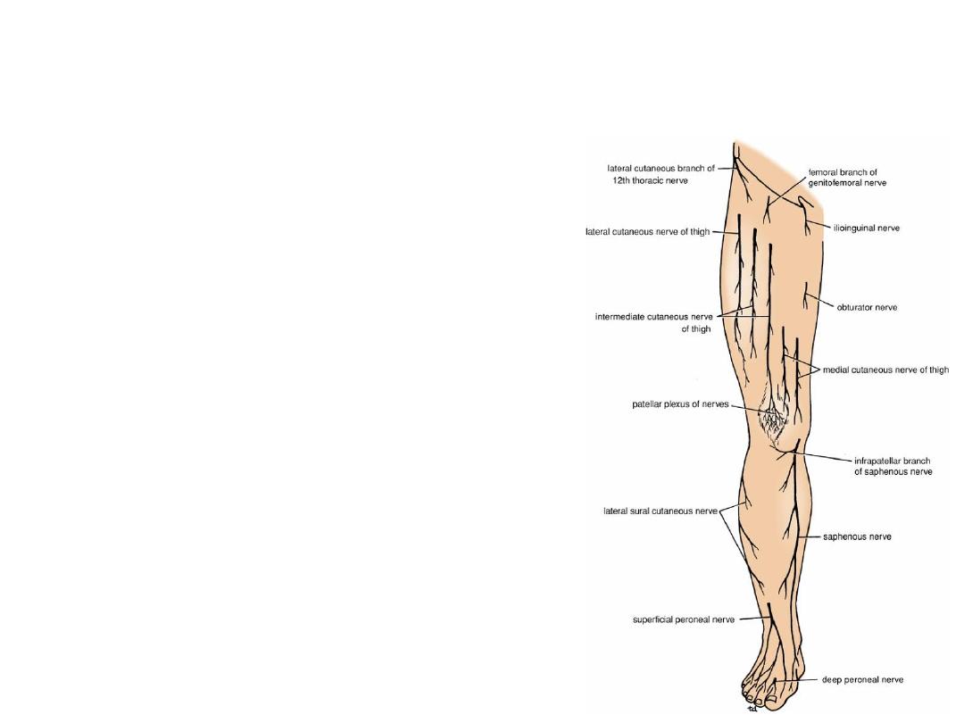
Cutaneous Innervation of Thigh
Front of the Thigh
1. Lateral cutaneous nerve of the thigh
2. Intermediate cutaneous nerve of the
thigh
3. Medial cutaneous nerve of the thigh
4. Femoral branch of the genitofemoral
nerve
5. Ilioinguinal nerve
6. Obturator nerve
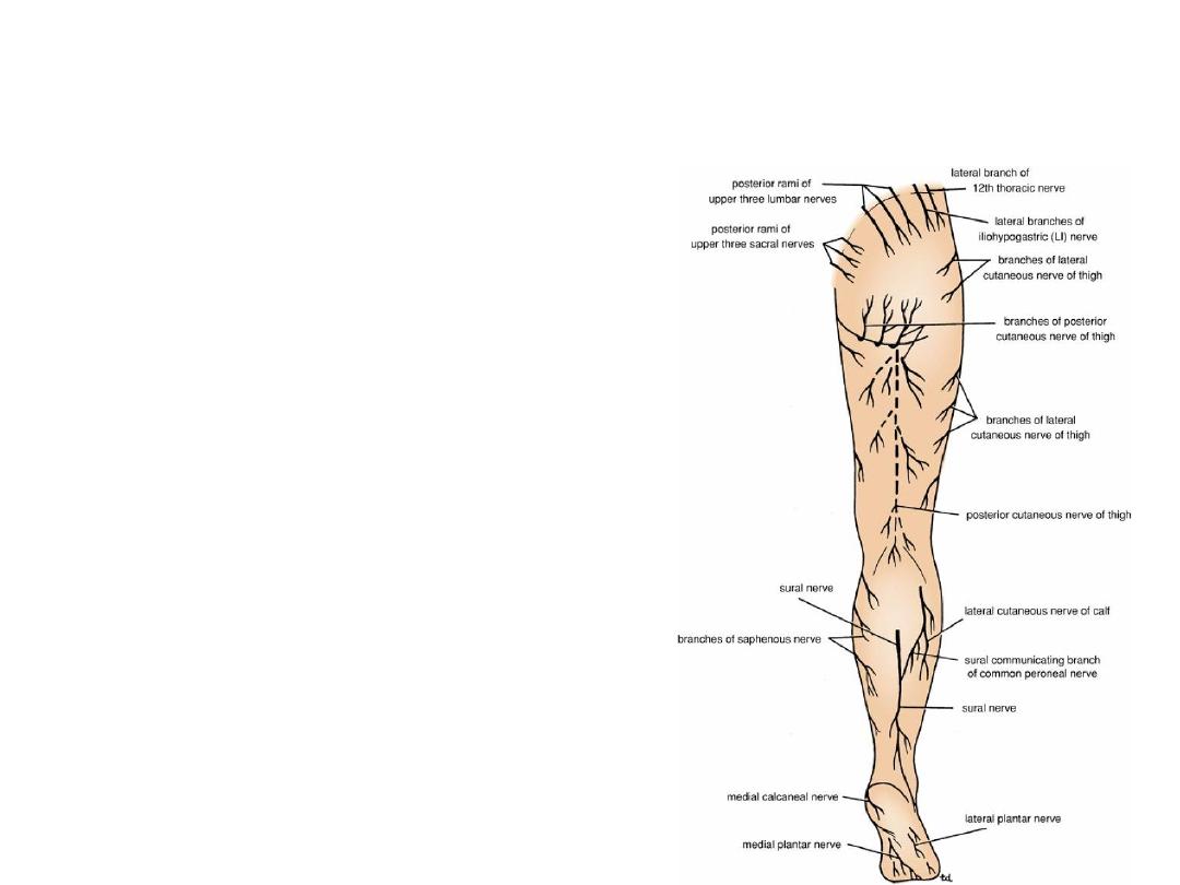
The back of the thigh
1.
Posterior cutaneous nerve of the thigh
2.
Branches from lumber nerves
3.
Branches from sacral nerves
4.
Lateral branch from the 12
th
thoracic
nerve
Cutaneous Innervation of Thigh
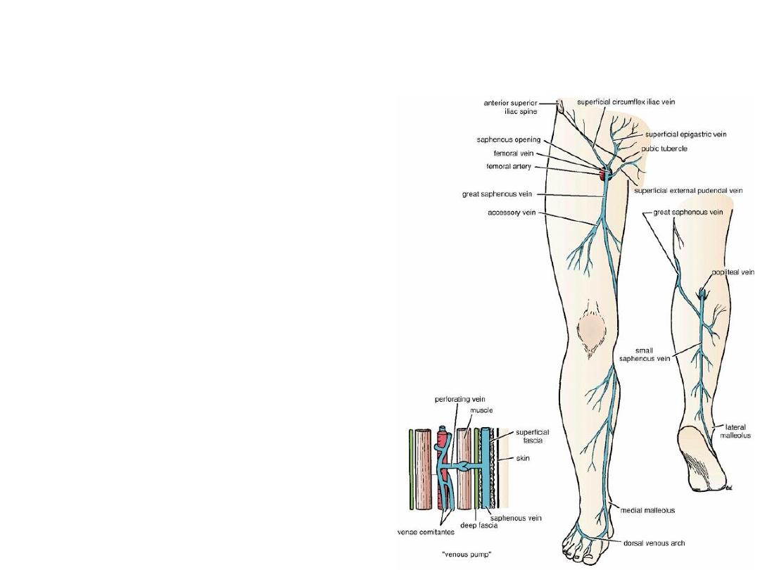
Superficial veins of the thigh
• Great Saphenous vein
• Lesser Saphenous vein
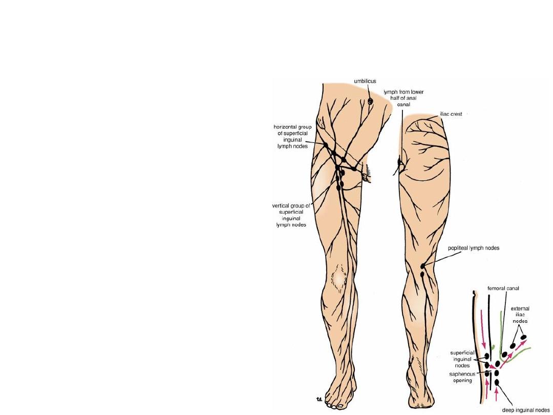
Lymph drainage of the lower limb
Inguinal Lymph Nodes:
1. Superficial Lymph
Nodes:
• Vertical group
• Horizontal group
2. Deep Lymph nodes
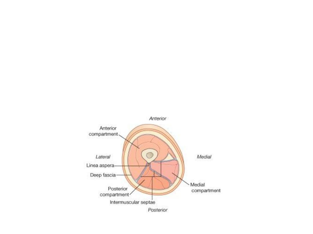
Fascial compartment of the thigh
• Anterior fascial compartment
• Medial fascial compartment
• Posterior fascial compartment
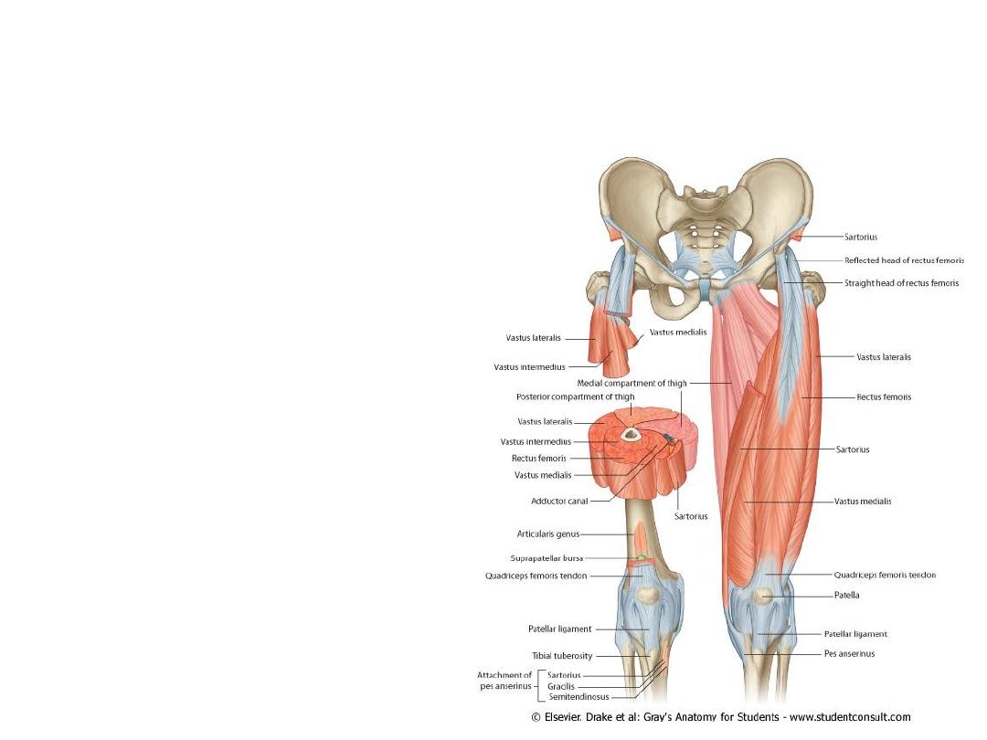
Anterior Fascial Compartment of the
thigh
1. Sartorius Muscle
2. Psoas Muscle
3. Iliacus Muscle
4. Pectineus Muscle
5. Quadriceps Femoris
Muscle that includes:
• Rectus Femoris (2 heads)
• Vastus Medialis
• Vastus Lateralis
• Vastus Intermedialis
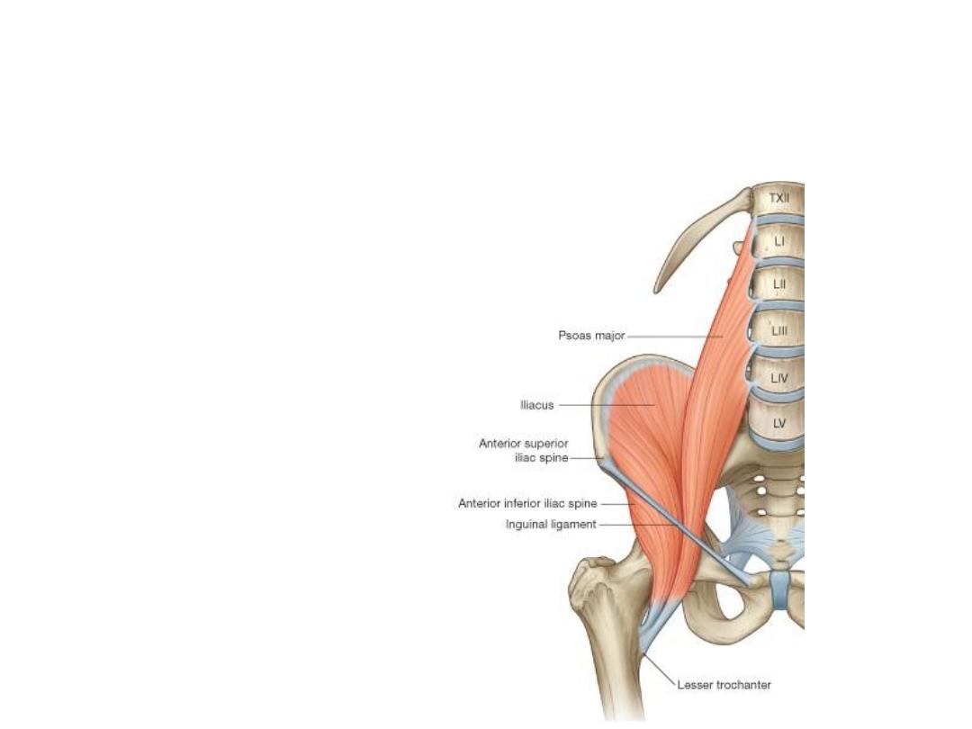
Ilio-Psoas Muscle
Ilio-Psoas Muscle is
formed of:
• Iliacus Muscle
• Psoas Muscle
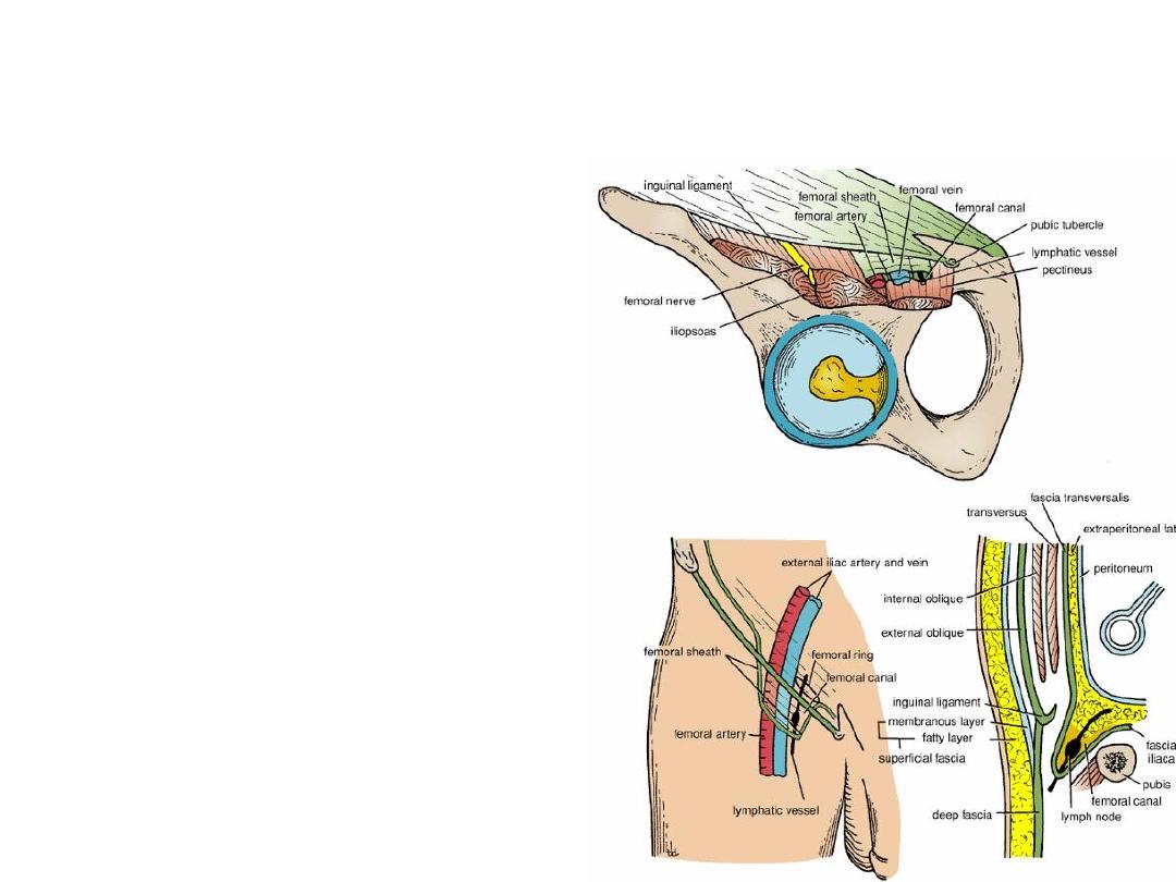
Femoral Sheath & Contents
• Protrusion of the deep fascia
enveloping the abdomen
• It has 3 compartments separated by
septa:
1. Lateral compartment (femoral artery)
2. Intermediate compartment (femoral
vein)
3. Medial Compartment = femoral canal
= (Lymphatics)
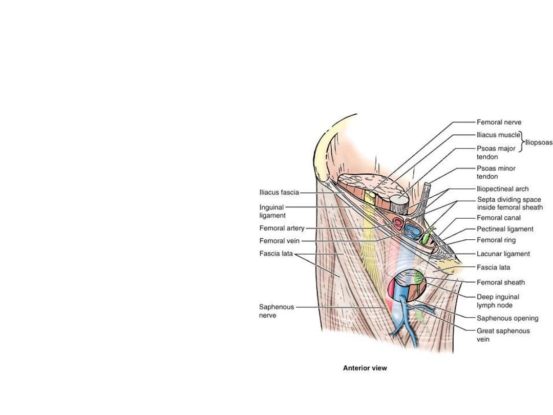
Femoral Sheath & Contents
• Femoral ring has relations:
1. Anteriorly –inguinal ligament
2. Posteriorly – superior pubic
ramus
3. Laterally – femoral vein
4. Medially – Lacunar Ligament
• Clinical Importance:
The Femoral Hernia
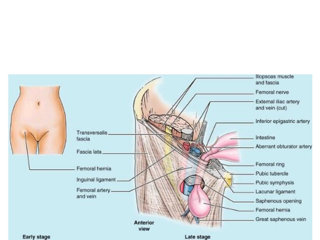
Femoral Hernia
Protrusion of the abdominal contents through the femoral ring
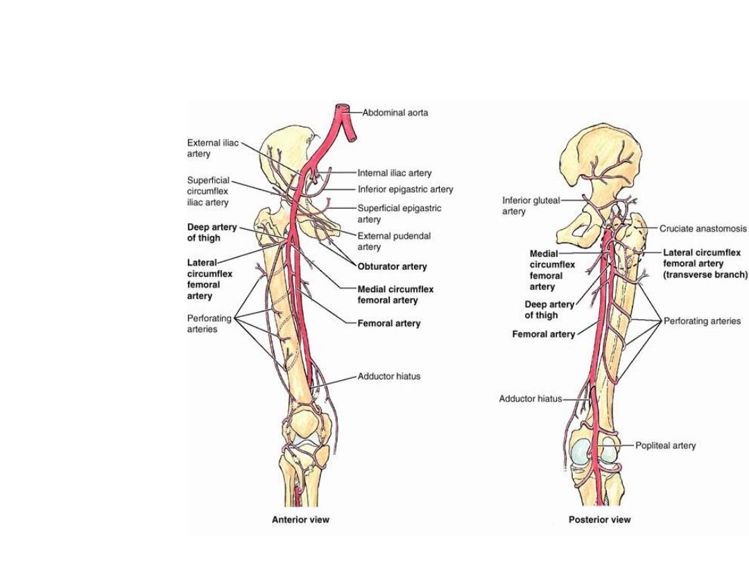
Arteries of the Anterior Compartment
• Femoral Artery
• Branches:
1. Superficial
circumflex iliac
artery
2. Superficial
epigastric artery
3. Superficial external
pudendal artery
4. Deep external
pudendal artery
5. Profunda Femoris
artery
6. Descending
Genicular artery
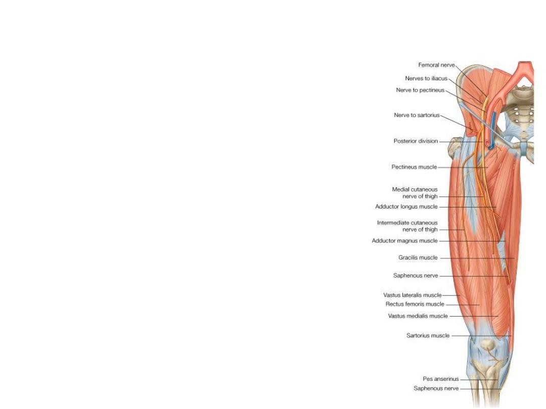
Femoral nerve
•
The largest branch of the lumbar plexus (L2, 3, and 4).
•
It emerges from the lateral border of the psoas muscle and
passes downward in the interval between the psoas and
iliacus.
•
it terminates by dividing into anterior and posterior
divisions.
•
Branches
•
Anterior Division
(2 cutaneous branches: medial & intermediate cutaneous
nerves of the thigh)
(2 muscular branches: nerve to sartorius and the pectineus.)
•
Posterior Division
(1 cutaneous branch: the saphenous nerve)
(1 muscular branches to the quadriceps muscle).

Thank You