49
ukaryotic chromosomes and the cell cycle
E
2
-
9
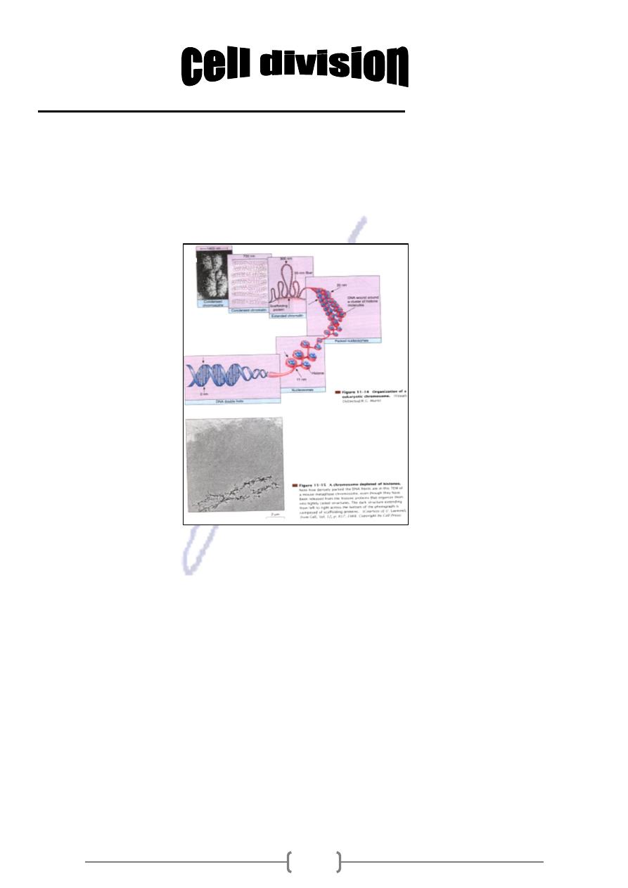
The DNA in the chromosomes of eukaryotes is associated with various proteins including
histone proteins that are especially involved in organizing chromosomes. When an
eukaryotic cell is not undergoing division, the DNA(associated proteins) within a nucleus
is a tangled mass of thin threads called chromatin. at the time of division, chromatin
becomes highly coiled and condensed, and it easy to see the individual chromosomes.
Cell division in eukaryotes involves nuclear division (karyokinesis) and cyto kinesis, which
is division of the cytoplasm. The nuclei of somatic or body cells undergo mitosis (nuclear
division in which the chromosome number stays constant). A 2n nucleus divides to
produce daughter nuclei that are also 2n. Mitosis is the type of nuclear division that is
involved in development, growth and repair of multicellular organisms. Before nuclear
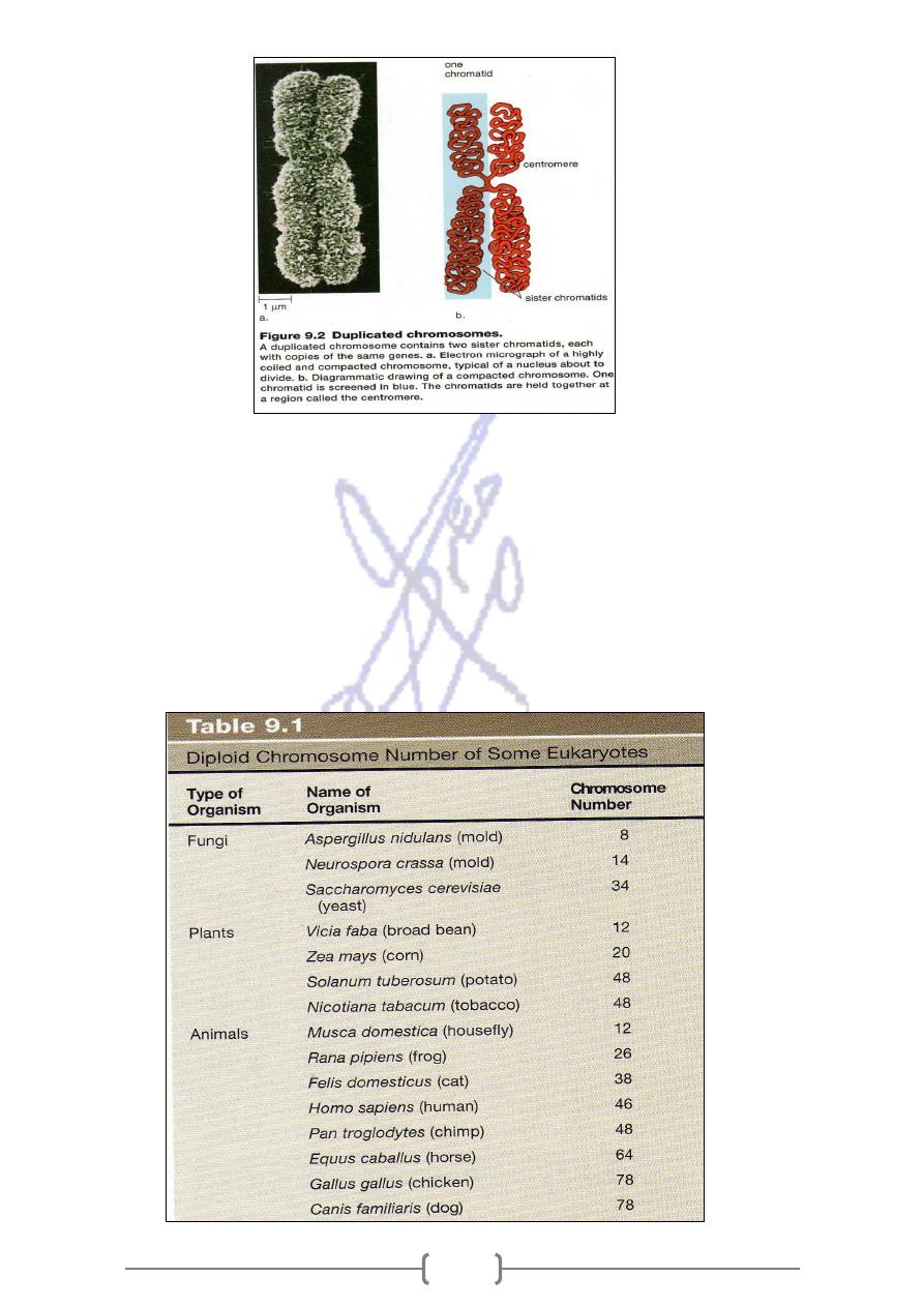
division takes place, DNA replicates, duplicating the chromosomes. Each chromosome
now has two identical parts called sister chromatids (fig.9-2). Sister chromatids are
genetically identical; that is, they contain exactly the same genes. Sister chromatids are
constricted and attached to each other at a region called centromere. During nuclear
division the two sister chromatids separate at the centromeres, and in this way each
duplicated chromosome gives rise to two daughter chromosomes. These chromosomes,
which consist of only one chromatid, are distributed equally to the daughter cells. In this
way, each daughter cell gets a copy of each chromosome.
51
When the chromosomes are visible it is possible to photograph and count them. Each
species has a characteristic chromosome number (table 9-1); for instance, human cells
contain 46 chromosomes, corn has 20 chromosomes and a crayfish has 200 this is called
the full or diploid (2n) number of chromosomes that is found in all cells of the body. The
diploid number includes two chromosomes of each kind. Half the diploid number is
called the haploid (n) number of chromosomes, contains only one of each kind of
chromosome. In the life cycle of many animals, only sperm and eggs have the haploid
number of chromosomes.

51
Cell division or mitosis
* During this process, the parent cell divides and each of the daughter cells receive a
chromosomal set identical to that of the parent cell.
* A longitudinal duplication of the chromosomes takes place and these chromosomes
are distributed to the daughter cells.
* The phase between two mitosis is called interphase, during which the nucleus as it
is normally observed in microscopic preparations.
* the process of mitosis is subdivided into phases to facilitate it.
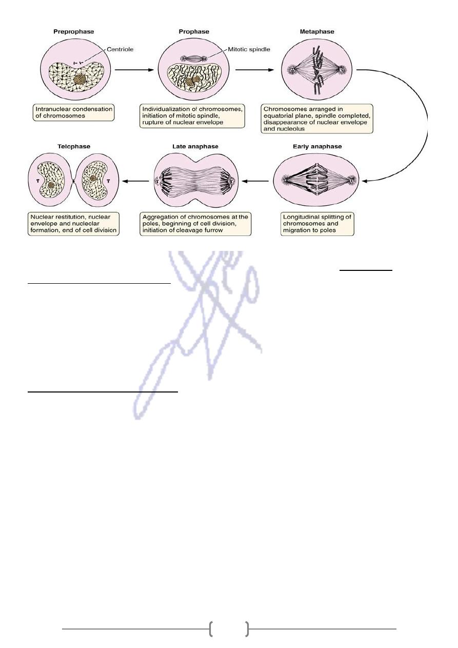
The prophase
-
1
* Is characterized by the gradual coiling of nuclear chromatin (coiled chromosomes) that
stains intensely. At the end of prophase:
1) the nuclear envelope is broken by phosphorylation (addition of PO4) of the nuclear
lamina proteins, originating vesicles that remain in the cytoplasm.
2) the centrosomes with their centrioles separate and a centrosome migrates to each pole
of the cell.
The duplication of the centrosomes and centrioles starts in the interphase, before
*
Simultaneously with centrosome migration, the microtubules of the mitotic
mitosis.
spindle appear between the two centrosomes, and the nucleolus disintegrates.
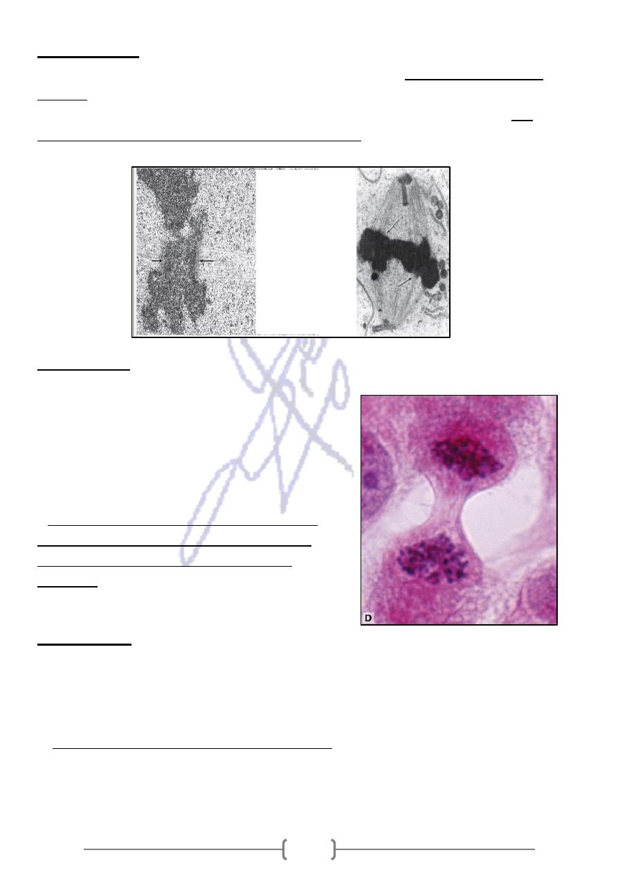
magnification. A: Interphase nuclei.
Note the chromatin and nucleoli
inside each nucleus. B: Prophase. No
distinct nuclear envelope, no
nucleoli. Condensed chromosomes.
C:
Metaphase
. The chromosomes are
located in a plate at the cell equator
B
C
52
2)metaphase
the equatorial plane of
Chromosomes, due to the activity of microtubules, migrate to
where each divides longitudinally to form two chromosomes called sister
the cell,
an
spindle, at
chromatids. The chromatids attach to the microtubules of the mitotic
chora,
moving, +
kinetos,
(Gr.
dense, DNA “protein plaque, the kinetochore
-
ron
elect
central region), located close to the centromere of each chromatid.
3)Anaphase
* The sister chromatids separate from each
other and migrate toward the opposite poles of the
cell, pulled by microtubules. Throughout this
process, the centromeres move away from the
center, pulling the remainder of the chromosome
along.
is the constricted region of a
The centromere
*
mitotic chromosome that holds the two sister
chromatids together until the beginning of
anaphase.
4)Telophase
Is characterized by
1-the reappearance of nuclei in the daughter cells.
2- The chromosomes revert to their semidispersed state.
3- and the nucleoli, chromatin, and nuclear envelope reappear.
of the parent cell and progresses until
A constriction develops at the equatorial plane
-
4
the cytoplasm and its organelles are divided in two.
* This constriction is produced by microfilaments of actin associated with myosin that
accumulate in a belt like shape beneath the cell membrane.
53
Figure 3—15.
Phases of mitosis.
rapid in the
he turnover rate of cells varies greatly from one tissue to another, a
* T
and the epidermis of skin cells, and slow in the pancreas
epithelium of the digestive tract
and the thyroid gland
* The nervous system, contains a few stem cells and a few dividing cells, but most
neurons, once differentiated, can live for more than 100 years without dividing again.
* Like stem cells, fibroblasts of the connective tissue, are typically nondividing, but they
can be stimulated to enter the cell cycle following wounding or other stimuli
How Eukaryotic cells cycle
* by the 1870s, microscopy could provide detailed and accurate description of
chromosomal movements during mitosis, but there was no knowledge of cellular events
between divisions. Because there was little visible activity between divisions, this period
of time is dismissed as a resting state termed interphase. When it was discovered in the
1950s that DNA replication occurs during interphase, the cell cycle concept was
proposed.
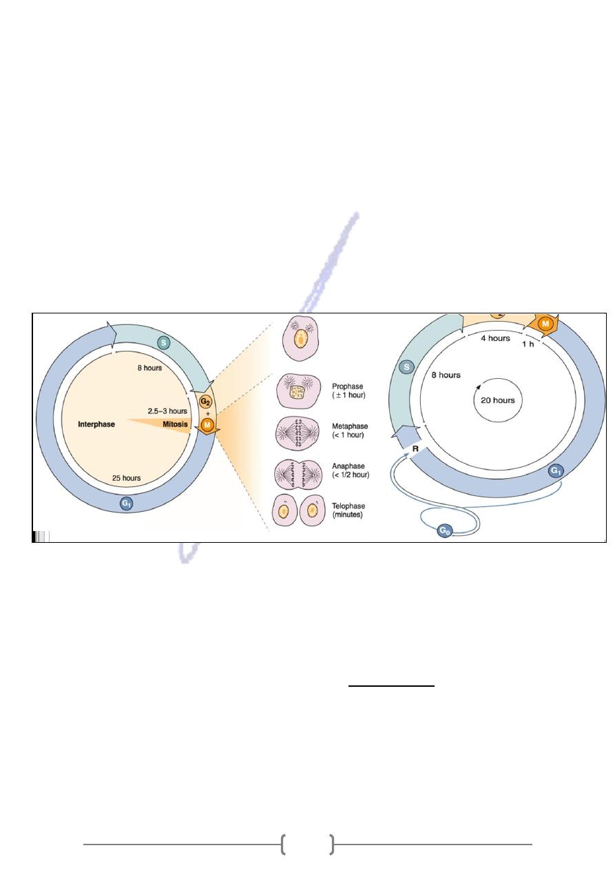
Cell grows and divides during a cycle that has four stages (fig.9-3). The entire cell
division stage, including both mitosis and cytokinesis, is termed the M stage (m=mitosis).
The period of DNA synthesis when replication occurs is termed the S stage (S=synthesis)
of the cycle. The proteins associated with DNA in eukaryotic chromosomes are also
synthesized during this stage. There are two other stages of the cycle. The prior of time
prior to the S stage is termed G1 stage, and the period of time prior to the M stage is
termed G2 stage. At first, not much known about these stages and they were thought of
54
as G=gap stages. Now we know that during the G1 stage, the cell grow in size and
cellular organelles increase in number. During the G2 stage, various metabolic events
occur in preparation for mitosis. Some biologists today prefer the designation G=growth
for these G stages. In any case, interphase consist of G1,S and G2 stages.
* Some cells such as skin cells, divide continuously throughout the life of the organism.
Other cells such as skeletal muscle cells and nerve cells are arrested in the G1 stage. If
the nucleus from one of these cells is placed in the cytoplasm of an S stage cell, it
finishes the cell cycle. Cardiac muscle cells are arrested in the G2 stage. If an arrested
cell is fused with a cell undergoing mitosis, it too starts to undergo mitosis. It appears,
then, there are stimulatory substances that cause the cell to proceed through two
critical checkpoints: G1 stage ---------------- S stage
G2 stage ---------------- M stage
* In cells that are not continuously dividing, the activities of the cell cycle may be
temporarily or permanently suspended. Cells in such a state (eg, muscle, nerve) are
referred to as being in the G
0
phase.
* Regulation of the mammalian cell cycle is complex. It is known that cultured cells
deprived of serum stop proliferating and arrest in G
0
. The essential components
, which are
growth factors
provided by serum are highly specific proteins called
required only in very low concentrations. There are some enzymes involved in cell
cycle
* A Kinase is an enzyme that removes a phosphate group from ATP (The form of
chemical energy used by cells) and adds it to a protein. Phosphorylated molecules are a
common way for the cell to turn on metabolic pathways. Notice in figure 9.4 that
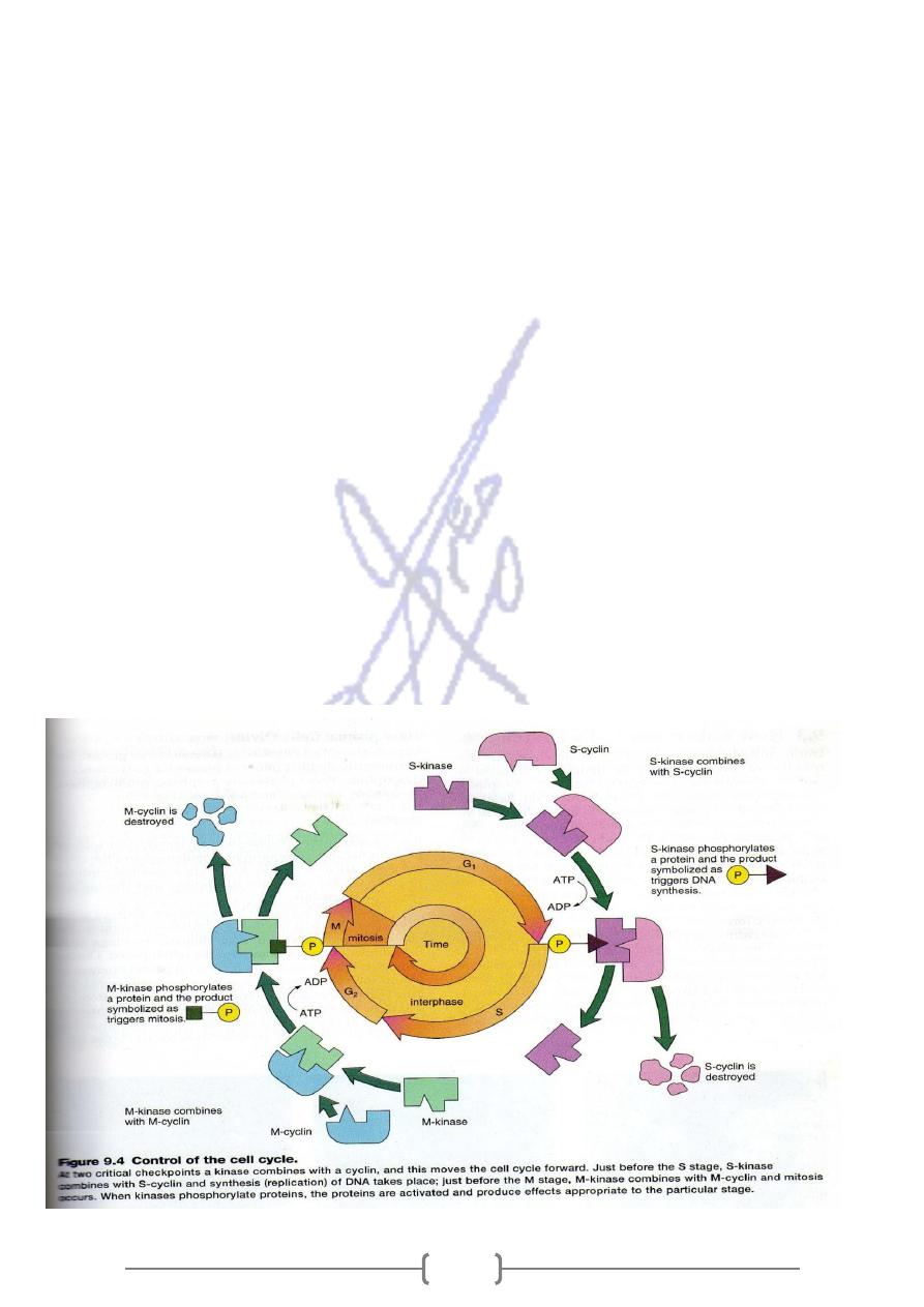
55
phosphorylation of a protein precedes the S stage and M stage of the cell cycle.
The kinases involved in the cell cycle are called cyclin dependent because they are
activated when they combine with a protein called a cyclin.
Figure 9.4 shows how the entire process works. In the diagram, S-Kinase is capable
of phosphorylating the protein that triggers DNA replication after it has combined with
S-cyclin. S-cyclin is now destroyed and S-Kinase is no longer active. M-Kinase is capable
of phosphorylating the protein that turns on mitosis after it has combined with M-cyclin.
It is known that this particular phosphorylated protein induces the process of
(1) chromosome condensation (2) nuclear envelope breakdown (3) spindle assembly.
(The spindle is the structure involved in chromosome movement during mitosis).
* Ordinary, a cyclin might combine with its kinase only when a growth factor is present.
But a cyclin that has gone awry might combine with its kinase even when a growth
factor is not present. The result would be uncontrolled cell growth resulting in a tumor.
On the other hand, tumor suppressor genes usually function to prevent cancer from
occuring. It has been shown that the product of one major tumor-suppressor gene (p53)
brings about the production of a protein that can combine with a cyclin-kinase complex
and prevent that kinase from becoming active. p53 can also help induce apoptosis in
cancerous cells. Apoptosis is a process of programmed cell death involving a cascade of
specific cellular events leading to the death and destruction of the cell.
56
checkpoints
* The cell cycle is highly regulated, and checkpoints control transitions between cell-
cycle stages.
Checkpoints are biochemical circuits that detect external or internal problems and send
.
cycle system
-
inhibitory signals to the cell
There are four major types of checkpoints
MEDICAL APPLICATION
erythropoietin, which
* Some growth factors are being used in medicine. One example is
stimulates proliferation, differentiation, and survival of red blood cell precursors in the
bone marrow.
* The cell cycle is also regulated by a variety of signals that inhibit progression through
the cycle.
* DNA damage arrests the cell cycle not only in G
2
but also at a checkpoint in G
1
.
G
1
arrest may permit the damage to be repaired before the cell enters S phase
* In mammalian cells, arrest at the G1 checkpoint is mediated by the action of a protein
p53.
the cell's
The gene encoding p53 is often mutated in human cancers, thus reducing
*
.
ability to repair damaged DNA
Inheritance of damaged DNA by daughter cells results in an increased frequency of
mutations and general instability of the genome, which may contribute to the
development of cancer
Processes that occur during the G2 phase include:
1-the accumulation of energy to be used during mitosis
2- the synthesis of tubulin to be assembled in mitotic microtubules
3- and the synthesis of chromosomal non histone proteins.
4- In G
2
there is also a checkpoint at which the cell remains until all DNA synthesized
with defects is corrected.
complex maturation promoting factor
cumulation of the protein
there is an ac
2
In G
-
5
(MPF) that induces the beginning of mitosis, the condensation of the chromosomes, the
rupture of the nuclear envelope, and other events related to mitosis
Prokaryotes and Eukaryotes
9.4 Comparing
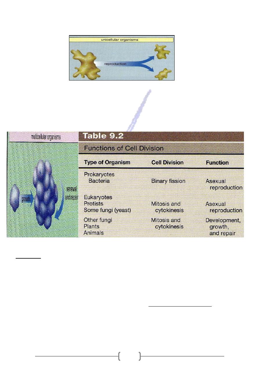
Binary fission and mitosis ensure that each daughter cell is genetically identical to the
parent cell. The genes consist of DNA found in the chromosomes.
57
Bacteria and protists such as amoeboids and paramecia, are unicellular. Cell
division is unicellular organisms produces two new individuals:
This is a form of a sexual reproduction because one parent has produces identical
offspring (table 9.2).
In multicellular forms such as most fungi, plants and animals, cell division is
part of the growth process that produces the multicellular form we recognize as the
organism. Cell division is also important in multicellular forms for renewal and repair:
Meiosis
* Meiosis, which requires two nuclear divisions, results in four daughter cells, each having
one of each kind of chromosome and therefore half the number of chromosomes as the
parental cell.
* The parental cell has the 2n number of chromosomes, while the daughter cells have the n
number of chromosomes. Therefore, meiosis is often called reduction division. The
daughter cells that result from meiosis go on to become the gametes.
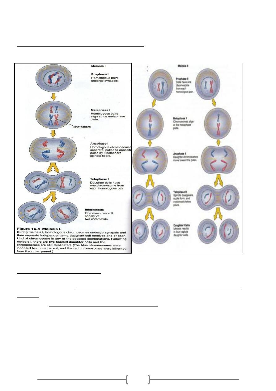
* Stages of Meiosis
The same four stages of mitosis—prophase, metaphase, anaphase, and telophase—occur
during both meiosis I and meiosis II.
58
The First Division
.During prophase I, the spindle appears while the nuclear envelope fragments and the
nucleolus disappear.
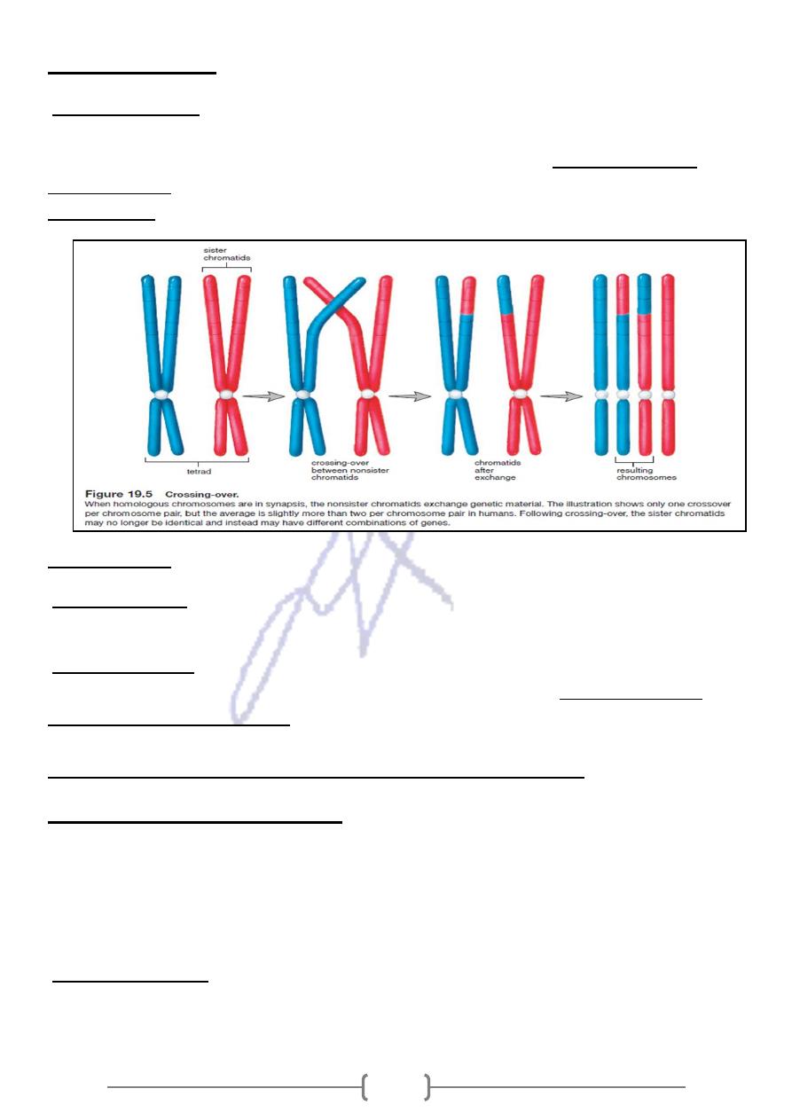
The homologous chromosomes, each having two sister chromatids, undergo synapsis,
forming tetrads.
Crossing-over occurs now.
In metaphase I, tetrads line up at the equator of the spindle.
During anaphase I, homologous chromosomes of each pair separate and move to opposite
poles of the spindle.
During telophase I, nucleoli appear, and nuclear envelopes form as the spindle disappears.
During cytokinesis, the plasma membrane furrows to give two cells. Each daughter cell
contains only one chromosome from each homologous pair.
The chromosomes are dyads, and each has two sister chromatids.
No replication of DNA occurs during a period of time called interkinesis
The Second Division meiosis II
At the beginning of prophase II, a spindle appears while the nuclear envelope disassembles
and the nucleolus disappears.
Dyads (one dyad from each pair of homologous chromosomes) are present, and each
attaches to the spindle independently.
During metaphase II, the dyads are lined up at the equator.
Anaphase II, the centromeres split. The sister chromatids of each dyad separate and move
toward the poles. Each pole receives the same number of chromosomes
59
In telophase II, the spindle disappears as nuclear envelopes form.
During cytokinesis, the plasma membrane furrows to give two complete cells, each of which
has the haploid, or n, number of chromosomes. Since each cell from meiosis I undergoes
meiosis II, there are four daughter cells altogether
The importance of meiosis
the chromosomal number stays constant in each generation of
* Because of meiosis,
humans.
during the production of the
curs in the testes and ovaries
, meiosis oc
In humans
*
gametes. When a haploid sperm fertilizes a haploid egg, the new individual has the
diploid number of chromosomes.
There are three ways the new individual is assured a different combination of genes
than either parent has:
1. Crossing-over recombines the genes on the sister chromatids of homologous pairs of
chromosomes.
61
2. Following meiosis, gametes have all possible combinations of chromosomes.
3. At fertilization, recombination of chromosomes occurs because the sperm and egg
carry varied combinations of chromosomes.
10.4 Viewing the human life cycle
Mammals, including, humans, have a life cycle that requires both meiosis and mitosis
(fig.10.6). In males, meiosis is a part of spermatogenesis which occurs in the testes and
produces sperm. In females, meiosis is a part of oogenesis which occurs in the ovaries
and produces eggs. A sperm and egg join at fertilization and the resulting zygote
undergoes mitosis during development of the fetus, which is the stage of development
before birth. After birth, mitosis is involved in the continued growth of the child and
repair of tissues at any time.
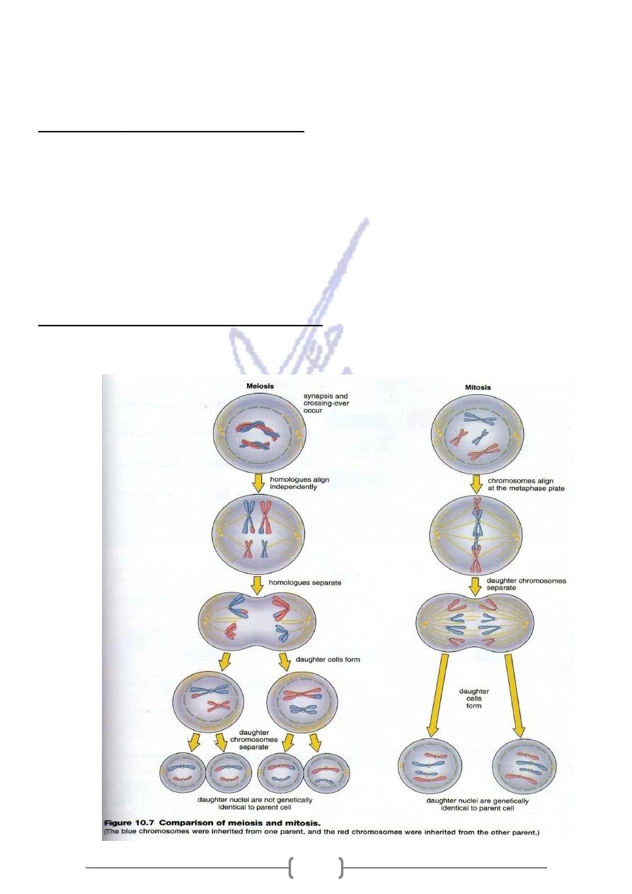
Comparison of Meiosis with Mitosis
the following lists and figure 10.7 will allow you to compare meiosis to mitosis.
61
Occurrence
Meiosis occurs only at certain times in the life cycle of sexually reproducing
organisms. In humans, meiosis occurs only in the sex organs and produces gametes.
Mitosis is more common because it allows growth and repair of body tissues in
multicellular organisms, including humans.
process
The following are distinctive differences between the process of meiosis and mitosis.
1) DNA is erplicated only once before both meiosis and mitosis; but there are two
nuclear divisions during meiosis and only one nuclear division during mitosis.
2) Homologous chromosomes pair and undergo crossing-over during prophase I of
meiosis but not during mitosis.
3) Paired homologous chromosomes (bivalents) align at the metaphase plate during
metaphase I in meiosis; individual (duplicated) chromosomes align at the metaphase
plate during metaphase in mitosis.
4) Homologous chromosomes (with centromeres intact) separate and move to opposite
poles during anaphase I in meiosis; and daughter chromosomes move to opposite
poles during anaphase in mitosis.
5) The events of meiosis II are just like those of mitosis except that meiosis II nuclei are
always haploid.
Daughter Nuclei and Cells
The genetic consequences of meiosis and mitosis are quite different as well:
1) Four daughter cells are produced by meiosis; mitosis results in two daughter cells.
2) The four daughter cells formed by meiosis are haploid; the daughter cells produced by
mitosis have the same chromosome number as the parent cell.
3) The daughter cells from meiosis are not genetically identical to each other or to the
parent cell. The daughter cells from mitosis are genetically identical to each other and
to parent cell.
Spermatogenesis and Oogenesis produce the gametes
Figure 10.8 contrasts spermatogenesis with oogenesis, processes that produce
the gametes in mammals, including humans. In the testes of males, primary
spermatocyte with 46 chromosomes divides to form two secondary spermatocytes. Each
with 23 duplicated chromosomes. Secondary spermatocytes divide to produce four
spermatids, also with 23 daughter chromosomes. Spermatids then differentiate into
sperm (spermatozoa).The process of meiosis in males always results in four cells that
become sperm. In the ovaries of females, a primary oocyte has 46 chromosomes and
divides meiotically into two cells, each having 23 chromosomes. One of these cells,
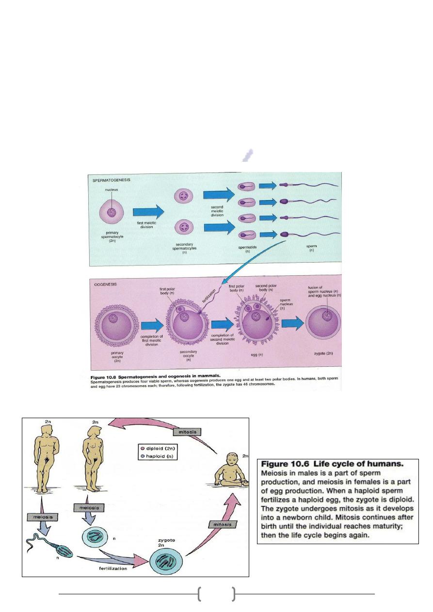
62
termed the secondary oocyte, receives almost all the cytoplasm (fig.10.8). The other is
polar body that may disintegrate or may divide again. The secondary oocyte begins
meiosis II and then stops at metaphase II. Then at ovulation, it leaves the ovary and
enters an oviduct where it may be approached by a sperm. If a sperm does enter the
oocyte, the oocyte is activated to continue meiosis II to completion. The mature egg has
23 chromosomes. Meiosis in females produces only one egg and possibly three polar
bodies. The polar bodies are a way to discard unnecessary chromosomes while retaining
much of the cytoplasm in the egg. The cytoplasm serves as a source of nutrients for the
developing embryo.