128
Abnormalities of Chromosomal Number
Cytogenesis: Is the study of chromosomes and their abnormalities.
1. Polyploidy:
chromosomes in its nucleus. Thus haploid
multiple of 23
A cell that contains a
Euploid:
gametes and diploid somatic cells are euploid.
Polyploidy: Is the presence of a complete set of extra chromosomes in a cell is seen
commonly in plants and often improve their agriculture value, also occurs in humans,
although much less frequently.
Polyploidy conditions that have been observed in humans are:
A. Triploidy: 69 chromosomes in the nucleus of each cell, the karyotype for this
condition is 69, XXX (assuming that all of the sex chromosomes were X, other
combinations of the X and Y chromosomes may be seen).
B. Tetraploidy: 92 chromosomes in each cell nucleus, the karyotype of this condition is
92,XXXX.
Because of the number of chromosomes present in each of these conditions is a
multiple of 23, the cells are euploid in each case.
However, the additional chromosomes encode a large amount of surplus gene
product, causing multiple anomalies such as defects of the heart and central nervous
system.
A. Triploidy: the incidence about 1 in 10,000 live births, and estimated about 15% of the
chromosomal abnormalities.
Causes:
1. Fertilization of an egg by two sperm (dispermy). The resulting zygote receives 23
chromosomes from the egg and 23 chromosomes from each of the two sperm cells (most
common)
2. Fusion of an ovum and a polar body, each containing 23 chromosomes, and subsequent
fertilization by a sperm cell.
3. Meiotic failure, in which diploid sperm or egg cell is produced.
Thus the vast majority of triploid conceptions are spontaneously aborted, and those
that survive to term die shortly after birth.
129
B. Tetraploidy is much rare than triploidy.
It has been recorded in few live births, and those infants that survive for a short period.
Causes:
1. May be due to Mitotic failure in early embryo in which all of the duplicated
chromosomes migrate to one of the two daughter cells
2. Also result from fusion of two diploid zygotes.
Autosomal Aneuploidy:
2.
Are among the most clinically important of the chromosome abnormalities.
Aneuploid: are cells that do not contain a multiple of 23 chromosomes, but instead
they contain missing or additional chromosomes. Usually only one chromosome is
affected, but it is possible for more than one chromosome.
:
They consist of
A. monosomy: the presence of only one copy of a chromosome in diploid cell (n- 1)
B. trisomy: presence of 3 copies of a chromosome in diploid cell (n+ 1)
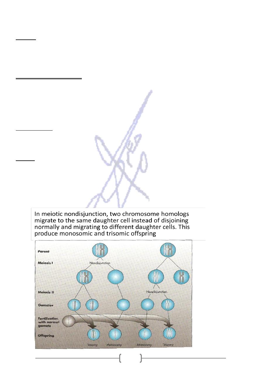
The most common cause is nondisjunction, the failure of chromosomes to
Causes:
disjoin normally during meiosis. It can be occur during meiosis I or meiosis II. The
resulting gamete either lacks a chromosome or has two copies of it.
Autosomal monosomies are almost always die before term (lethal). In contrast, some
trisomies are seen among live births
131
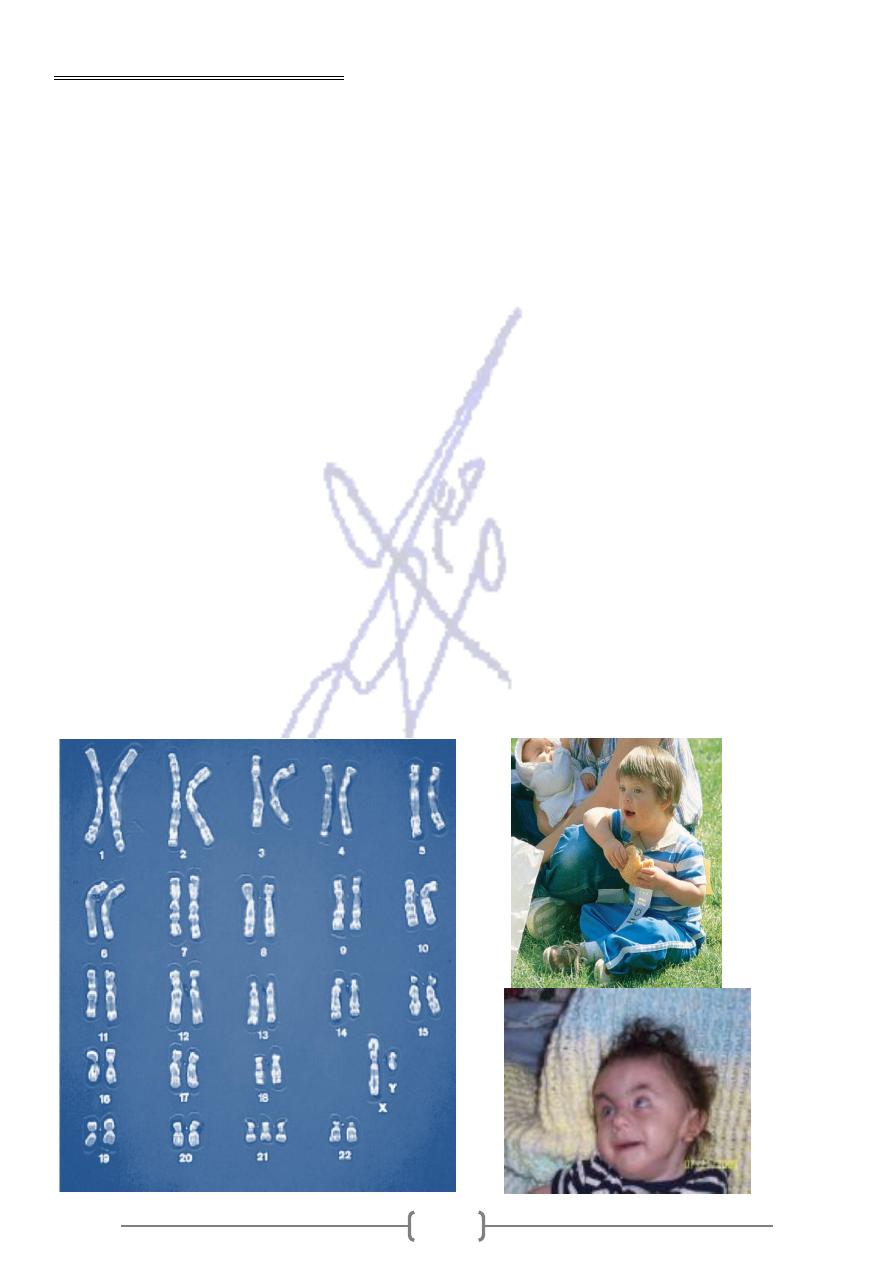
Trisomy 21 (Down’s syndrome)
Karyotype (47, XY,+ 21 or 47,XX,+21)
Down syndrome is also called trisomy 21 because the individual usually has three copies of
chromosome 21.
It is the most common autosomal aneuploidy seen among live births.
In most instances (80%), the egg had two copies instead of one of this chromosome.
(In 23% of the cases studied, however, the sperm had the extra chromosome 21.)
The chance of a woman having a Down syndrome child increases rapidly with age,
starting at about age 40.
The frequency of Down syndrome is 1 in 800 live births for mothers under 40 years of
age and 1 in 80 for mothers over 40 years of age
This syndrom is called “mongolism” in earlier literature, but this term is inappropriate.
The most significant problems include mental retardation, gastrointestinal tract
obstruction , congenital heart defects (atrioventricular canal), respiratory infections,
and leukemia (15-20 times higher than in general population).
Males are nearly always sterile.
About 80% of children survive to age 10 years and about half survive to age 50 years.
Mosaicism is seen in approximately 2% to 4% of trisomy 21 live births often results in
milder clinical expression of a chromosomal abnormality.
These individuals have some normal somatic cells and some cells with trisomy 21.
This is designated in male 47, XY, +21/ 46, XY.
131
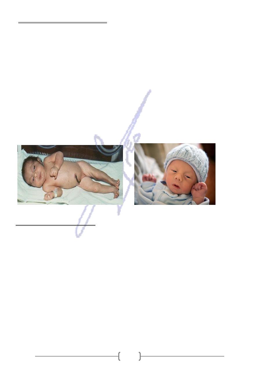
Trisomy 18 (Edwards syndrome)
Karyotype (47, XY, +18)
Edwards syndrome is the second most common autosomal trisomy, with a prevalence rate
of 1 per 6000 live births.
Infants are produced a more seriously affected phenotype with prenatal growth
deficiency (weight is low for gestation age).
Infants have characteristic facial features, hand abnormalities, short sternum and
short big toes. The major malformation is congenital heart defects.
90% of trisomy 18 cases are the result of a maternally contributed extra chromosome
More than 95% of infants have complete trisomy 18, and small percentage have
mosaicism.
About 50% of infants die within the first several weeks of life, and only 5% are still
alive at 12 months of age.
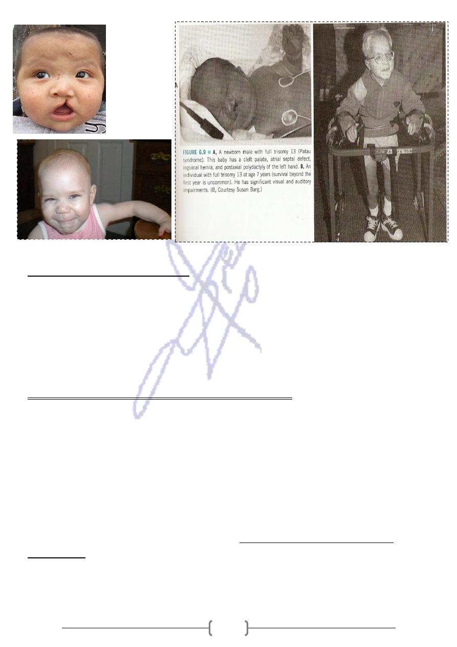
Trisomy 13 (Patau syndrome)
Karyotype (47, XY, +13)
Prevalence rate about 1per 10000 births.
It is much less common at birth than trisomy 21.
The malformation primarily includes oral- facial clefts, and postaxial polydactyly.
Also malformation of Central nervous system (CNS) and heart defects.
The risk of bearing a child with this condition increases with advanced maternal age.
About 95% of live- born infants dying during the first year of life.
Children who survive have significant developmental retardation in vision and hearing
About 80% of patients with this syndrom have full trisomy 13. Most of the remaining
patients have trisomy of the long arm of chromosome 13 due to a translocation.
132
Sex chromosome Aneuploidy
Among live- born infants about 1 in 400 males and 1 in 650 females have some form
of sex chromosome aneuploidy.
Primarily because of X inactivation, the consequences of this class of aneuploidy are
less severe than those of autosomal aneuploidy.
With the exception of a complete absence of X chromosome, all of the sex
chromosome aneuploidies are compatible with survival.
A. Monosomy of the X chromosome (Turner syndrome)
Karyotype (45, X)
Prevalence rate about 1 per 2000 to 1per 3ooo live- born females.
It is relatively rare among live births, reflecting a very high rate of spontaneous
abortion.
Females with turner syndrome are short stature (mature height is reduced by
approximately 20 cm) with sexual infantilism and lack of ovaries.
Lymphedema of the hands and feet is observed at birth, with the heart defect,
but females with this syndrome usually have normal intelligence.
absence of a paternally derived sex
of monosomy X cases are caused by the
Most
, so the offspring receives an X chromosome only from the mother.
chromosome
About 10% to 20% of patients with this syndrome have structural X chromosome
abnormalities involving a deletion of some or all the short arm, this variation in
chromosome abnormality leads to phenotypic variation seen in this syndrome.
133
Klinefelter Syndrome
• Karyotype (47,XXY)
• Seen in approximately 1/500 to 1/1000 male births.
• Males tend to be taller than average with long arms and legs, and the body shape may
be somewhat feminine.
• They have small testes and are usually sterile as a result of atrophy of the seminiferous
tubules.
• Testosterone level are low.
• Gynecomastia (breast development) is seen in approximately one third of affected
males and leads to increased the risk of breast cancer.
• They may have a reduction in IQ.
• The extra X chromosome is derived maternally in about 50% of klienfelter cases, and the
syndrome increases with advanced maternal age.
• Mosaicism seen in about 15% of patients and increases the viable sperm production.
• Individuals with the 48,XXXY and 49,XXXXY karyotypes have also be reported, the degree
of mental deficiency and physical abnormality increases with additional X chromosome.
B. Trisomy X
• Karyotype (47,XXX)
• Prevalence rate is 1/ 1000 females.
• Physical abnormalities are rarely seen, but these females sometimes suffer from sterility
or mild menal retardation (slight reduction in IQ)
• Females have also been seen with four, five, or even more X chromosomes.
• Each additional X chromosome is accompanied by increased mental retardation and
physical abnormalities.
47,XYY Syndrome
• Karyotype (47,XYY)
• Prevalence rate 1/1000 males
• Males are taller than average with
• few physical problems but
• they have 10- to 15- points reduction in average IQ and learning disabilities.
• The prevalence rate of this syndrome among prison population was discovered to be
1/30, suggested that this karyotype might confer a predisposition to violent and criminal
behavior
134
Abnormalities of chromosomal structure
Parts of chromosomes can be lost or duplicated and the arrangement of portions of
.
gamete formation
chromosomes can be altered during the
These abnormalities often do not produce serious structural abnormalities (if the
rearrangement does not produce a loss or gain of chromosome material), or
can produce serious disease in individuals or their offspring (if the rearrangement causes
loss or gain of chromosomal material).
Mechanisms exist to repair these breaks, and usually the break is repaired perfectly
with no damage to the daughter cell. Sometimes, the break remain, or they heal in a
fusion that alters the structure of the chromosome.
Chromosomal breakage may be increased in the presence of certain harmful agents
include ionizing radiation, some viral infections, and some chemicals.
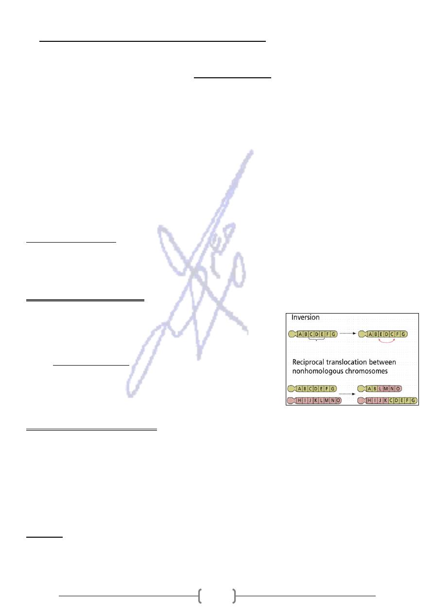
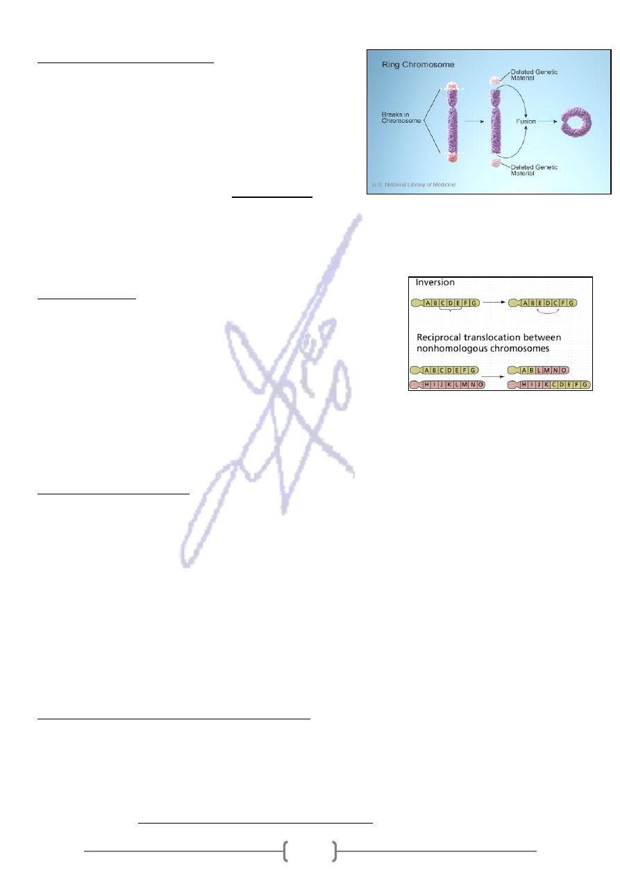
1. Translocation:
It is the interchange of genetic material between nonhomologous chromosomes.
There are two basic types: A. Reciprocal B. Robertsonian
A. Reciprocal Translocation:
When breaks occur in two different chromosomes with
subsequent exchange of material. The resulting are called
derivative chromosomes (der).
of this translocation usually
individual carriers
While
appears with normal phenotype, their offspring may have
a partial trisomy or partial monosomy and an
abnormal phenotype
B. Robertsonian Translocation;
The short arms of two acrocentric nonhomologous chromosomes are lost and the long
arms fuse at the centromere to form a single chromosome.
This type of translocation occurs in chromosomes 13, 14, 15, 21, and 22, because the short
arms of these acrocentric chromosomes are very small and contain no essential genetic
material.
The carrier of this abnormality can produce offspring with monosomy or trisomy.
hromosomes 14 and 21.
Fusion of the long arms of c
:
Example
The karyotype of a male carrier of this translocation is 45,XY,der(14;21).
135
Robertsonian translocation are responsible for approximately 5% of Down syndrome
cases.
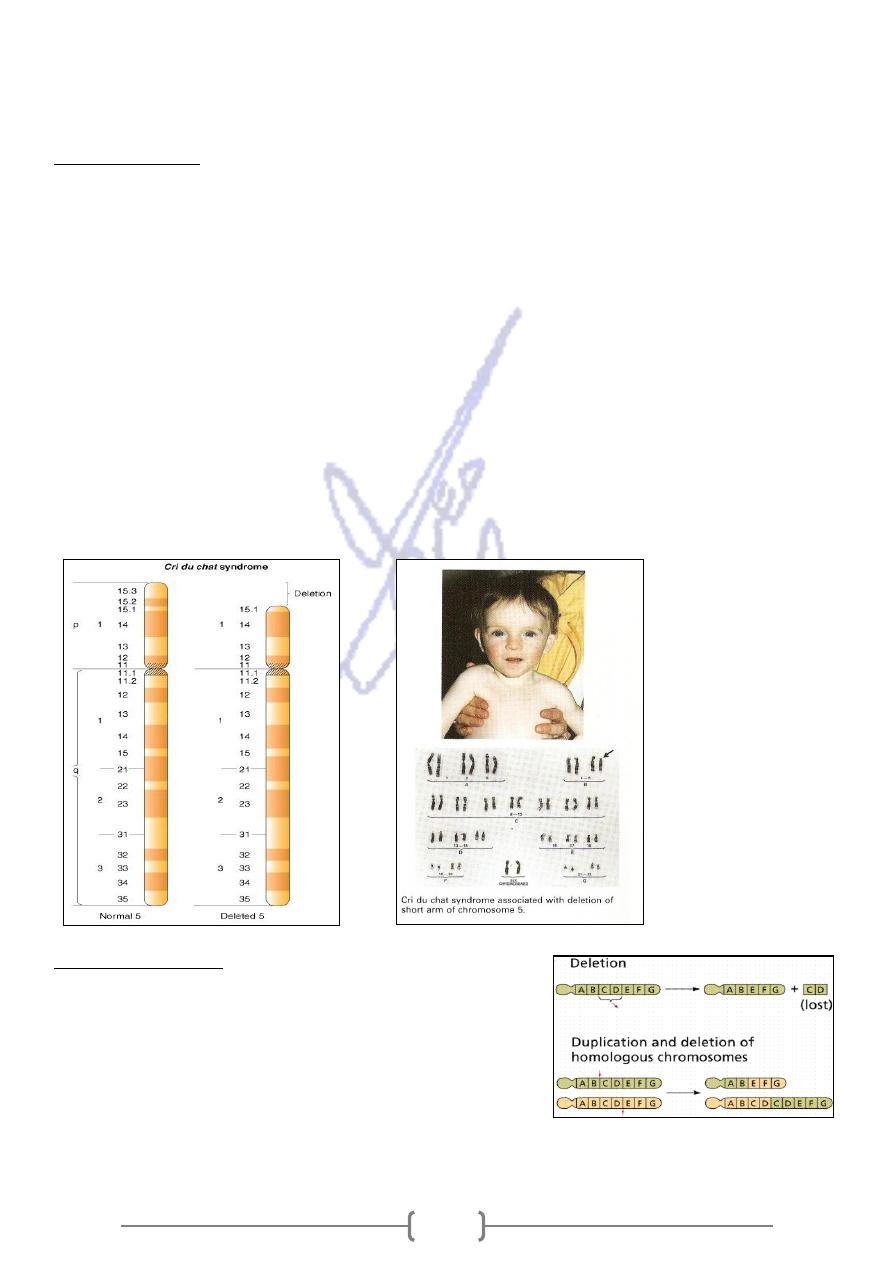
2. Deletions:
Is caused by a chromosomal break and subsequent loss of genetic material.
A single break leading to a loss the chromosome’s tip is called a termination deletion,
(figure) When two breaks occur and the material between the breaks is lost, an
interstitial deletion occurs.
This abnormality usually affect large number of genes and produce recognizable
syndromes.
A well known example is the cri-du-chat syndrome. This term (French, “cry of the cat”)
describe the distinctive cry of the child, a cat-like high-pitched cry during infancy with
mental retardation and small head.
Survival to adulthood has been observed but it is not common.
This syndrome is caused by a deletion of the distal short arm of chromosome 5:
the karyotype is 46,XY,del(5p).
3. Duplications
Generally produce less serious consequences than
deletions do.
It can arise from: Unequal crossover, during meiosis, or
Occur among the offspring of reciprocal translocation
carriers
136
4. Ring Chromosomes:
Deletions occur at both tips of a chromosome.
The remaining chromosome ends can then
fuse, forming a ring chromosome.
The karyotype of a female with a ring X
chromosome is 46,Xr(X).
, it can
a centromere
If the ring chromosome includes
proceed through cell division, but its structure can create difficulties.
Ring chromosomes are often lost, resulting in monosomy for the chromosome in at
least some cells.
5. Inversion:
It is common structural abnormalities. Is the result of two
breaks on a chromosome followed by the reinsertion of the
missing fragment at its original site but in inverted order.
May either pericentric (if the inversion includes the
centromere), or paracentric (not including the centromere).
Parents with inversion are usually normal in phenotype but can produce offsprings
with deletions or duplications.
Cancer Cytogenesis:
Chromosome rearrangement in somatic cells are responsible for a number of important
cancers in humans.
Examples:
Reciprocal translocation between chromosome 9 and 22 seen in patients with chronic
myelogenous leukemia (CML).
The Philadelphia chromosome, as this location is known, consists of a translocation of
most chromosome 22 onto the long arm of chromosome 9. A small distal portion of 9q in
turn translocated to chromosome 22.
This will interrupting or altering genes or their regulatory sequences.
Chromosome Instability Syndromes
Several autosomal recessive disease condition exhibit an increased incidence of
chromosome breaks under specific conditions.
For example, Xeroderma pigmentosum.
Each syndrome is associated with a significant increase in cancer risk. All are thought to
tem.
faulty DNA replication or repair sys
be a result of