187
Is characterized by an extracellular matrix enriched with glycosaminoglycans GAG &
proteoglycans ,macomolecules that interact with collagen & elastic fibers .
There are three types of cartilage according the variations in the composition of these matrix:
Hyaline ,Elastic ,& Fibro cartilage.
The firm of the extracellular matrix allow the tissue to bear mechanical stresses without
permanent distortion .
Support soft tissues .
It is a shock – absorbing and sliding area for joints and facilitates bone movements .
Is essential for the development and growth of long bones both before and after birth .
Consists of cells called Chondrocytes and extracellular matrix composed Of fibers and
ground substance
Chondrocytes ,synthesize and secrete the extracellular matrix .
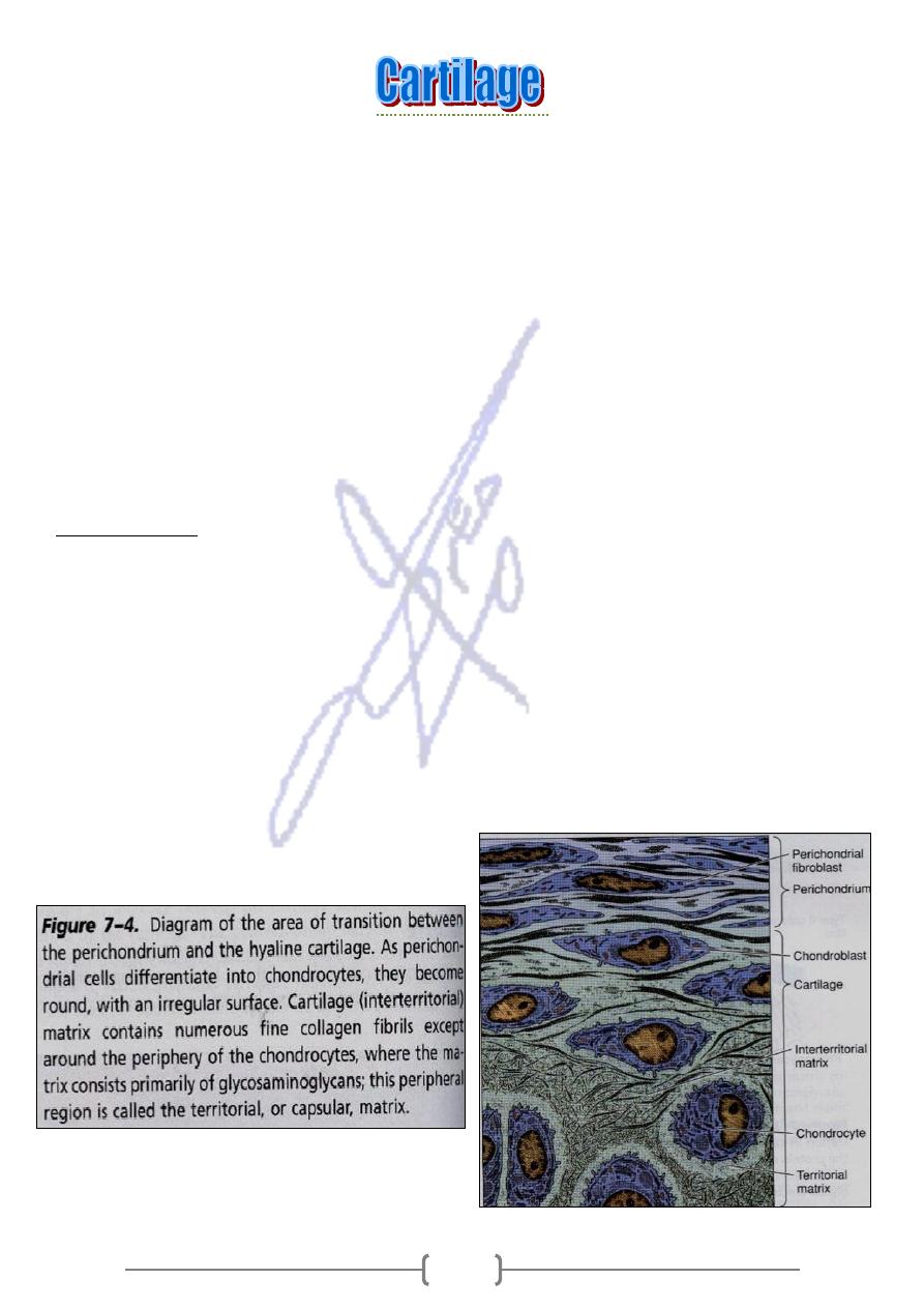
Perichondrium
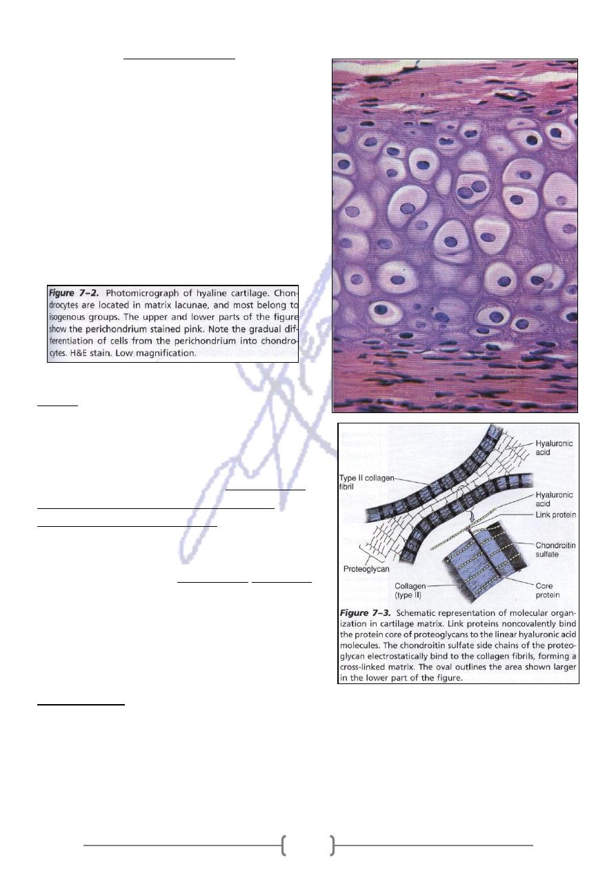
Chondrocytes are located in matrix called Isogenous .
The principle macromolecules present in all types of cartilage are collagen, hyaluronic acid,
proteoglycns, and small amounts of several glycoprotein.
Cartilage is a vascular and is nourished by tissue ((Perichodrium) ,or by synovial fluid
from joint cavities .Cartilage has no lymphatic vessels or nerves
Perichodrium is a sheath of dense connective tissue that surrounds cartilage & the
vascular supply for the a vascular cartilage & contains nerves, lymphatic vessels .
Reticular cartilage ,which covers the surfaces of the bones of the movable joints ,is
devoid of perichondrium and is sustained by the diffusion of O2 and nutrients from
synovial fluid .
188
Hyalinn cartilage
Is the most common and best studied of the
three forms
Fresh hyaline c. t. Is blush –white & translucent.
In embryo ,it is served as a temporary skeleton
until it is gradually replaced by bone
In adult ,hyaline cartilage is located in the
articular surfaces of movable ,bronchi ) ,in the
ventral ends of ribs .
In the epiphyseal plate ,where it is responsible
for the longitudinal growth of bone .
Matrix
40% of dry weight of h. c.t. consists of
collagen embedded in a firm hydrated gel of
proteoglycans and glycoprotein .
-
chondroitin 4
Cartilage proteoglycans contain
sulfate ,and keratan,
-
sulfate , chondroitin 6
.
covalently linked to core proteins
More than 200 of these proteoglycan are
nonconvalently associated with long molecules of
)
aggregates
proteoglycan
hyaluronic acid , forming
.that interact with collagen, resemble bottlebrushes.
Chondronectin: A macromolecule of
structural glycoprotein that binds to GAG and
collagen type II, mediating the adherence of
chondrocytes to the extracellular matrix.
Perichondrium
All hyaline cartilage is covered by a layer of dense connective tissue except articular cartilage.
It is essential for the growth and maintenance of cartilage .
It is rich in collagen type I fibers and numerous of fibroblasts .
In the inner layer of the perichodrium, there are a chodroblasts and easily differentiate into
chondrocytes.
189
Chondrocytes
They are found in the periphery of hyaline cartilage ,have an elliptic or round shape, and may
appear in groups of up to eight cells ,originating from mitotic divisions of single chondrocytes
called (Isogenous).
They synthesize collagens & other matrix molecules .
They metabolize glucose by anaerobic glycolysis to produce lactic acid .
Because cartilage is devoid of blood capillaries, chondrocytes respire under low oxygen
tension.
GAG
sulfated
The synthesis of
.
ds on a proper hormonal balance
function depen
Chondrocytes
hormone thyroxin ,& testosterone and is slowed by cortisone,
is accelerated by growth
.
hydrocortisone
Cartilage growth depends on hormone somatotropin, which does not act directly on cartilage
somatomedin C in liver .
t promotes the synthesis of
bu
.
e cells, promoting their growth
acts on cartilag
Somatomedin C
Medical application
Cartilage cells can give rise to benign (chondroma) or malignant (chodrosarcoma) tumors
Hyaline cartilage is more susceptible to degenerative aging processes.
Calcification of the matrix, which lead to increase in size and volume of chondrocytes and
followed death .
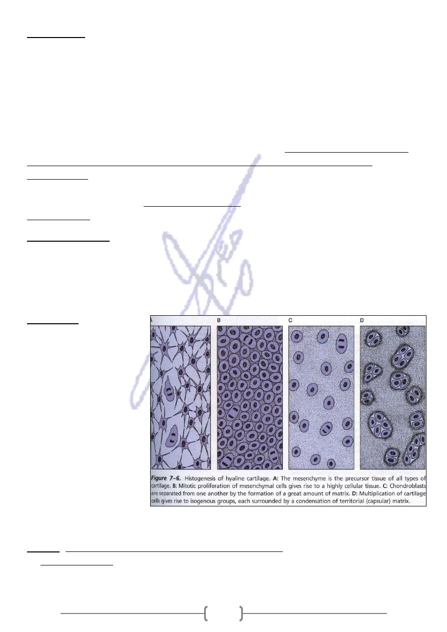
Histogenesis
Cartilage drives from the
mesenchyme
Mesenchyme cells
multiply rapidly , and form
mesenchymal condensation
of chondroblasts, which
differentiate, now called
chondroblasts.
Chodroblasts are separated
from one another by the
formation of a great amount
of matrix.
During development ,the
differentiation of cartilage takes place from the center outward; whereas the peripheral cells are
typical chondroblasts.
growth of cartilage is attributable to two processes:
The
:
Growth
Resulting from mitotic division of preexisting chondrocytes .
:
Interstitial growth
It occurs during the early phase of cartilage formation & occurs in epiphyseal plates of long
bones and within articular cartilage since there is no perichondrium there.
191
Appositional growth: Resulting from differentiation of perichondrium cells. (chondroblasts
proliferate & become chodrocyte) once they have surrounded themselves with cartilaginous
matrix
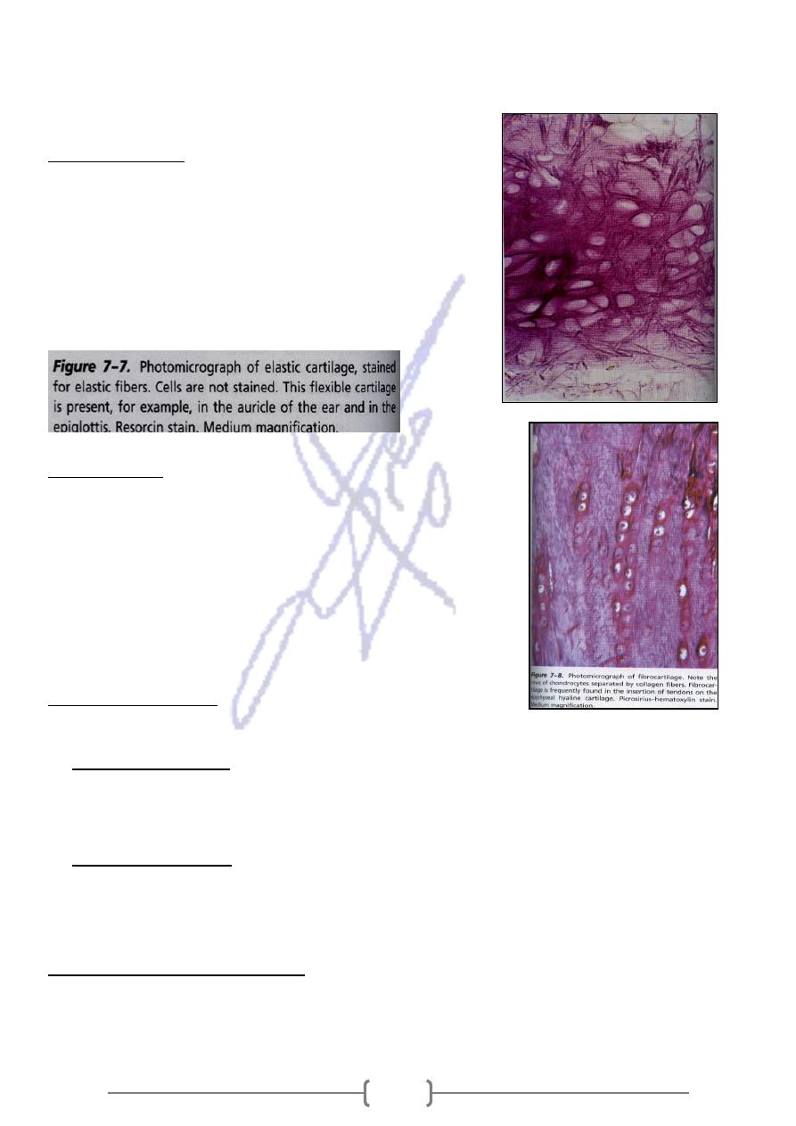
Elastic cartilage
Found in the auricle of the ear , the wall of the external
auditory canals and auditory tubes, the epiglottis & cuneiform
cartilage in the larynx
It is identical to hyaline cartilage except that it contains an
abundant network of elastic fibers.
Has a yellowish color owing to the presence of elastin in
elastic fibers
Fibrocartilage
It is intermediate between dense connective tissue and
hyaline cartilage .
It is found in intervertebral disks , in attachments of certain
ligament to the cartilagenous surface of bones ,and in the
symphysis pubis .
Always associated with dense connective tissue.
Contains chondrocytes ,either singly or in isogenous groups,
arranged in long rows separated collagen type
Intervertebral disks
Is situated between 2 vertebrate & is held to them by ligaments. Have two components :
of
Has an external layer of dense connective tissue or composed
The annulus fibrosis:
overlapping laminae of fibrocartilage .The multiple lamellae, with collagen fibers in adjacent
layers provide the disk with annual resilienc that enables it to withstand the pressures generated
by impinging vertebrae.
, derived from
annulus fibrosis
r of the
Is situated in the cente
:
The nucleus pulposus
embryonic notochord , consists of a few rounded cells embedded in a viscous matrix, rich in
hyaluronic acid & collagens fibril .It becomes smaller with age and is partially replaced by
fibrocartilage.
e itervertebral disk
Hernation of th
Rupture of annulus, results in expulsion of nucleus pulpous and a concomitants flattening of the
disk, so the disk slips from its position between the vertebrae . If it moves toward the spinal
cord, it can compress the nerves & results in severe pain.