
List of contents
1- Cytology (Page: 2-17)
Lab 1: The Microscope (Page: 3-5)
Lab 2: Cells (Page: 6-7)
Lab 3: Types of Cells according to the shape (Page: 8-9)
Lab 4: Cellular Structure (Page: 10-12)
Lab 5: Locomotor Organelles (Page: 13-17)
2- Genetics (Page: 18-40)
Lab 6: DNA Isolation (Page: 19-21)
Lab 7: Detection & Measurement of Genetic Variation (Page: 22-27)
Lab 8: Cytogenesis & Karyotype (Page: 28-33)
Lab 9: Chromosomal Abnormalities (Page: 34-40)
3- Part of Histology (Page: 41-60)
Lab 10: Epithelial Tissue (Page: 42-46)
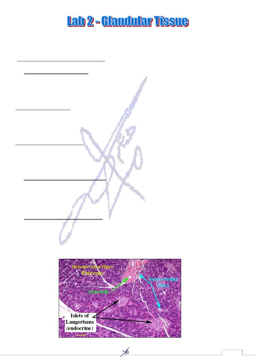
Lab 11: Glandular Tissue (Page: 47-50)
Lab 12: Connective Tissue (Page: 51-52)
Lab 13: Cartilage & Bone (Page: 53-56)
Lab 14: Muscular Tissue (Page: 57-58)
Lab 15: Nervous Tissue (Page: 59-60)
1

2
3
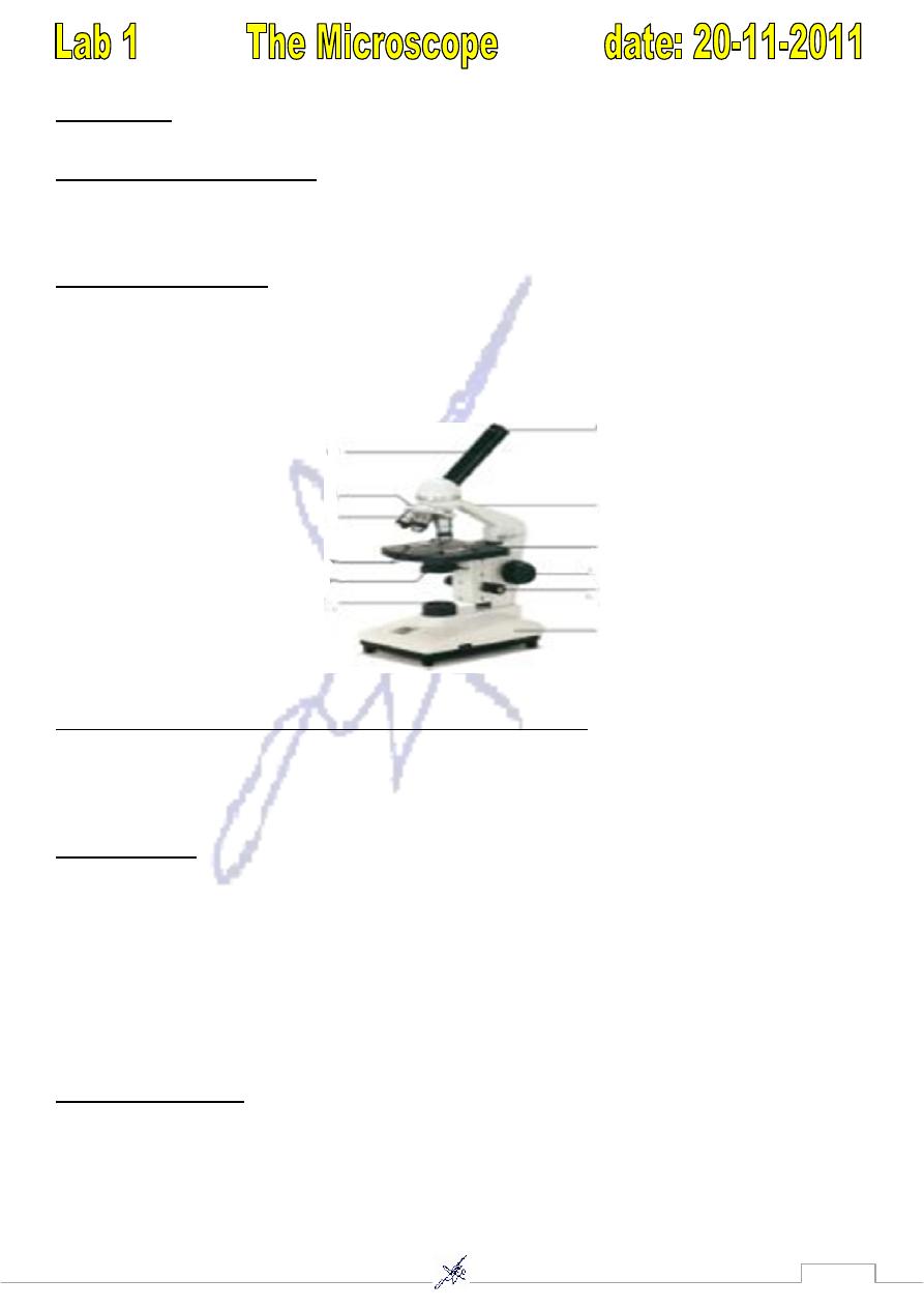
Microscope :
is Definition of an optical instrument used to magnify small objects so that they can
be seen much better than with your eye.
*Aim to use the microscope?
1) To see things usually living things that we cannot see them by the naked eye.
2) To examine things and studies the properties, size, number, shapes…..etc.
Types of Microscope
1) The compound microscope (Light microscope).
2) Stereoscopic Dissecting microscope.
3) Electron microscope (E.M)
A- Transmission (T.E.M.) B- Scanning microscope (S.E.M.)
1) Compound microscope (Light microscope):
The light microscope is based on the interaction of photons ( light unite ) & tissue components.
Parts of compound microscope:
1- optical parts:
A- eye lenses (ocular lenses) : the lenses at the top that you look through. They are usually
5x,7x,10x,20x magnification power.
B- Objective Lenses: Usually you will find 3 or 4 objective lenses on a microscope. They almost
always consist of 4X (very low ), 10X (low), 40X (high )and 100X (very high or oil immersion)
magnification powers.
C- condenser: it collect & focus the illumination to produce a cone of light that illuminates the
object to be observed.
2- Mechanical parts :
A-Stage: The flat platform where you place your slides. Stage clips hold the slides in place. If your
microscope has a mechanical stage, you will be able to move the slide around by turning two
Knobs. One moves it left & right; the other moves it up & down.

4
2- Revolving Nosepiece : This is the part that holds two or more objective lenses and can be
rotated to easily change power.
3- diaphragm : many microscopes have rotating disk under the stage. This diaphragm has different
sized holes & is used to vary the intensity & size of the cone of light that is projected upward into
slide.
4-body tube : connects the eye lenses to the objective lenses.
5- arm: supports the tube & connects it to the base.
6- base: the bottom of the microscope, used for support.
7- light source : contain on/off switch & adjustable lamps intensities with colored filter.
8-coarse adjustment: used to move the stage up & down, it must used only with low power lenses
9- fine adjustment: it used only with high power lenses.
Avoiding hazards in microscopy:
1- always carry the microscope with one hand on the arm & other hand on the base, carry it close
to your body.
2- always clean the lenses & stage of the microscope before & after use.
3- place slide on the microscope stage.
4- always start & end with low power lenses.
*Efficiency of Microscope depend on the resolution power & magnification power:
Resolution power :
the ability of a microscope to show fine detail, defined as the minimum
distance between two points at which they can be seen as separate images. It depends on the
wavelength of the light. As the wavelength decrease , the resolution increases( reverse correlation)
.the resolution power of L.M. is 0.1 µm , wavelength of light (400- 700 nm) this power permits
good images magnified 1000 -1500 times. Objects smaller than 0.1 µm cannot be distinguished
with this instrument.
Magnification power :
the total Magnification is obtained by multiplying the Magnification
power of the objective lenses by the Magnification power of eye lenses.
2) stereoscopic dissecting M.S :
the magnification power of this microscope is (4x- 50x ).
According to the type of it , the specimens used are the dissecting specimens , the light source is
external & form above. These are the differences from the compound microscope.
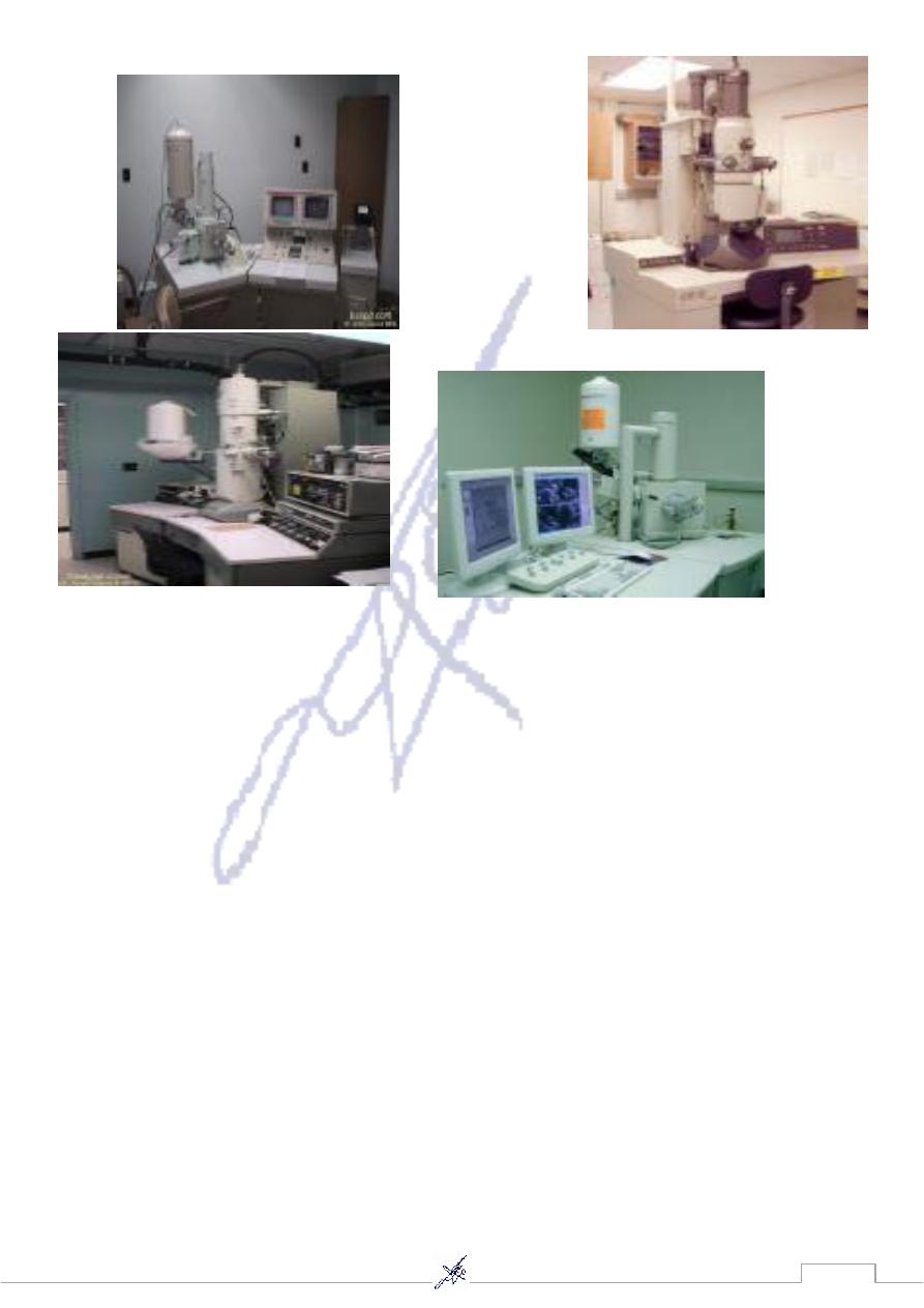
3)Electron microscope (E.M)
Is based on interaction of electron and tissue components , We use this microscope (E.M.) to
things that we cannot see them by light microscope or to see more details of these things because
the wave length of electron is 0.005 nm therefore it gives high resolution (reverse correlation
between wave length and resolution) , the maximum resolution power of E .M. Is 0.1 nm . This
show that the E.M. gives hundred thousand of resolution than L.M. .
5
6
its of all living organisms.
are the structural & functional un
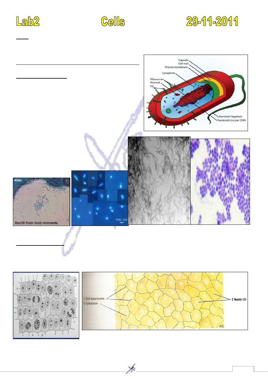
Cells
*Cells can be divided according to many characteristics:
Types of cell according to origin:
1)
Prokaryotic cell :
-
A
( Gr. Pro : before, karyotic : nucleus) cell is found
only in bacteria ( unicellular). these cells are small
(1 – 5 µm long) usually have a cell wall , lack a
nuclear envelope & lack a membrane – bound
components, called organelles.
Ex: Bacilli (rod shape) *Cocci (sphere shape)
B- Eukaryotic cell :
(Gr. Eu : good , karyotic : nucleus ) cells are larger than prokaryotic( multicellular) , have a distinct
nucleus surrounded by a nuclear envelope & numerous organelles are found in cytoplasm.
Ex: Plant cell (onion cell) Ex : animal cell (buccal cavity)
7
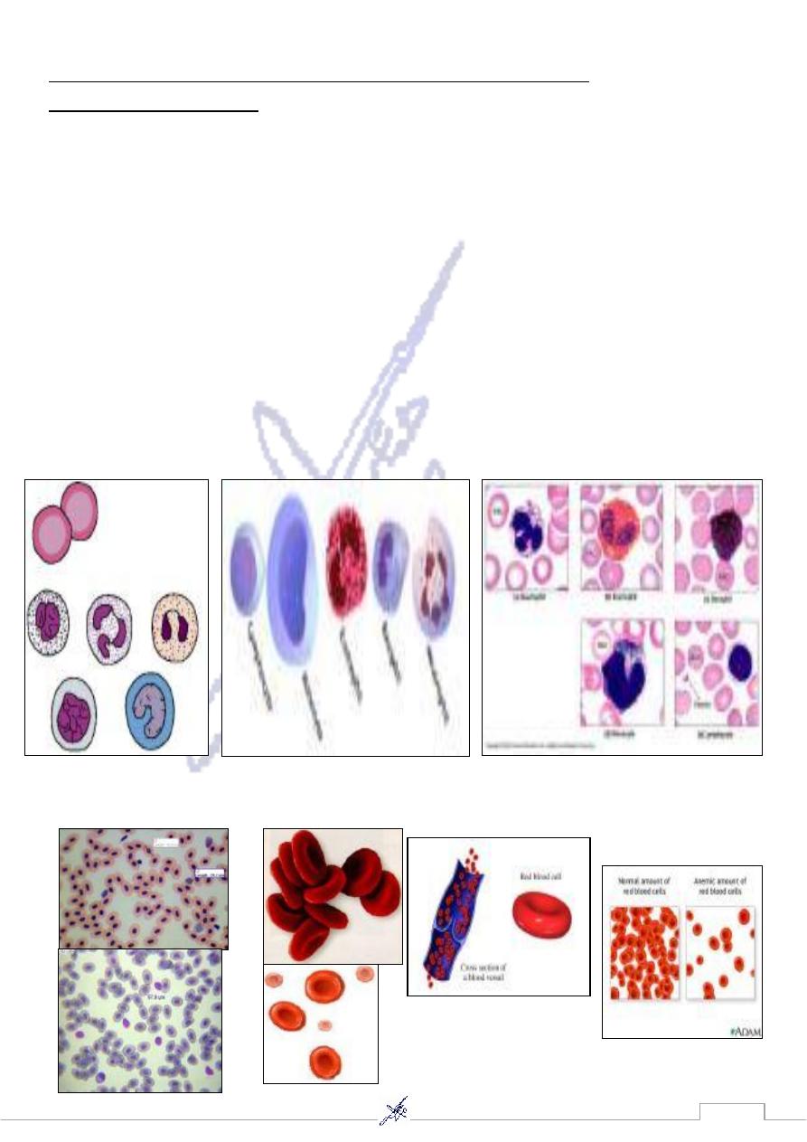
2)Types of cell according to the presence of nucleus
A- Nucleated cell ex:
1) White Blood Cells (W.B.C)
1- Neutrophils : The neutrophils with granules and lobulated nucleus are the polymorph nuclear
granulocytes. the cytoplasm contain the fine violet or pink granules, the nucleus consist of several
lobes that are connected by narrow chromatin strand.
2-Eosinophils : The eosinophils are identified in blood smear by their cytoplasm , which is filled
with distinct large , eosinophils ( bright – pink ) granules , the nucleus is bilobed.
3- Basophils : The granules in basophils are not as numerous as in eosinophils , however , they are
more vary in size , & are less densely packed & stain dark blue or brown . The nucleus is not
lobulated & stain pale basophilic , but it is usually obscured by the density of the granules.
4- Lymphocyte : A granulated leukocytes have round to horseshoe _shape nucleus ,the nucleus
occupy most of the cytoplasm which appears as a thin basophilic rim around the nucleus.
5- Monocyte : are the largest a granular leukocytes. the nucleus varies from round or oval to
horseshoe-shaped & it stain lighter than lymphocyte nucleus.
2) Red Blood cells of frog
Red blood cells of human ( R.B.C )
8
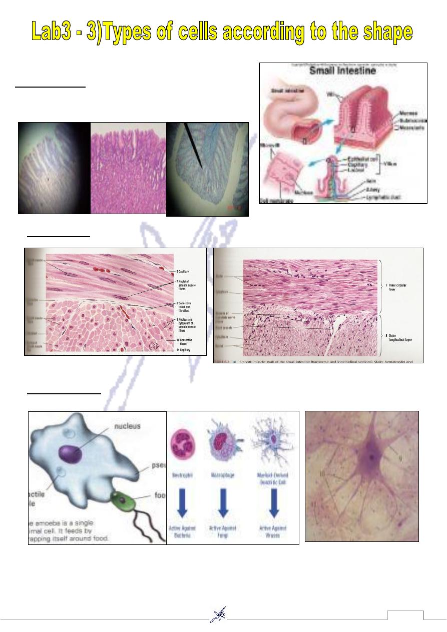
stomach, small intestine.
ex :
Columnar cell
-
1
stomach
smooth muscle
ex:
Spindle cell
-
2
3- Star shape cell
ex: neuron (nerve cell) in spinal cord.
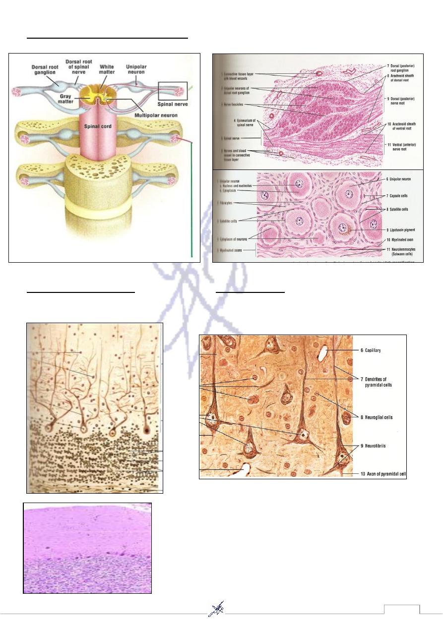
9
4- Round shape cell (pseudounipolar)
ex: Neuron in dorsal root ganglia.
5-Pyriform(purkinje cell)
6-Pyramidal cell
ex: neuron in Cerebellum. ex: neuron in Cerebral cortex.
11
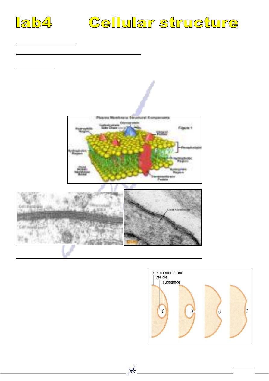
1) Cell membrane
A- Electron micrograph membrane (2 layers)
Cell membrane The cytoplasm of cell is surrounded by cell membrane or plasma membrane .
It serves to separate & protect cell from environment. Plasma membrane range from 7.5 – 10 nm
in thickness & consequently are visible only in the E. M. ; plasma membrane consist of a bilayer or
double layer of phospholipids ( hydrophobic (non polar ) chains directed toward the center of the
membrane & their hydrophilic (polar) heads directed outward embedded into protein molecules .
Plasma membrane is semi – permeable .
B- Electron micrograph of the main process in plasma membrane:-
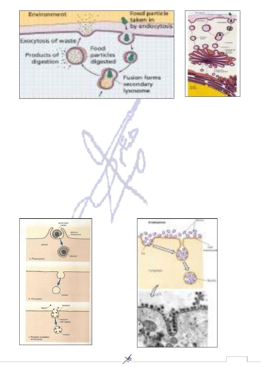
1- Exocytosis:
During exocytosis, vesicles often formed by the Golgi
apparatus and carrying a specific molecule, fuse with
the plasma membrane as secretion occurs. This is the
way that insulin leaves insulin-secreting cells, for
instance.
Notice that the membrane of the vesicle becomes a
part of the plasma membrane. During cell growth,
exocytosis is probably used as a means to enlarge the
plasma membrane, whether or not secretion is also taking place.
11
2- Endocytosis ( Phagocytosis & Pinocytosis)
During endocytosis,cells take in substances by vesicle formation (Fig. 5,11). A portion of the plasma
membrane invaginates to envelope the substance, and then membrane pinches off to form an
intercellular vesicle.
* When the material taken in by endocytosis is large, such as a food particle or another cell, the
peocess is called phagocytosis. Phagocytosis is common in unicllular organisms like amoebas and
ameboid-type cells like macrophages, which are large cells that engulf bacteria and worn-out red
blood cells in mammals. When the endoctic vesicle fuses with a lysosome, digestion occurs.
* Pinocytosis occurs when vesicles form around a liquid or very small particles. Blood cells, cells
that line the kidney tubules or intestinal wall, and plant root cells all use this method of ingestion
substances. Whereas phagocytosis can be seen with the light microscope, the electron microscope
must be used to observe pinocytic vesicles, which are no longer than 1-2 µm.
12
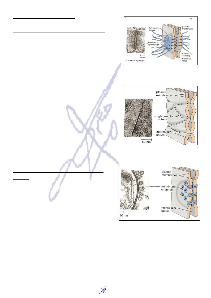
C- Junctions in animal cells:-
1) Desmosomes (Adhesion junction),
(between epithelial cells) ex: heart & stomach.
In adhesion junction, internal cytoplasmic plaques, firmly
attached to the cytoskeleton within each cell, are joined
by intracellular filaments. In some organs like the heart,
stomach and bladder, where tissues get stretched.
Adhesion junction hold the cells together.
2) Tight junction,
(between epithelial cells) ex: intestine & kidney .
Adjacent cells are even more closely joined by tight
junctions, in which plasma membrane proteins actually
attach to each other, producing a zipperlike fastening,
The cells of tissues that serve as barriers are held
together by tight junctions; in the intestine the digestive
juices stay out of the body, and in the kidneys the urine
stays within kidney tubules, because the cells are joined
by tight junctions.
3) Gap junction
(between embryonic cells) ex: liver & cardiac
muscle .
A gap junction allows cells to communicate. A gap
junction is formed when two identical plasma
membrane channels join. The channel of each cell is
lined by six plasma membrane proteins. A gap
junction lends strength to the cells, but it also allows
small molecules and ions to pass between them. Gap
junctions are important in heart muscle and smooth
muscle because they permit a flow of ions that is required for the cells to contract.
13
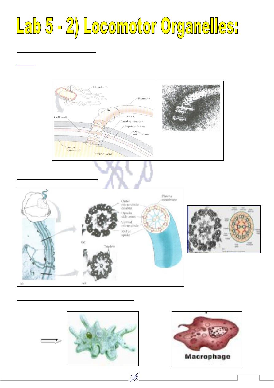
A-Flagella ex: Human sperm .
Flagella
are the organelles of mobility. They arise from cytoplasm and extrude through the cell
wall. They are long and thick thread like appendages, protein in nature, formed of flagellin protein
B- Cilia (9+2) ex: Paramecium .
c-Pseudopodia ex: Amoeba , Macrophage .
Amoeba
14
The cell organelles
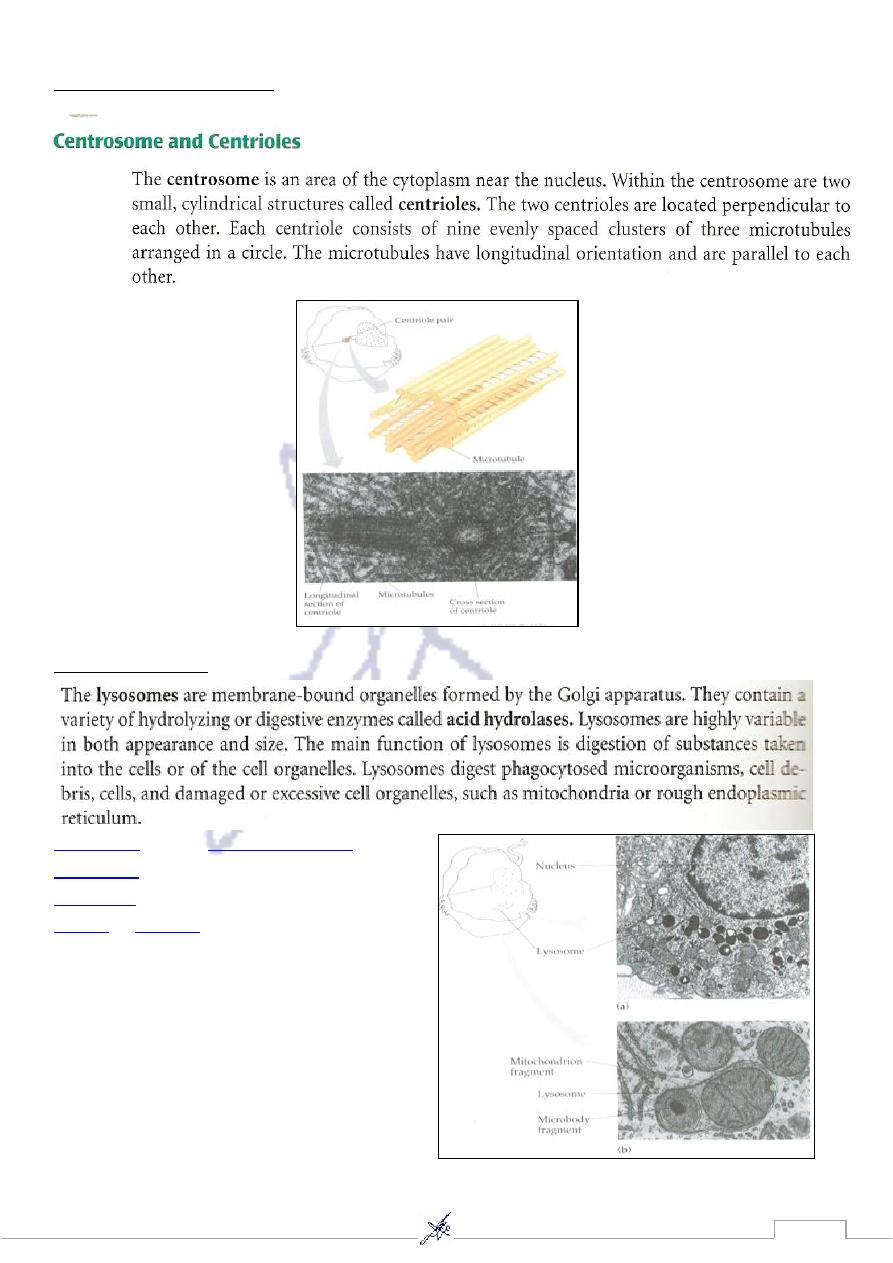
3) Mitochondria:
The mitochondria are elongated, rod-shaped structures
that often vary in size and shape, depending on the cell
function. Each mitochondrion (plural, mitochondria)
consists of an outer and inner membrane. The inner
membrane exhibits numerous folds in the form of cristae.
In protein secreting cells, these cristae project into the
interior of the organelle in the form of shelves. In
contrast, mitochondria exhibit tubular cristae in steroid-
secreting cells of the adrenal cortex or interstitial cells in
the testes. Surrounding the mitochondrial cristae is an
amorphous mitochondrial matrix.
Mitochondria are self-replicating organelles that occur in various numbers, shapes, and sizes in the
cytoplasm of all eukaryotic cells .Mitochondria play a critical role in generating energy in the
eukaryotic cell .Mitochondria generate the cell's energy by the process of
oxygen
, utilizing
oxidative phosphorylation
to release energy stored in cellular nutrients
( typically pertaining to
glucose
)to generate
AT
P.
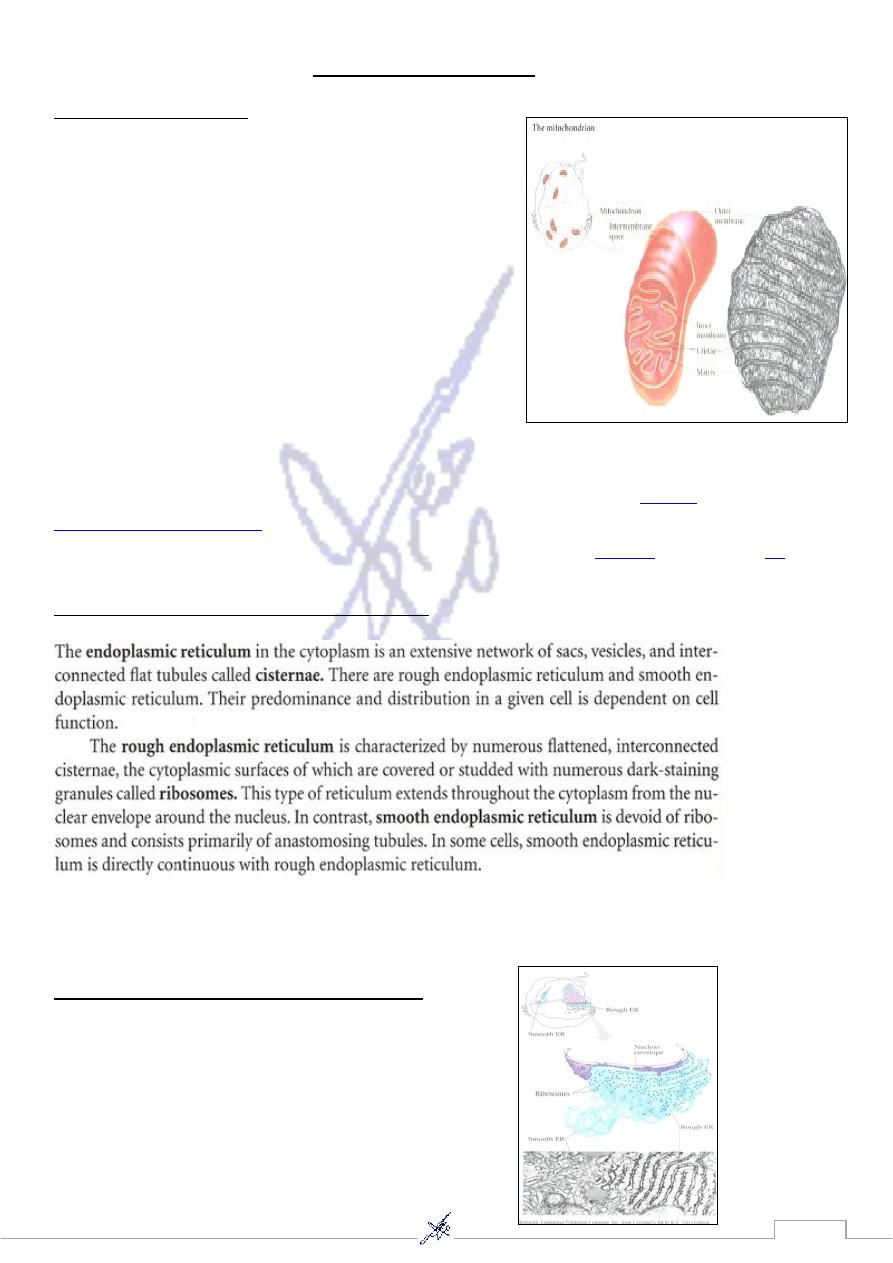
4) Endoplasmic reticulum (E.R.)
The ER has two forms: the rough ER, which has ribosomes on its surface and secretes proteins into
the cytoplasm, and the smooth ER, which lacks them. Smooth ER plays a role in calcium
sequestration and release.
A- Smooth endoplasmic reticulum (S.E.R.).
15
B- Rough endoplasmic reticulum (R.E.R.)
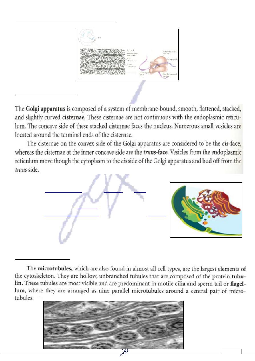
5) Golgi apparatus
The primary function of the Golgi apparatus is to process and
package the
macromolecules
,such as
proteins
and
lipids
that are
synthesized by the cell. It is particularly important in the
processing of proteins for
secretion
. The Golgi apparatus forms a
part of the
endomembrane system
of eukaryotic cells.
Vesicles
that enter the Golgi apparatus are processed in a cis to trans
direction, meaning they coalesce on the cis side of the apparatus
and after processing pinch off on the opposite (trans) side to form
a new vesicle in the animal cell.
6) Microtubules.
16
7) Centrioles(9+0)
8) Lysosome:
Lysosomes
contain
digestive enzymes
(acid
hydrolases
). They digest excess or worn-out
organelles
, food particles, and engulfed
viruses
or
bacteria
. The cell could not house
these destructive enzymes if they were not
contained in a membrane-bound system.
These organelles are often called a "suicide
bag" because of their ability to detonate and
destroy the cell.
17
9) Lipid droplets
Note…..
3,4,5,6,7,8,9 are included in the endomembranous system
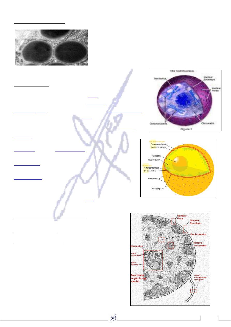
10) Nucleus
The nucleus (pl .nuclei ;from
Latin
nucleus or nuculeus ,
or kernel) is a membrane-enclosed
organelle
found in all
eukaryotic
cells
.It contains most of the cell's
genetic material
,
organized as multiple long linear
DNA
molecules .
The main structures making up the nucleus are the
nuclear
envelope
,a double membrane that encloses the entire
organelle and separates its contents from the cellular
cytoplasm
,and the
nuclear lamina
,a meshwork within the
nucleus that adds mechanical support, much like the
cytoskeleton
supports the cell as a whole. Because the
nuclear membrane is impermeable to most molecules ,
nuclear pores
are required to allow movement of
molecules across the envelope. These pores cross both of
the membranes, providing a channel that allows free
movement of small molecules and
ions
.
Types of chromatin
A- Euochromatin (light stained and active).
B-Heterochromatin (dark stained and inactive)
18
19
Deoxyribonucleic acid (DNA) isolation is an extraction process of DNA from
various sources.
The aim to separate DNA present in the nucleus of the cell from other cellular
components.
Application of DNA isolation:
It is needed for genetic analysis which used for:
, such as introduction of DNA into
use DNA in number of Applications
scientific
-
1
cells & animals or plants for diagnostic purposes.
is the most common.
Medicine
-
2
needs to recover DNA for identification of individuals ,( for
science
forensic
-
3
example rapists, petty thieves, accident , or war victims) , paternity determination.
:
Sample collection
A- Sample can be isolated from any living or dead organism . Common sources for
DNA isolation include whole blood , hair, sperm , bones , nails , tissues , blood
stains , saliva , buccal (cheek) swabs , epithelial cells , urine , paper cards used for
sample collection .
B- Sample size may be small ( for example sperm , or single hair ) the method has to
be different to the method used in isolating DNA from a couple of milligrams of
tissue or milliliters of blood.
C- Sample age may be fresh or has been stored . Stored sample can come from
archived tissue samples , frozen blood or tissue , exhumed bones or tissues &
ancient human sample.
Diversity of DNA isolation method :
.
lysis or breakdown , of tissue or cells
The isolation of DNA usually begins with
-
1
This process is essential for destruction of protein structures & allows for release of
nucleic acids from the nucleus .
lysis is carried out in a salt solution, containing detergents to denature proteins or
proteases (enzymes digesting proteins) such as proteinase K , or in some cases both .
It results in the breakdown of cells & dissolving of membranes.
DNA may be isolated from hard bones such as bone & wood
Example ;bones are highly mineralized & the ions have to be removed from the
samples before extraction so they do not later interfere with PCR. They homogenized
in lysis buffer using a mechanical homogenizer.

21
commercial DNA purification kits:
-
2
Commercial DNA purification kits use the common lysis solutions contain : sodium chloride
, tromethamine (also known as tris ) , which is a buffer to retain constant pH;
ethylendiaminetetraacetic (EDTA) , which binds metal ions , & sodium dodecyl sulfate
(SDS) which is a detergent . A common enzyme used in
.
DNA extraction is protienase K
alkaline denaturation:
-
3
When sample is small size like buccal swabs & occasionally blood stain using this method
,alkaline denaturation of the sample is used to release DNA from the cells. Samples can be
placed in small
plastic tubes (eppendorfs) subjected)
4- heart denaturation :
Achieved by boiling samples .heating of a sample to 100c releases DNA into the solution but
also denatures it by separating the two strand , there are remaining inhibitors in the form of
degraded proteins other organic compound or ions .
5- another method used commonly in forensic : laboratories
This method utilizes chelex ion exchange resin that bind multivalent metal ions & is
particularly useful in removing inhibitors form DNA , it can be used with any type of
sample , including whole blood ,blood stains , seminal stains , buccal swabs or hair.
6- paramagnetic beads with DNA binding capacity :
Samples are lyses & then the solid material is treated with proteinase K ; the lysates are
then applied to the beads. Resin is subsequently washed & DNA is eluted of it at 65c ;
magnetic beads are separated from the sample on a magnetic stand .
Summary of DNA extraction :
There are three basic & two optional steps in a DNA extraction :
1- breaking the cells open , commonly referred to as cell disruption or cell lysis , to expose
the DNA within . This is commonly achieved by chemical & physical methods – blending ,
grinding or sonicating the sample
2- removing membrane lipids by adding a detergents or surfactants .
3- removing proteins by adding a protease (optional but almost always done ) .
4- removing RNA by adding an RNase (often done ).
5- precipitating the DNA with alcohol- usually ice cold ethanol or isopropanol since DNA is
insoluble
In these alcohols , it will aggregate together, giving a pellet upon centrifugation . This step
also removes alcohol- soluble salt

21
Detecting DNA:
DNA concentration can be determined measuring the intensity of absorbance of the solution
at the 600 nm with a spectrophotometers & comparing to a standard curve of known DNA
concentration
Measurement of purity:
Measuring the intensity of absorbance of the DNA solution at wavelength 260nm & 280nm
is used as a measure of DNA purity . DNA absorbs uv light at 260 &280 nm & aromatic
proteins absorb uv light at 280 nm , Apure sample of DNA has the 260/280 ratio at 1.8 & is
relatively free from protein contamination. A DNA preparation that is contaminated with
protein will have a 260/280 ratio lower than 1.8
22
1. Blood Groups:
Each of the blood group system is determined by a different gene or set of genes.
The various antigens that can be expressed within a system are the result of different
DNA sequences in these genes.
Two blood systems that have special medical significance, the ABO and Rh systems.
The ABO system consist s of two major antigens, labeled A and B located on the
surface of erythrocytes. Individuals can have one of four blood types depends on
presence or absence of one or both antigens.
Blood Groups:
The ABO system which is encoded by a single gene on chromosome 9, consists of three
primary alleles, labeled IA, IB, and IO. Individuals with IA allele have the A antigen on
their erythrocyte surface (blood type A), those with IB have the B antigen on their cell
surface (blood type B). Those with both alleles express both antigens (blood type AB), and
those with two copies of IO allele have neither antigen (type O blood), because the IO allele
produces no antigens.
Relationship between ABO Genotype and blood type
Blood type
Genotype
A
I
A
I
A
A
I
A
I
O
B
I
B
I
B
B
I
B
I
O
AB
I
A
I
B
O
I
O
I
O
Applications
1. Blood group system used extensively in studies of genetic variation among individuals
and populations.
2. Determining the compatibility of blood transfusions and tissue grafts.
3. Some combinations of these systems can produce maternal- fetal incompatibility with
serous results for the fetus.

23
2. Protein electrophoresis
s technique is
The principle of thi
to detect variations in DNA, RNA, and variations in genes that encode certain
serum proteins.
These variations are observable because all (DNA, RNA, Protein) can be separated
by means of an electric field.
n:
Clinical Applicatio
To determine whether an individual has normal hemoglobin (HbA) or the mutation
that causes Sickle cell disease (HbS). The replacement of glutamic acid with valine
in the ß- globin chain produces a difference in electrical charge.
The hemoglobin is placed in an electrically charged gel composed of starch or
agarose. The slight difference in charge resulting from amino acid replacement causes
the HbA and HbS forms to migrate at different rates through the gel.
After several hours of migration, the protein then stained with chemical solutions so
that their positions can be seen.
So polymorphism can detected if the HbA is homozygote or HbS homozygote, or
having a heterozygote HbA on one chromosome and HbS on the other.
3. Molecular Techniques:
The rate of variation in human DNA occurs at an average of 1 in every 300 to 500
base pair (bp).
Thus, approximately 10 million polymorphisms may exist among the 3 billion bp that
comprise the human genome.
Fortunately, molecular techniques developed during the past 30 years enable the
detection of thousands of new polymorphisms at the DNA level.
These techniques includes:
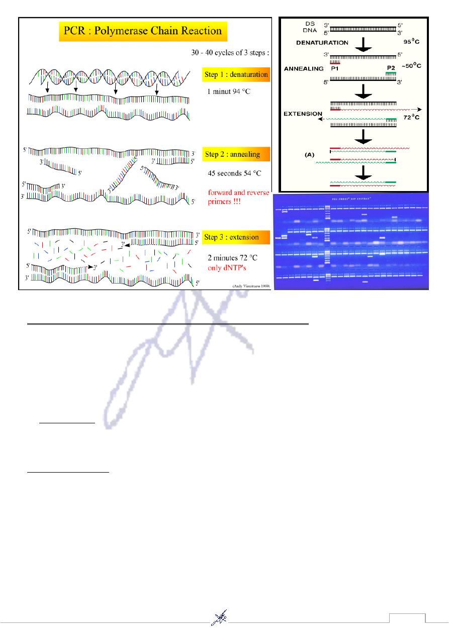
A. Polymerase Chain Reaction (PCR)
Principle:
PCR making millions of copies of a short, specific DNA sequence very quickly.
Heating- cooling cycles are used to denature DNA and then build new copies of a
specific, primer- bounded sequence.
Clinical Application:
Because of its speed and ease of use, this technique is now widely used for assessing
genetic variation for diagnosis genetic diseases , forensic purpose, detection and diagnosis
of infectious diseases, and used as fingerprints to identify genetic relationship between
individuals, such as parent- child or between siblings, and are used in paternity testing

24
PCR process requires four components:
1. Two primers: each consisting of 15-20 bases of DNA, containing sequences
complementary to the 3’ end of target region of DNA that contains the polymorphism
or a mutation that causes disease.
2. Heat- stable DNA polymerase enzyme: originally isolated from the bacterium
Thermus aquaticus with a temperature optimum at round 70 C.
free DNA nucleotides (dNTPs).
A large number of
3.
4. Small quantity of Genomic DNA from an individual act as a template.
PCR steps:
Typically, PCR consists of a series of 20- 40 repeated temperature changes called cycles,
with each cycle commonly consisting of 2-3 discrete temperature steps usually three.
1. Denaturation step:
The genomic DNA is first heated to a temperature of 94-98 C for 20-30 seconds. It
causes melting of the DNA template by disrupting the hydrogen bonds between
complementary bases, yielding single- stranded DNA molecules.
2. Annealing step:
The reaction temperature is lowered to 50-65 C for 20-40 seconds allowing annealing
of the primers to the single –stranded DNA template. Stable DNA-DNA hydrogen
bonds are only formed when primer sequence very closely matches the template
sequence. The polymerase enzyme binds to the primer- template hybrid and begins
DNA synthesis.
3. Extension/ elongation step:
Taq polymerase has its optimum activity temperature at 75- 80 C, and commonly a
temperature of 72 C is used with this enzyme. At this step the DNA polymerase
synthsizes a new strand complementary to the DNA template strand by adding dNTPs
in 5’to3’ direction. The DNA polymerase will polymerize a thousand bases per minute.
4. Final elongation:
This single step is occasionally performed at a temperature of 70- 74 C for 5- 15
minutes after last PCR cycle to ensure that any remaining single- stranded DNA is
fully extended.
Finally agarose gel electrophoresis is employed for size separation of the PCR
products. The size(s) of PCR products is determined by comparison with a DNA
ladder (a molecular weight marker) which contains fragments of known size, run on a
gel alongside the PCR products.
25
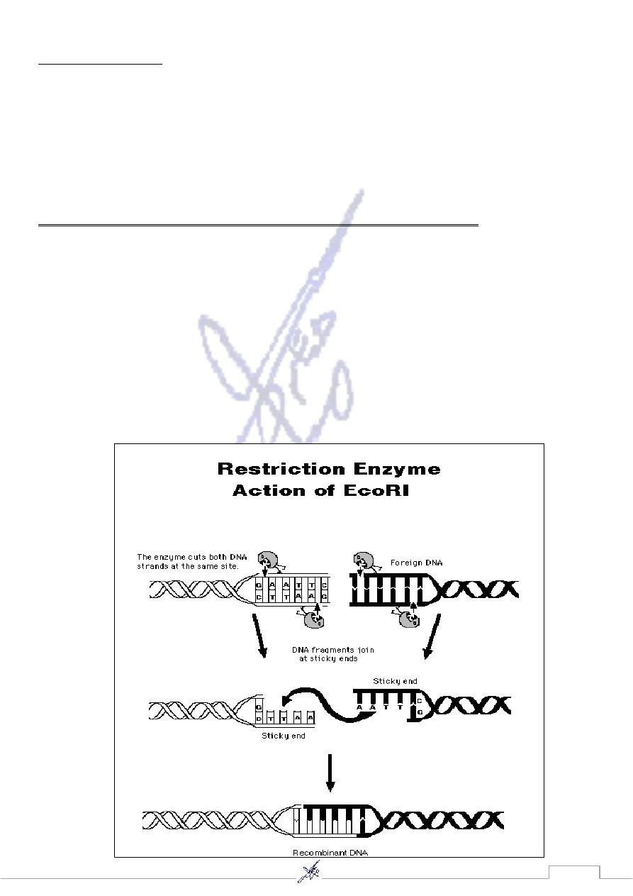
B. Restriction Fragment length Polymorphism (RFLP)
It took advantage of the existence of bacterial enzymes known as restriction
endonucleases or restriction enzymes. These enzymes are produced by various
bacterial species to “restrict” the entry of foreign DNA into the bacterium by cutting
or cleaving the DNA at specifically recognized sequences. These sequences are
called restriction sites.
that
1,
EcoR
produces a restriction enzyme called
Escherichia coli
,
For example
recognizes the DNA sequence GAATTC so this enzyme cleaves the sequence
between the G and the A, this produces DNA restriction fragments.
The RFLP process:
First DNA is extracted from blood samples
then digested by a restriction enzyme
and then loaded on a gel.
Electrophoresis separate the DNA fragments according to their size.
The DNA is denaturated and transferred to a solid membrane
and hybridized with a radioactive probe.
Exposure to x-ray film appears specific DNA fragments (bands) of different sizes in
individuals.
26
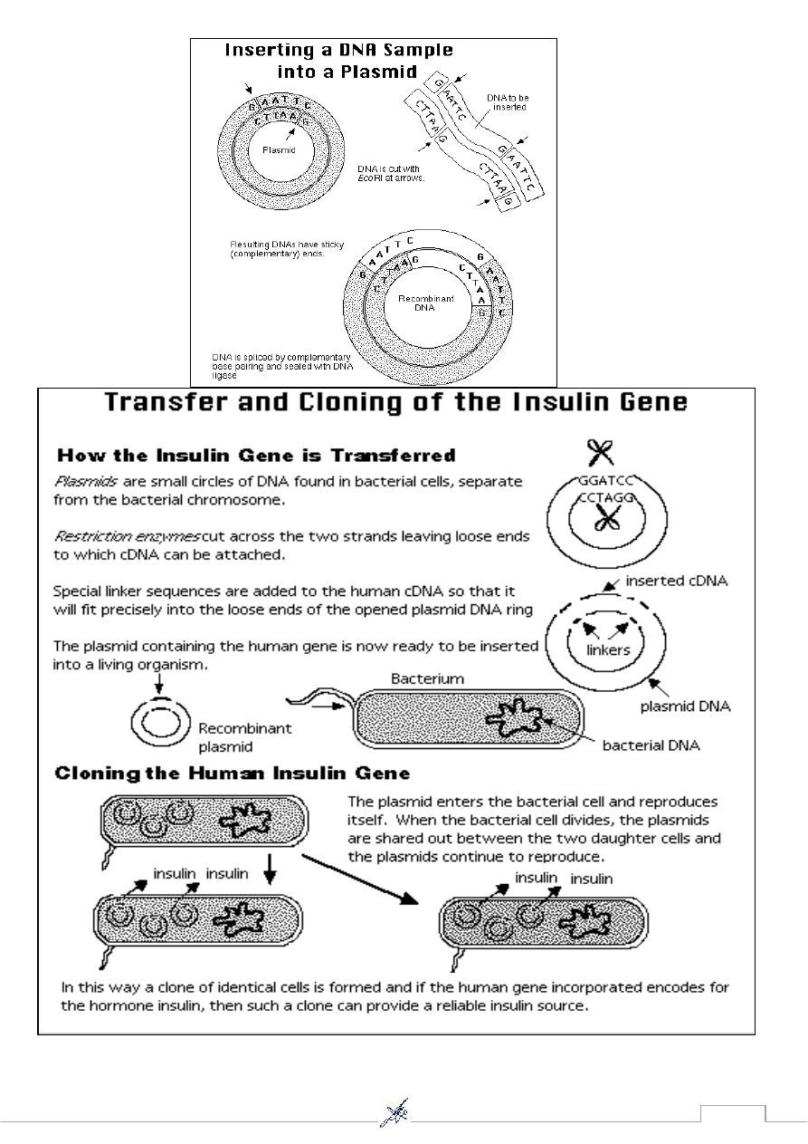
4. Gene cloning
The cloning of a gene produce many identical copies. Recombinant DNA technology
is used when a very large quantity of the gene is required.
Recombinant DNA (rDNA) contains DNA from two or more different sources. It
required a vector to introduce the rDNA into a host cell. One common type of vector is
plasmid. Plasmids are small accessory rings of DNA from bacteria. The ring is not part
of the bacterial chromosome and replicates on its own.
Two enzymes are needed to introduce foreign DNA into vector DNA.
The first enzyme, called a restriction enzyme, cleaves the vector’s DNA,
and the second, called DNA ligase, seals foreign DNA into the opening created by the
restriction enzyme.
The single-stranded, but complementary, ends of the two DNA molecules are called
“sticky ends” because they can bind a piece of foreign DNA by complementary base
pairing
Bacterial cells take up recombinant plasmids.
Thereafter, if the inserted foreign gene is replicated and actively expressed, the
investigator can recover either the cloned gene or a protein product.
27
28
Cytogenesis: Is the study of chromosomes and their abnormalities.
Karyotypes: is the complete set of chromosomes in the cells of an organism.
It is most often studied when the cell is at metaphase of mitosis and all the
chromosomes are present as pairs.
Chromosome: is an organized structure of DNA and protein found in cells. It is a
single piece of coiled DNA containing many genes, regulatory elements and other
nucleotide sequences. Chromosomes also contain DNA-bound proteins, which
serve to package the DNA and control its functions.
Structure of a chromosome
1) Levels of chromosomal structure
a- DNA helix. b- Nucleosomes. c- Coiled nucleosomes (solenoids)
d- Looped chromatin. e- Condensed chromatin. f- Condensed chromosome
Nucleosomes
DNA is packed inside the nucleus in association
with a number of proteins, which are extensively coiled
and folded forming nucleosomes. Each nucleosome is
made up a histone octamer mainly made up of
histones H2A, H2B, H3 and H4. The DNA is wound
around a histone protein, and then interacting with
another histone H1 forming a thicker fiber called
solenoid consisting of six nucleosomes. The solenoids themselves are organized into
chromatin loops, which are attached to a protein scaffold
Each of these loops contains approximately 100,000 bp or
100 kb of DNA.
Patterns of DNA coiling. DNA is wound around
histones to form nucleosomes. These are organized into
solenoids, which in turn make up the chromatin loops
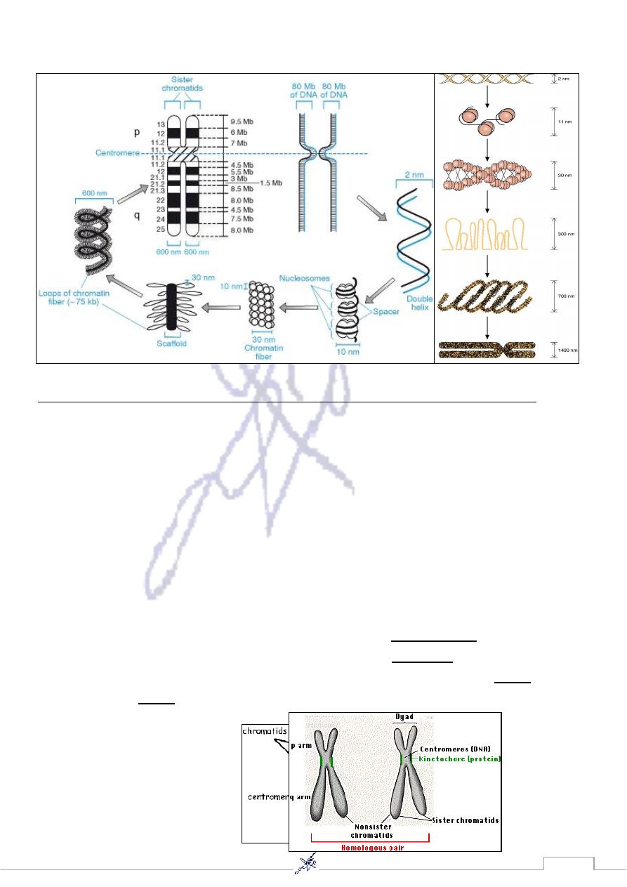
29
Patterns of DNA coiling.
Structure of a chromosome (Typical metaphase chromosome):
As mitosis begins, the duplicated chromosomes condense into short (~ 5 µm)
structures which can be stained and easily observed under the light microscope.
These duplicated chromosomes are called dyads.
The duplicates are held together at their centromeres.
In humans, the centromere contains 1–10 million base pairs of DNA. Most of this is
repetitive DNA: short sequences (e.g., 171 bp) repeated over and over in tandem
arrays.
While they are still attached, it is common to call the duplicated chromosomes
sister chromatids, but each chromosome is with a full complement of genes.
The kinetochore is a complex of >100 different proteins that forms at each
that will
spindle fibers
centromere and serves as the attachment point for the
anaphase.
separate the sister chromatids as mitosis proceeds into
;
p arm
The shorter of the two arms extending from the centromere is called the
.
q arm
the longer is the
31
zed according to:
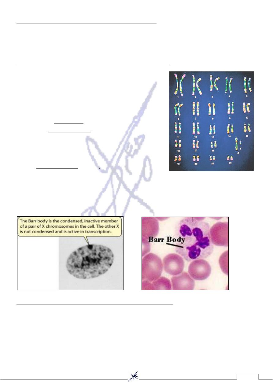
Individual chromosomes are recogni
1. their size
2. location of the centromere (a constriction)
3. and characteristic banding due to staining.
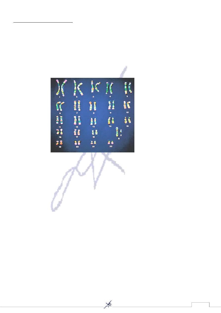
The Karyotype of human chromosome according to size
1.
The images of the 22 pairs of autosomes are
arranged according to: length, with the
sex
chromosomes in the right- hand corner.
The karyotype of the human female contains 23 pairs
of homologous chromosomes:
autosomes
22 pairs of
X chromosomes
1 pair of
The karyotype of the human male contains:
the same 22 pairs of autosomes
one X chromosome, larger
smaller
Y chromosome,
one
Barr body: the inactive X-chromosome of mammals
one of the 2 XX's becomes transcriptionally inactive,
which X is inactive seems to be randomized, in interphase cells the inactive X
chromosome can be visualized
2. Karyotyping according to the position of the centromere
Chromosomes are further classified according to the location of the centromere:
1. Metacentric chromosome: If the centromer occurs near the middle of the
chromosome.
2. Acrocentric chromosome has its centromere near the tip.
3. Submetacentric chromosomes have centromeres between the middle and the tip.
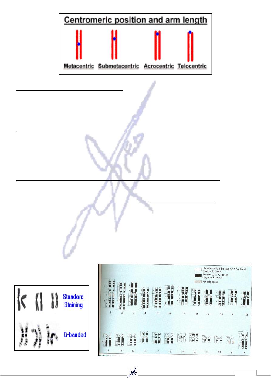
31
Characteristic banding due to staining
3.
Chromosome banding
Staining techniques were developed in the 1970 to produce the chromosome bands
characteristic of modern karyotypes.
Chromosome banding helps in detection:
the deletions, duplications, and other structural abnormalities.
* The major bands on each chromosome are numbered. Thus 14
q
32 refers to the second
band in the third region of the long arm of chromosome 14. Sub- band are designated by
decimal points following the band number (e.g. 14
q
32.3 is the third sub- band of band 2).
es
banding techniques are used in cytogenetics laboratori
-
Several chromosome
1. Quinacrine banding (Q- banding) was the first staining method used to produce
and is no
fluorescence microscope
specific banding pattern. This method requires a
longer as widely used as
2. Giemsa banding (G- banding). To produce G band, a Giemsa stain is applied after
the chromosomal proteins are partially digested by trypsin.
3. Reverse banding (R- banding) requires heat treatment and reverses the usual
white and black pattern that seen in G- bands and Q- bands. This method is
particularly helpful for staining the distal ends of chromosomes.

32
Other staining techniques specifically stain certain positions of the chromosome, include:
C- banding: stains the heterochromatin, which usually lies near the centromere.
Nucleolar organizing region stains (NOR stains) staining the satellites and stalks of
acrocentric chromosomes.
Staining with the trypsin-Giemsa method reveals a series of alternating light and dark bands
.
G bands
called
.
gene loci
G bands are numbered and provide "addresses" for the assignment of
Each band contains millions of DNA nucleotide pairs which do not correspond to any
functional structure.
Chromosome Numbers
called
body cells
All animals have a characteristic number of chromosomes in their
) number.
2n
(or
diploid
the
, one member of each pair having been acquired
homologous pairs
These occur as
from the gamete of one of the two parents of the individual whose cells are being
examined.
The gametes contain the haploid number (n) of chromosomes.
33
Stained chromosomes
Chromosomes are analyzed by collecting a living tissue (usually blood), (WBC)
culturing the tissue for the appropriate amount of time (usually 48 to 72 hours for
peripheral lymphocytes), adding colcemide or (concavalinin-A) to produce
metaphase arrest, harvesting the cells, placing the cell sediment on a slide, rupturing
the cell nucleus with a hypotonic saline solution, staining with a designated nuclear
stain, and photographing the metaphase “spread” of chromosomes on the slide.
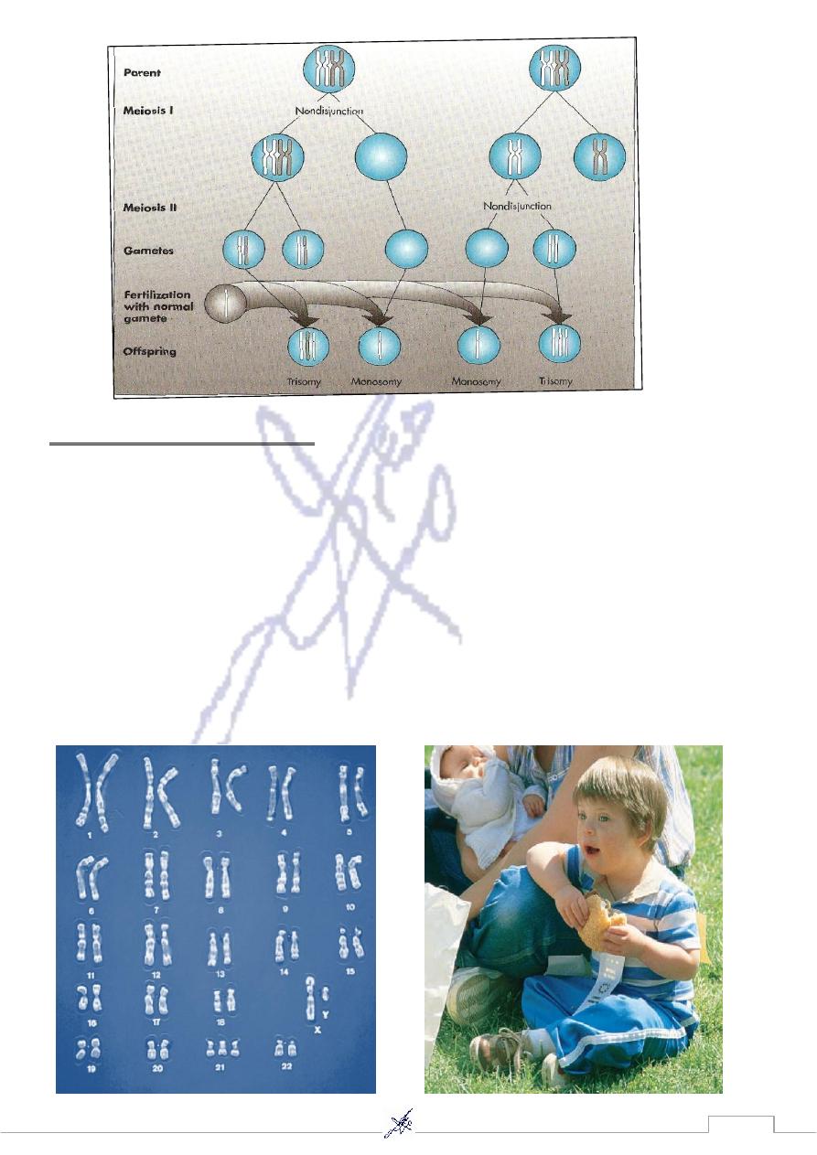
Non disjunction
An abnormal chromosomal makeup in an individual can be due to non disjunction.
Non disjunction is the failure of homologous chromosomes or sister chromatids to
separate during the formation of gametes.
Non disjunction occurs during meiosis when both members of a homologous pair
of chromosomes go into the same daughter cell, or during meiosis II when
daughter chromosomes fail to separate and both go into the same gamete
34
Abnormalities of Chromosomal Number
-
1
1. Polyploidy:
Polyploidy: Is the presence of a complete set of extra chromosomes in a cell is seen
commonly in plants and often improve their agriculture value, also occurs in humans,
although much less frequently.
chromosomes in its nucleus. Thus haploid
multiple of 23
A cell that contains a
Euploid:
gametes and diploid somatic cells are euploid.
d in humans are:
Polyploidy conditions that have been observe
A. Triploidy: 69 chromosomes in the nucleus of each cell, the karyotype for this
condition is 69, XXX (assuming that all of the sex chromosomes were X, other
combinations of the X and Y chromosomes may be seen).
It estimated about 15% of the chromosomal abnormalities.
, and those
eously aborted
spontan
Thus the vast majority of triploid conceptions are
that survive to term die shortly after birth.
B. Tetraploidy: 92 chromosomes in each cell nucleus, the karyotype of this condition is
92,XXXX.
Tetraploidy is much rare than triploidy.
It has been recorded in few live births, and those infants that survive for a short period.
multiple of 23 chromosomes,
are cells that do not contain a
Autosomal Aneuploidy:
2.
but instead they contain missing or additional chromosomes. Usually only one
chromosome is affected, but it is possible for more than one chromosome.
:
They consist of
A. monosomy: the presence of only one copy of a chromosome in diploid cell (n- 1)
B. trisomy: presence of 3 copies of a chromosome in diploid cell (n+ 1)
Autosomal monosomies are almost always die before term (lethal). In contrast, some
trisomies are seen among live births
:
Causes
Nondisjunction, In meiotic nondisjunction, two chromosome homologs migrate to the
same daughter cell instead of disjoining normally and migrating to different daughter
cells. This produce monosomic and trisomic offspring
35
syndrome)
Trisomy 21 (Down’s
Karyotype (47, XY,+ 21 or 47,XX,+21)
Down syndrome is also called trisomy 21 because the individual usually has three copies of
chromosome 21.
It is the most common autosomal aneuploidy seen among live births.
In most instances (80%), the egg had two copies instead of one of this chromosome.
The most significant problems include mental retardation, gastrointestinal tract
obstruction , congenital heart defects (atrioventricular canal), respiratory infections,
and leukemia (15-20 times higher than in general population).
Males are nearly always sterile.
36
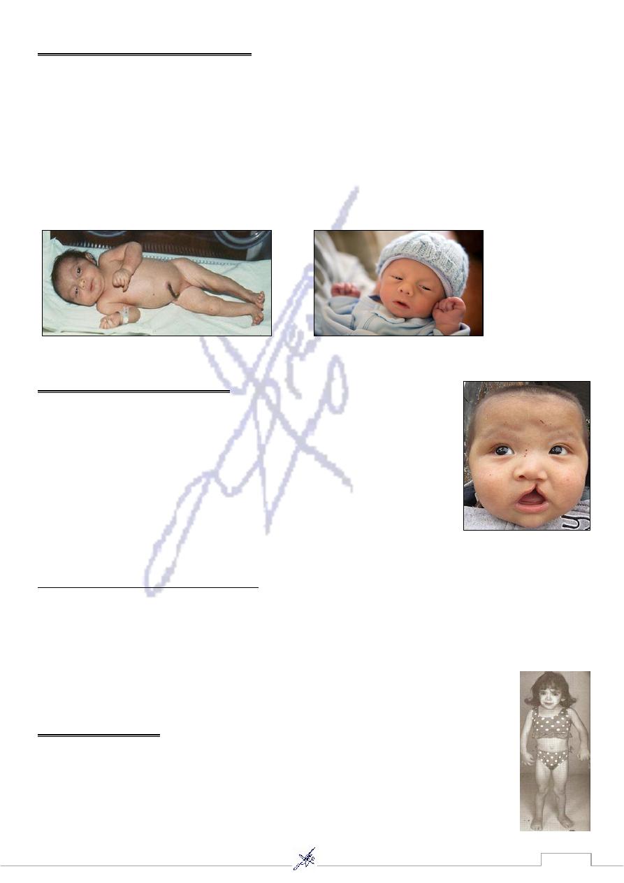
Trisomy 18 (Edwards syndrome)
Karyotype (47, XY, +18)
Edwards syndrome is the second most common autosomal trisomy.
Infants are produced a more seriously affected phenotype with prenatal growth
deficiency (weight is low for gestation age).
Infants have characteristic facial features, hand abnormalities, short sternum and
short big toes. The major malformation is congenital heart defects.
90% of trisomy 18 cases are the result of a maternally contributed extra chromosome
Trisomy 13 (Patau syndrome)
Karyotype (47, XY, +13)
Prevalence rate about 1per 10000 births.
It is much less common at birth than trisomy 21.
The malformation primarily includes oral- facial clefts, and
postaxial polydactyly. Also malformation of Central nervous
system (CNS) and heart defects.
The risk of bearing a child with this condition increases with advanced maternal age.
Sex chromosome Aneuploidy
Primarily because of X inactivation, the consequences of this class of aneuploidy are
less severe than those of autosomal aneuploidy.
With the exception of a complete absence of X chromosome, all of the sex
chromosome aneuploidies are compatible with survival.
Monosomy of the X chromosome
(45, X)
Karyotype
(Turner syndrome)
Females with turner syndrome are short stature (mature height is reduced
by approximately 20 cm) with sexual infantilism and lack of ovaries.
Lymphedema of the hands & feet is observed at birth, with the heart defect
but females with this syndrome usually have normal intelligence.
37
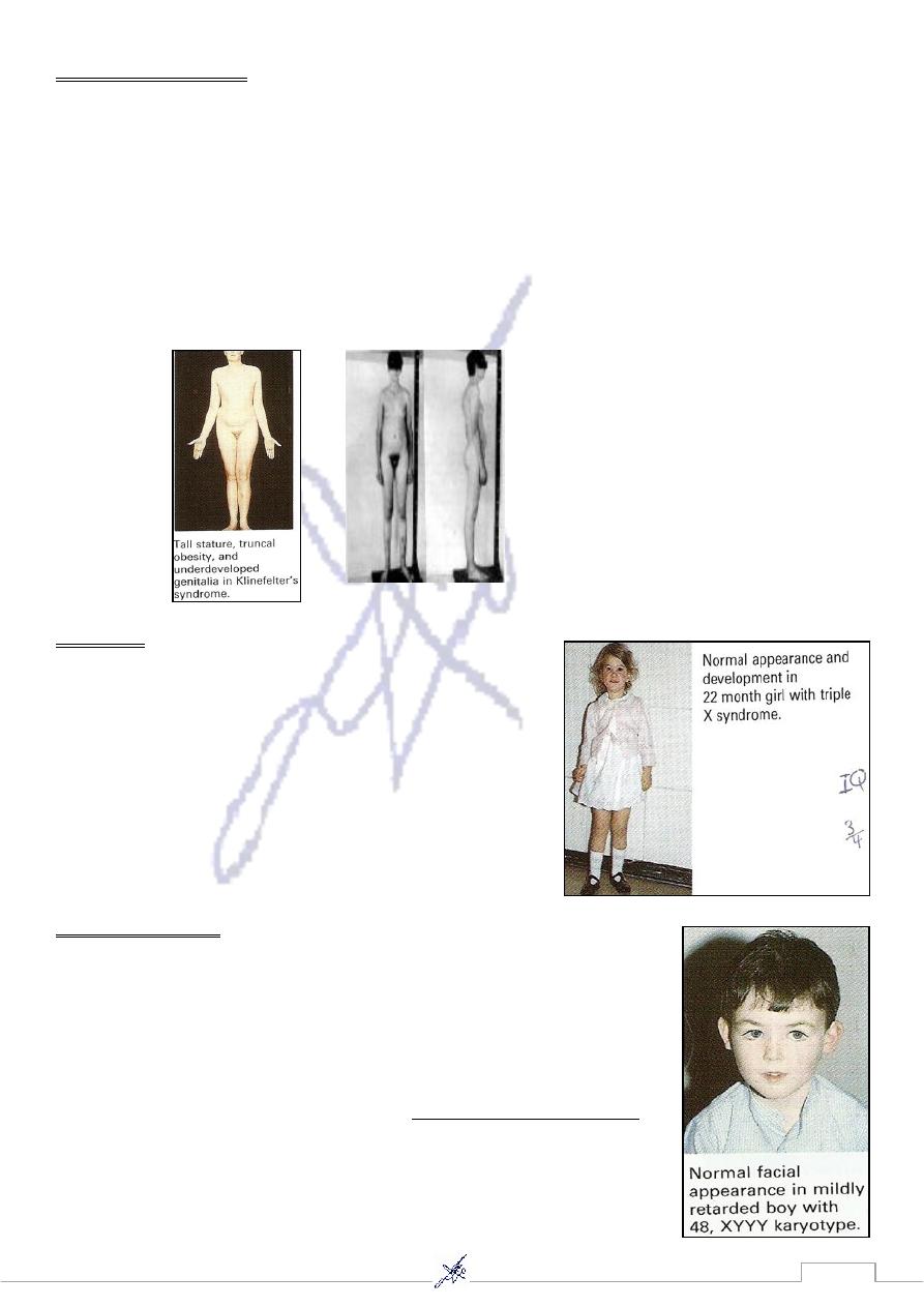
(47,XXY)
Karyotype
Klinefelter Syndrome
Males tend to be taller than average with long arms and legs, and the body shape may
be somewhat feminine.
They have small testes and are usually sterile
Gynecomastia (breast development) is seen in approximately one third of affected
males and leads to increased the risk of breast cancer.
They may have a reduction in IQ.
Individuals with the 48,XXXY and 49,XXXXY karyotypes have also be reported, the degree
of mental deficiency and physical abnormality increases with additional X chromosome.
(47,XXX)
Karyotype
Trisomy X
Physical abnormalities are rarely seen, but these
females sometimes suffer from sterility or mild
menal retardation (slight reduction in IQ)
Females have also been seen with four, five, or even
more X chromosomes.
Each additional X chromosome is accompanied by
increased mental retardation and physical
abnormalities.
47,XYY Syndrome
Karyotype (47,XYY)
Prevalence rate 1/1000 males
Males are taller than average with few physical problems but they
have 10- to 15- points reduction in average IQ and learning
disabilities.
among prison population
The prevalence rate of this syndrome
was discovered to be 1/30, suggested that this karyotype might
confer a predisposition to violent and criminal behavior
38
Abnormalities of chromosomal structure
-
2
Parts of chromosomes can be lost or duplicated and the arrangement of portions of
.
gamete formation
chromosomes can be altered during the
Mechanisms exist to repair these breaks, and usually the break is repaired perfectly
with no damage to the daughter cell. Sometimes, the break remain, or they heal in a
fusion that alters the structure of the chromosome.
Chromosomal breakage may be increased in the presence of certain harmful agents
include ionizing radiation, some viral infections, and some chemicals.
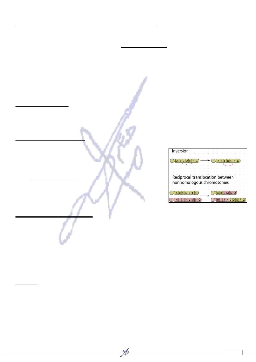
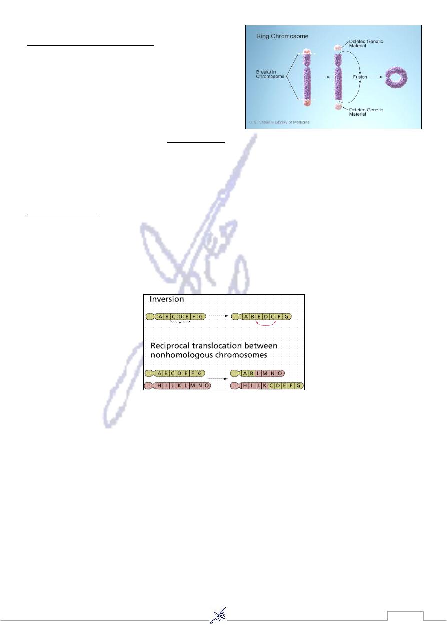
1. Translocation:
It is the interchange of genetic material between nonhomologous chromosomes.
There are two basic types: A. Reciprocal B. Robertsonian
A. Reciprocal Translocation:
When breaks occur in two different chromosomes with
subsequent exchange of material. The resulting are called
derivative chromosomes (der).
of this translocation usually
individual carriers
While
appears with normal phenotype, their offspring may have
a partial trisomy or partial monosomy and an
abnormal phenotype
B. Robertsonian Translocation;
The short arms of two acrocentric nonhomologous chromosomes are lost and the long
arms fuse at the centromere to form a single chromosome.
This type of translocation occurs in chromosomes 13, 14, 15, 21, and 22, because the short
arms of these acrocentric chromosomes are very small and contain no essential genetic
material.
The carrier of this abnormality can produce offspring with monosomy or trisomy.
Fusion of the long arms of chromosomes 14 and 21.
:
Example
The karyotype of a male carrier of this translocation is 45,XY,der(14;21).
Robertsonian translocation are responsible for approximately 5% of Down syndrome
cases.
39
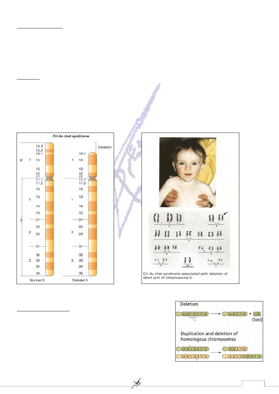
2. Deletions:
Is caused by a chromosomal break and subsequent loss of genetic material.
This abnormality usually affect large number of genes and produce recognizable
syndromes.
Figure: Termination deletion
This term (French, “cry of the cat”) describe the
syndrome.
chat
-
du
-
cri
is the
Example
distinctive cry of the child, a cat-like high-pitched cry during infancy with mental retardation
and small head.
Survival to adulthood has been observed but it is not common.
This syndrome is caused by a deletion of the distal short arm of chromosome 5:
3. Duplications
It can arise from: Unequal crossover, during meiosis, or
Occur among the offspring of reciprocal translocation
carriers
41
4. Ring Chromosomes:
Deletions occur at both tips of a chromosome.
The remaining chromosome ends can then
fuse, forming a ring chromosome.
The karyotype of a female with a ring X
chromosome is 46,Xr(X).
, it can proceed through cell division, but
a centromere
If the ring chromosome includes
its structure can create difficulties.
Ring chromosomes are often lost, resulting in monosomy for the chromosome in at
least some cells.
5. Inversion:
Is the result of two breaks on a chromosome followed by the reinsertion of the missing
fragment at its original site but in inverted order.
Parents with inversion are usually normal in phenotype but can produce offsprings
with deletions or duplications.

41
42
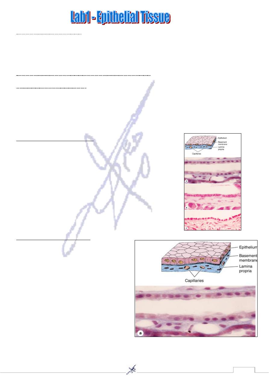
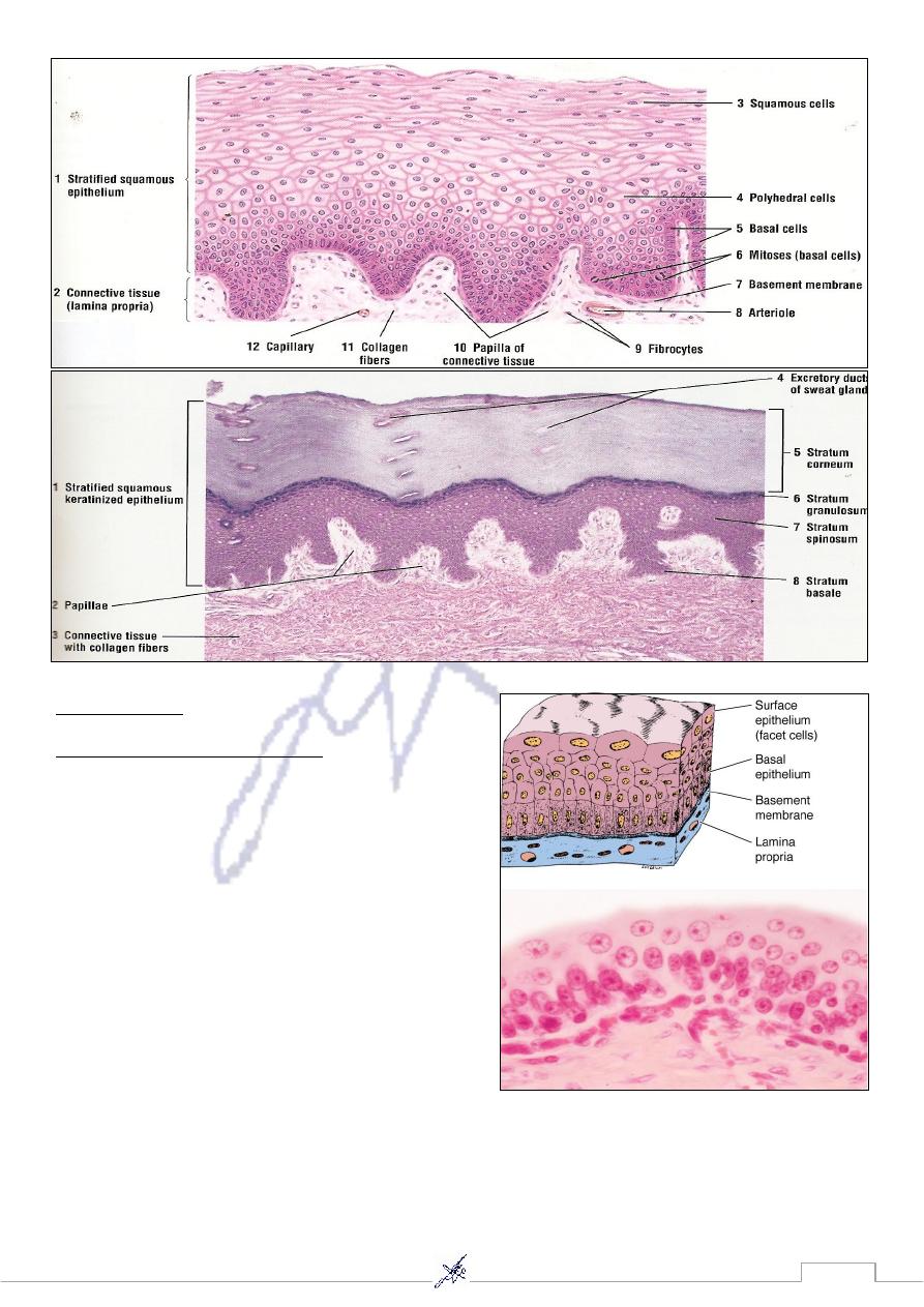
Simple epithelial tissue
-
1
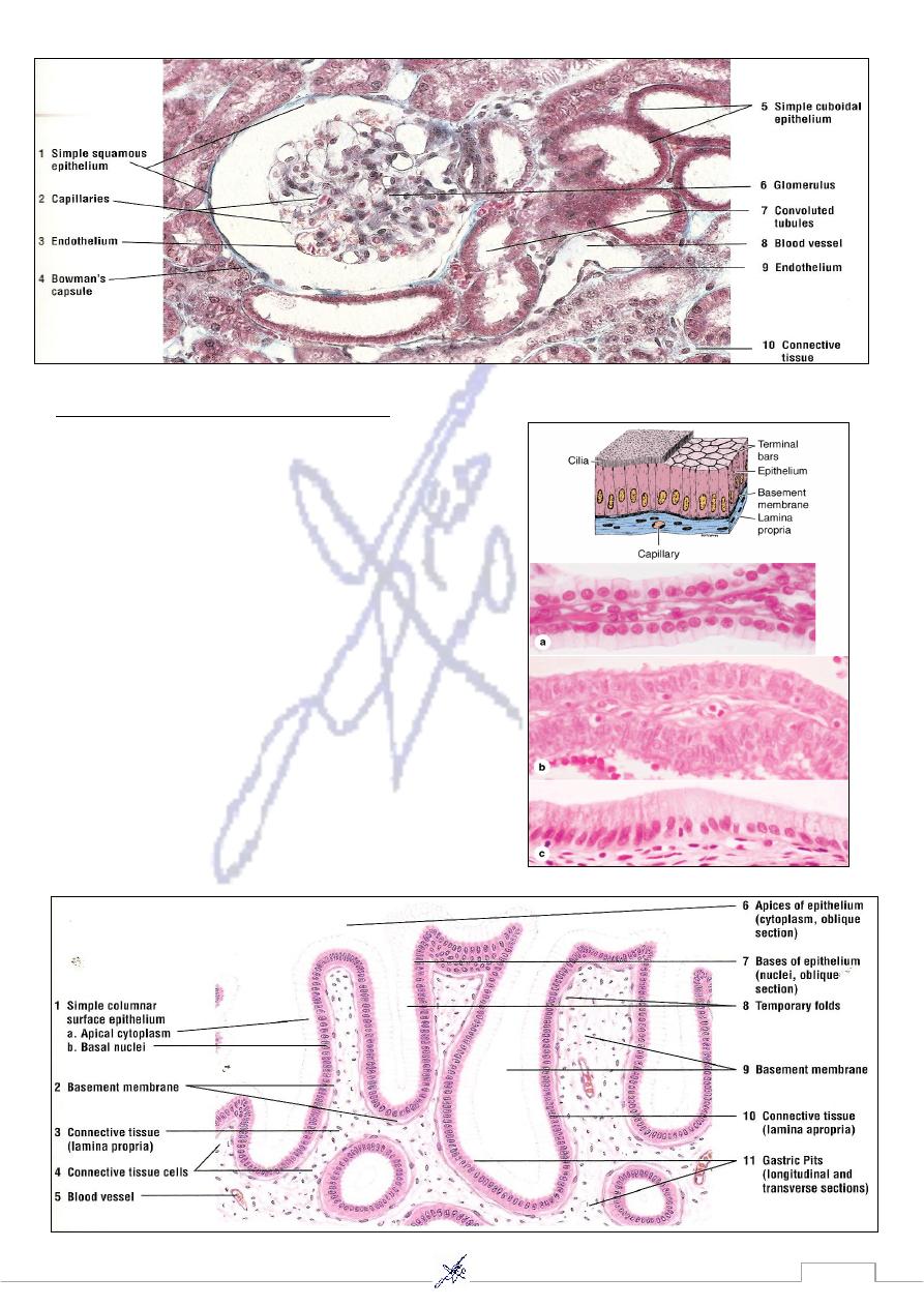
a) Simple squamous epithelial tissue ex: Kidney (glomerulus)
b) Simple cuboidal epithelial tissue ex: Proximal convoluted tubules
c) Simple columnar epithelial tissue ex: Stomach
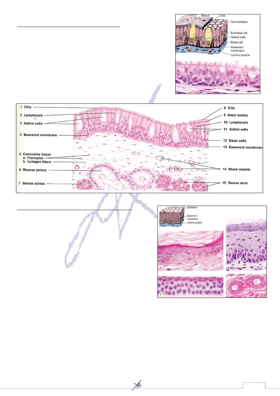
: Trachea
Ex
columnar epithelial tissue
Pseudostratified ciliated
-
2
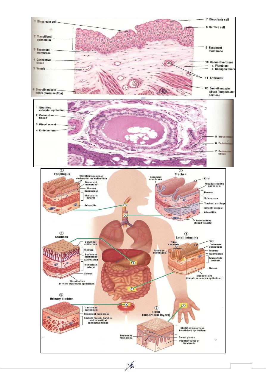
Stratified epithelial tissue
-
3
a) Transitional epithelium ex: Urinary bladder
b) Stratified squamous epithelium: ** (Non Keratinized) ex: Esophagus
** (Keratinized) ex: Skin.
c) Stratified cuboidal epithelium ex: Excretory duct of salivary gland.
mple squamous epithelia
Si
In simple squamous epithelium, cells of the single layer are flat and
usually very thin, with only the thicker cell nucleus appearing as a
bulge to denote the cell. Simple epithelia are typically specialized as
lining of vessels and cavities and regulate substances which can
enter underlying tissue from the vessel or cavity. The thin cells often
exhibit transcytosis. Examples shown here are those lining the renal
loops of Henle (a), the mesothelium lining a mesentery (b), and the
endothelium lining the inner surface of the cornea (c). Endothelium
and mesothelium are nearly always simple squamous
Simple cuboidal epithelial
Simple Cells of simple cuboidal epithelia vary
in their height but are roughly as tall as they are
wide. Their greater thickness often includes
cytoplasm rich in mitochondria providing
energy for a high level of active transport of
substances across the epithelium. Examples of
simple cuboidal epithelia shown here are from a
renal collecting tubule (a), a pancreatic duct..
43
columnar epithelial tissue
Simple
Cells of simple columnar epithelia are taller than they
are wide. Such cells are usually highly specialized for
absorption, with microvilli, and often have interspersed
secretary cells or ciliated cells. Such epithelial cells
always have tight and adherent junctional complexes at
their apical ends, but are often loosely associated in
more basolateral areas. This allows for rapid transfer of
absorbed material to the space between the cells rather
than transport the full length of the cells. The additional
cytoplasm in columnar cells allows additional
mitochondria and other organelles needed for
absorption and processing. The examples shown here
are from a renal collecting duct (a), the oviduct lining,
with both secretary and ciliated
cells (b), and the lining
of the gall bladder .(c), All
44
stratified epithelial tissue
-
pseudo
appear to be in layers, but the basal ends of the cells are all in
contact with the basement membrane, which is often very
thick in these epithelia. The best example of this epithelial
type is the pseudo stratified ciliated columnar epithelium of
the upper respiratory tract, which contains cell types with their
nuclei at different levels.
Stratified Squamous Epithelial tissue
Stratified squamous epithelia have protective
functions: protection against easy invasion of
underlying tissue by microorganisms and protection
against water loss. In the skin, protection against water
loss and desiccation is particularly important and the
epithelium is keratinized. As epidermal cells of the
skin (a) differentiate they become filled with keratin
and other substances and eventually lose their nuclei
and other organelles. The superficial flattened squames
form a layer which impedes water loss and eventually
slough off and are replaced from below. (b) covering the cornea (c) are considered nonkeratinize.
(d) where the double layer of cells apparently provides a more robust lining than a simple
epithelium.
45
Transitional
Stratified epithelial tissue
Stratified transitional epithelium lining the urinary
bladder has rounded or dome-shaped superficial
cells with two unusual features. The surface cells
have specialized membranes and are able to
withstand the hypertonic effects of urine and protect
underlying cells from this toxic solution. Cells of
transitional epithelium are also able to adjust their
relationships with one another as the bladder fills
and the wall is stretched, so that the transitional
epithelium of a full, distended bladder seems to have
fewer cell layers than that of an empty bladder.
46
47
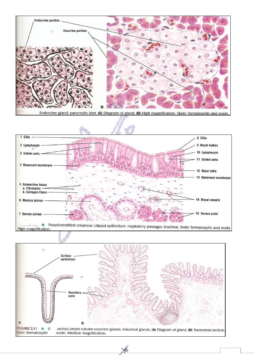
Exocrine Gland
Endocrine Gland ex: Pancreatic Islet
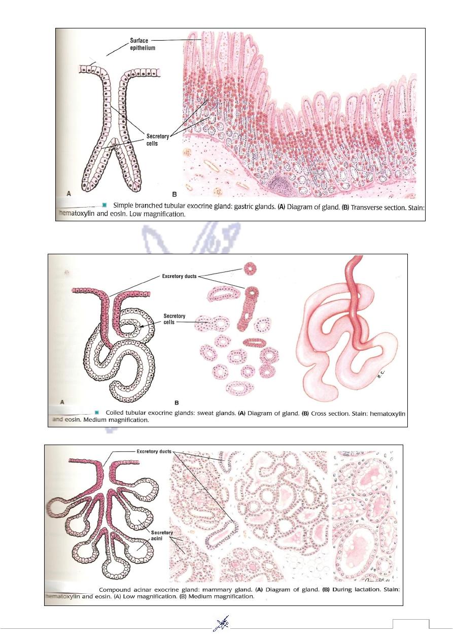
Classification of exocrine glands
According to number of cell
A)
Unicellular gland ex: Goblet cell
Multicellular gland can be classified :
Simple exocrine glan
-
1
a- Simple unbranched tubular gland ex: Intestinal gland
b- Simple branched tubular gland ex: Gastric gland
c- Coiled tubular gland ex: Sweat gland
Compound exocrine gland
-
2
a- Compound Acinar gland ex: Mammary gland
b- Compound Tubular gland
c- Compound Tubuloacinar gland ex: salivary, Submaxillary, sublingual & Parotid glands
According to the secretary products
B)
Mucous ex: Sublingual gland.
Serous ex: Pancreas , Parotid gland
Mixed ex: Submandibular gland.
According to the mode of secretion
C)
Merocrine ex: Pancreas, Gastric gland .
Holocrine ex: Sebaceous gland.
Apocrine ex: Mammary gland
48
49
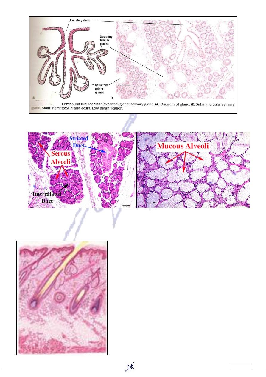
51
Sublingual gland
Parotid gland
Skin notice sebaceous gland
51
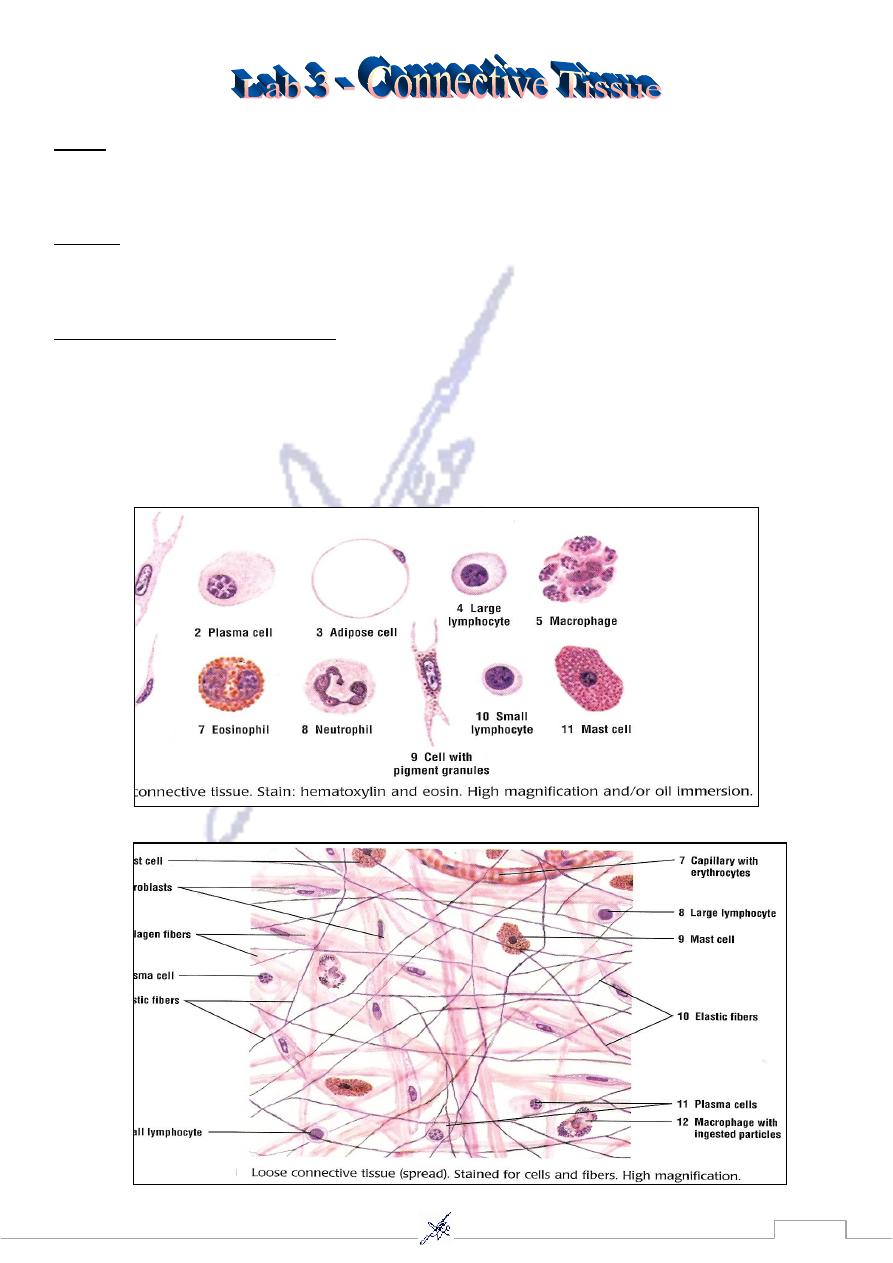
Cells
Fibroblast Plasma cell Adipose cell Macrophage Mast cell Leucocytes
Fibers
Collagen fibers Elastic fibers Reticular fibers
Types of Connective Tissue
Loose Connective Tissue(spread).
Embryonic connective Tissue
Dense Connective Tissue
a- Dense Regular Connective Tissue ex: Tendon
b- Dense Irregular Connective Tissue ex: Dermis of skin
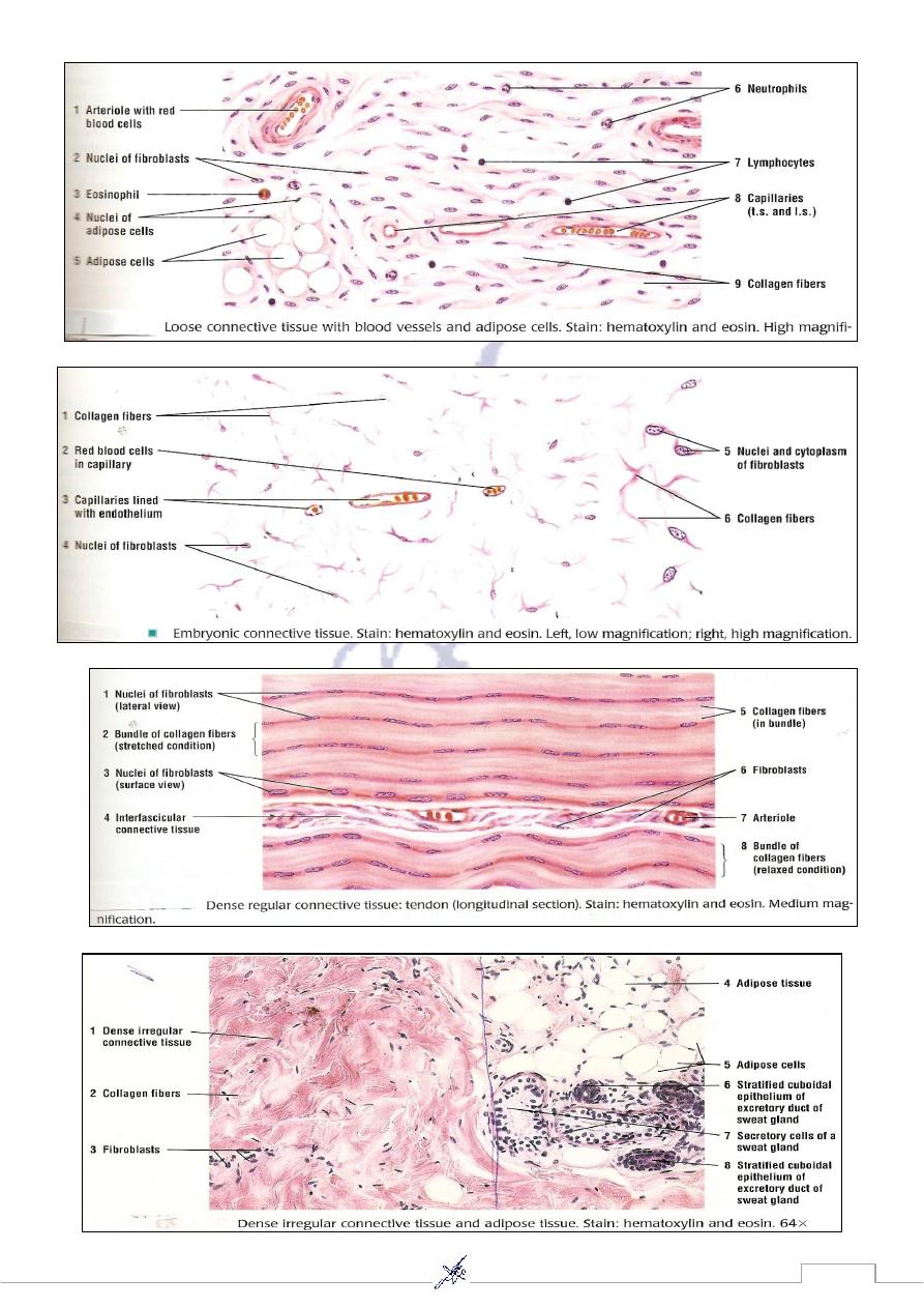
52
53
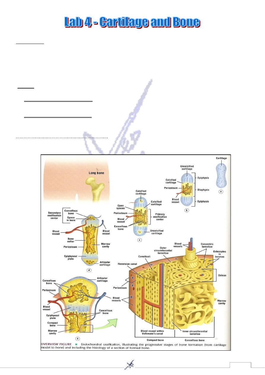
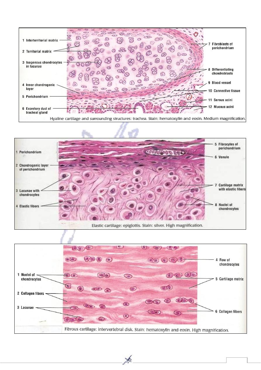
Cartilage
Hyaline cartilage ex : Trachea.
Elastic cartilage ex : Epiglottis.
Fibrous cartilage ex : Intervertebral disk.
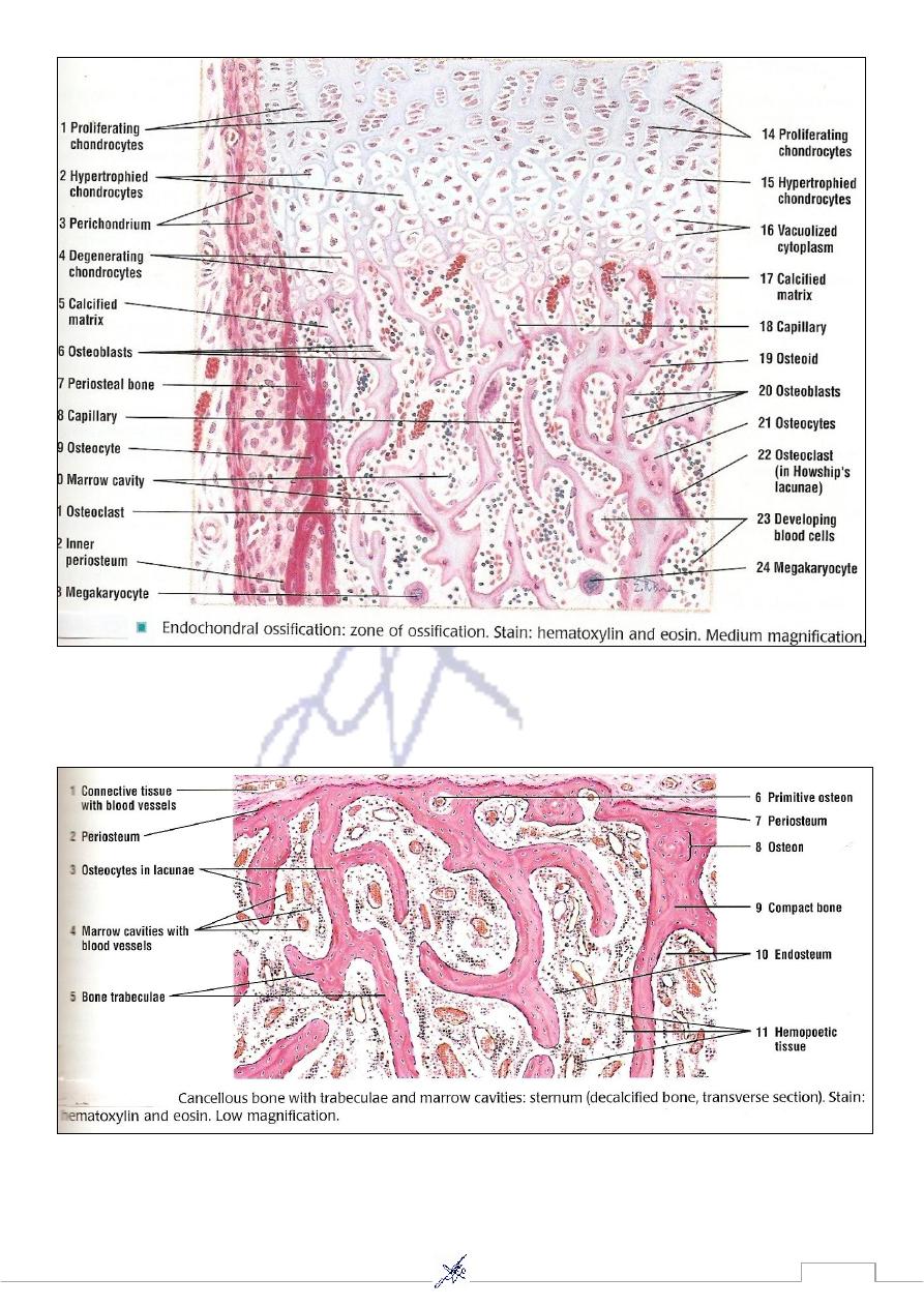
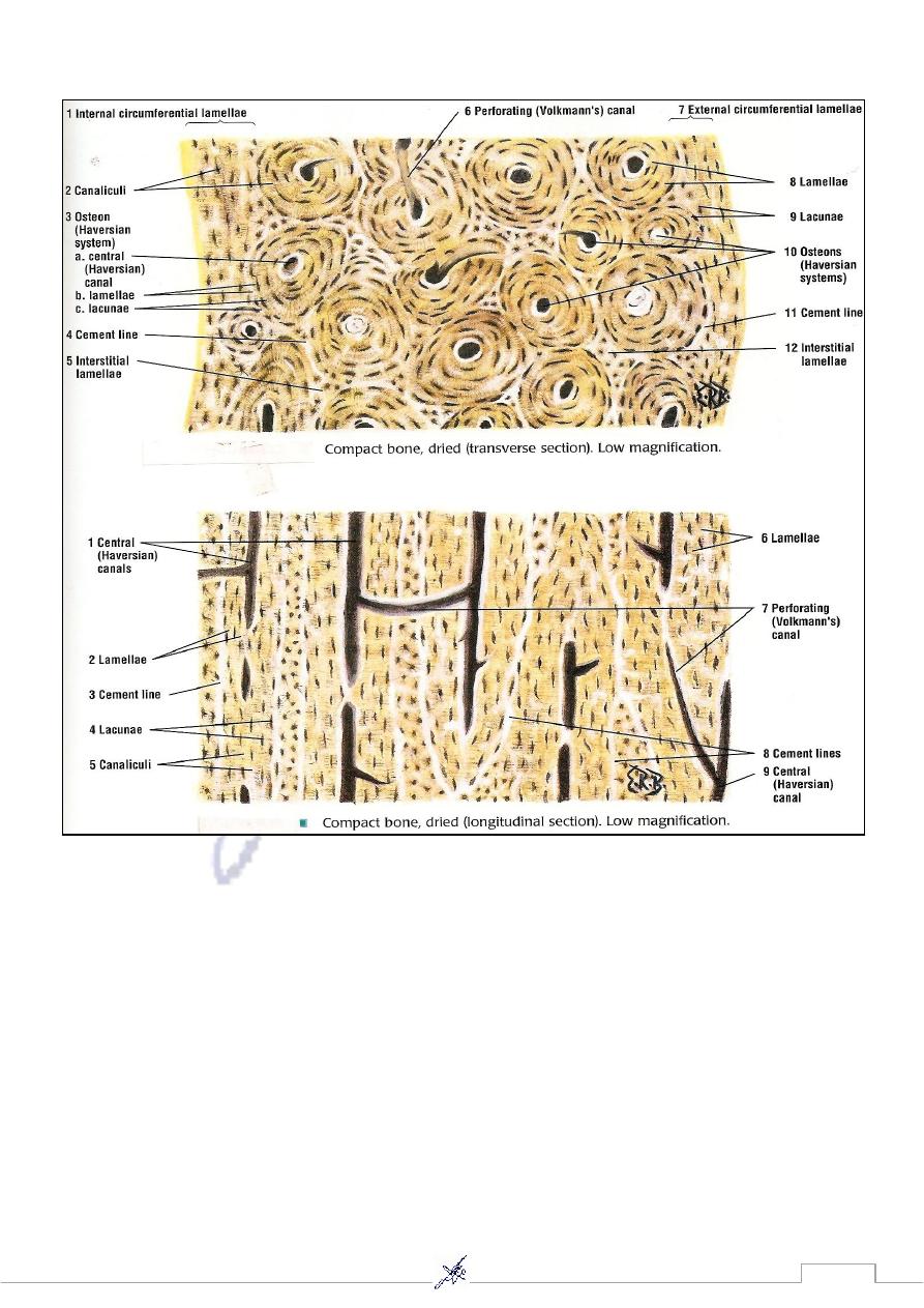
Bone
cement line, Haversian
Canaliculi,
,
Osteon : Osteocyte
; notice: (
Compact Bone (Long Bone)
canal, Volkmann's canal ).
Hemopoietic tissue,
,
e : Marrow cavities with marrow
;notic
Cancellous Bone (Sternum)
Periosteum).
Formation of bone (Bone ossification)
Endochondral ossification. Intramembranous ossification.
54
55
56
57
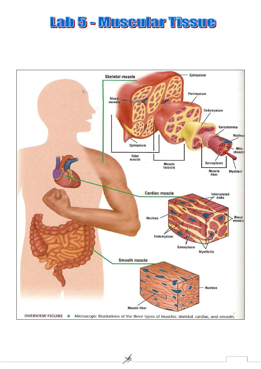
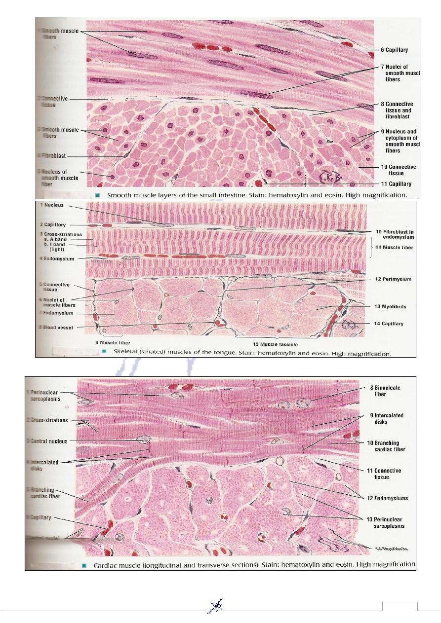
Smooth muscle (non-striated) ex: small intestine(L.S).
Skeletal muscle (striated) ex: Tongue (T.S. ,L.S).
Cardiac muscle (striated) ex: Heart.
58
59
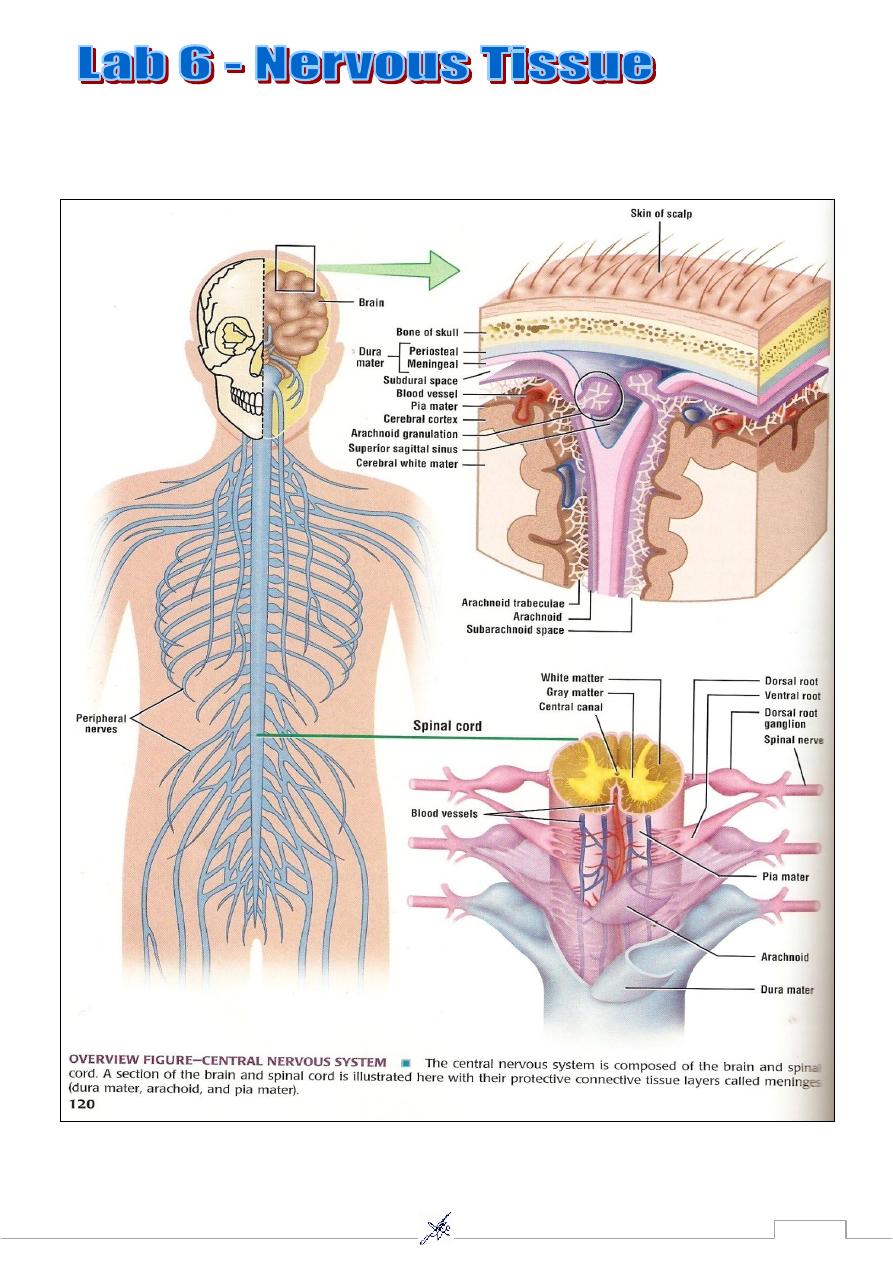
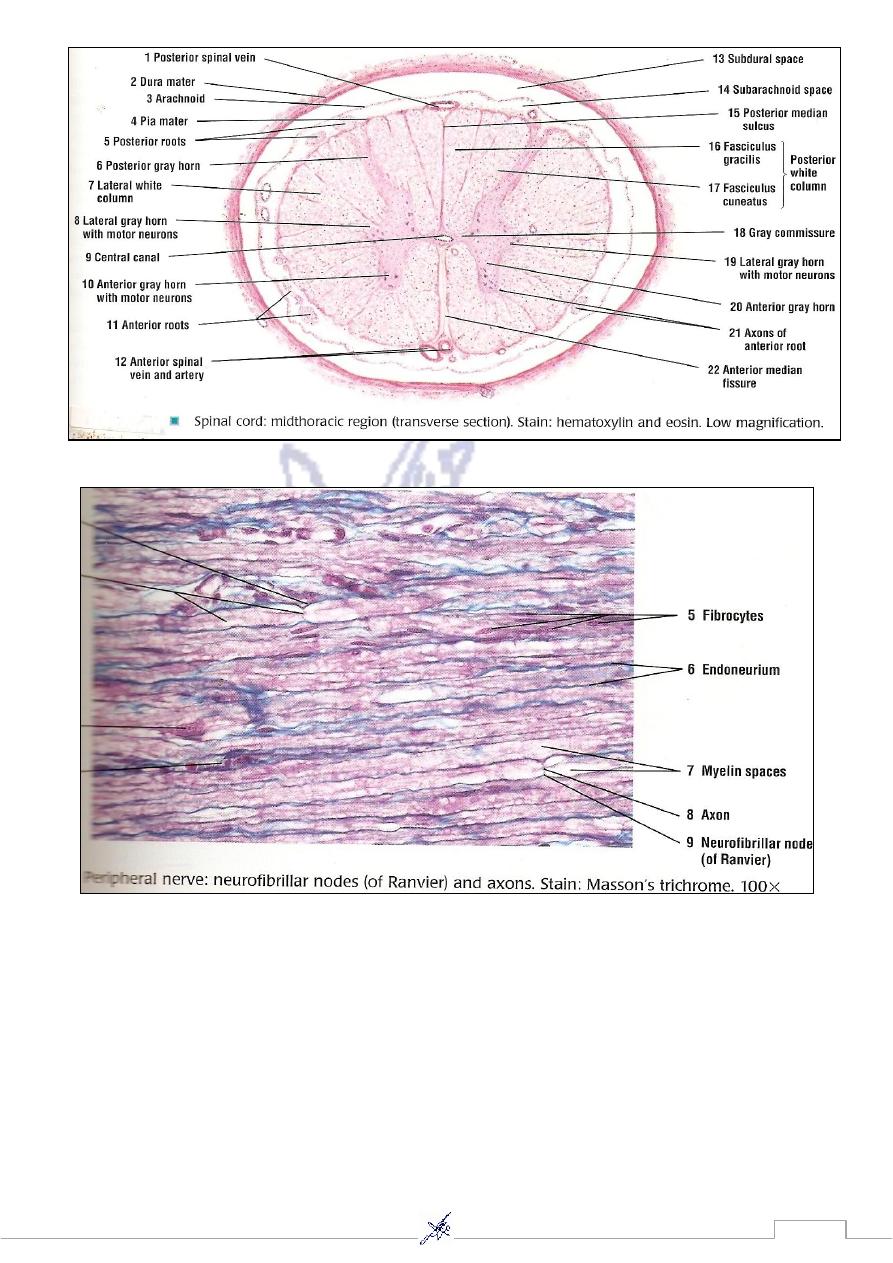
Central nervous system (Brain and Spinal cord ).
Peripheral nervous system . (Nerve , Ganglion).
61