Genetic disorders
Robbins Basic Pathology
Ch 6
P 215
Dec. 7
th
. 2015
Genetic disorders
Objectives
• Introduction
• Mutations
• Structure & No. variations in genes
• Epigenetic changes.
• Types of genetic disorders
• Genes encoding structural pr.
Marfan Syndrome “MFS”
Ehlers- Danlos Syndrome “EDSs”
• Genes encoding receptor proteins’ or channels.
Familial hypercholestremia.

Almost
all diseases
have a genetic
component.
Diseases cause
long term morbidity
are
genetically determined
50%
of spontaneous abortuses have a
genetic component.
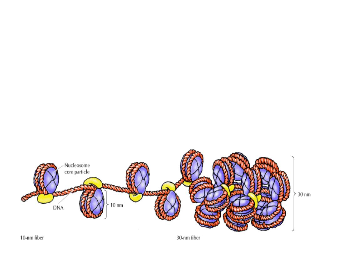
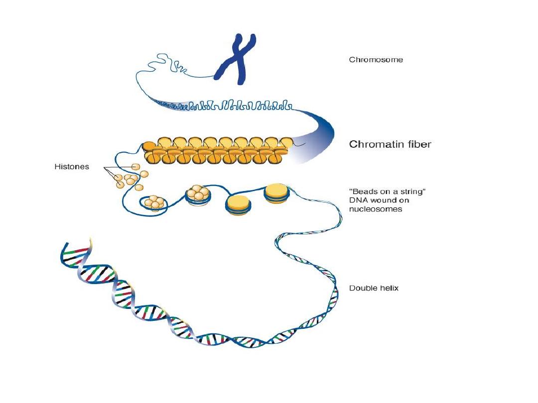
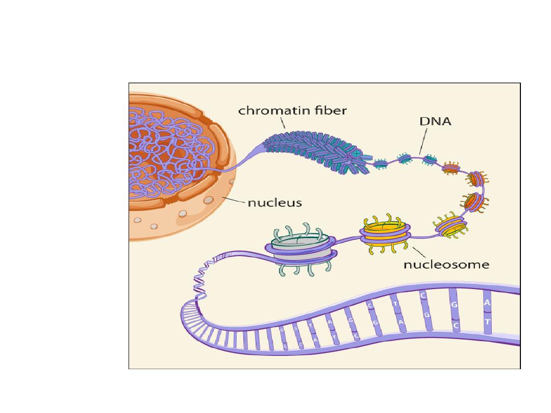
DNA, chromosomes and chromatin
• In the nucleus, the
DNA
double helix is packaged
by special proteins
(histones)
to form a complex
called
chromatin
.
• The chromatin undergoes further condensation
to form the
chromosome
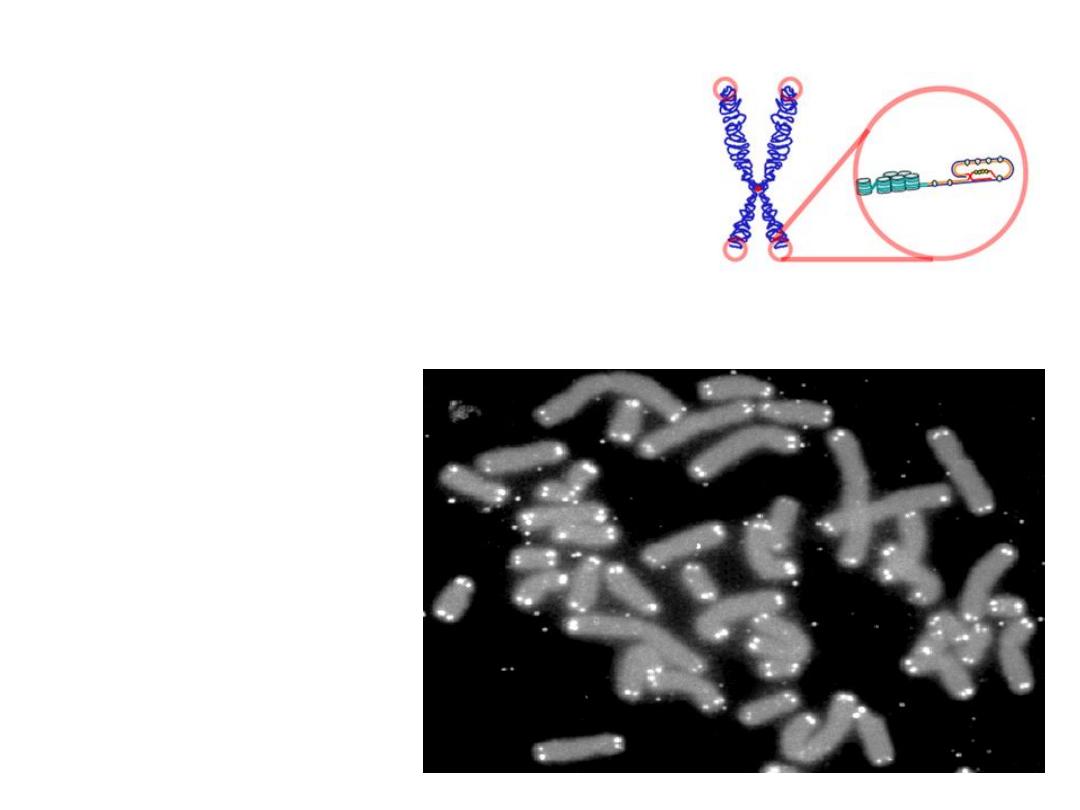
• DNA is double-stranded, with the
exception of the ends of
chromosomes, of single stranded
DNA, called
telomeres
• Telomeres
prevent degradation and
accidental fusion of chromosomal
DNA
.
Human ch. (grey) capped by
telomeres (white)
Telomere
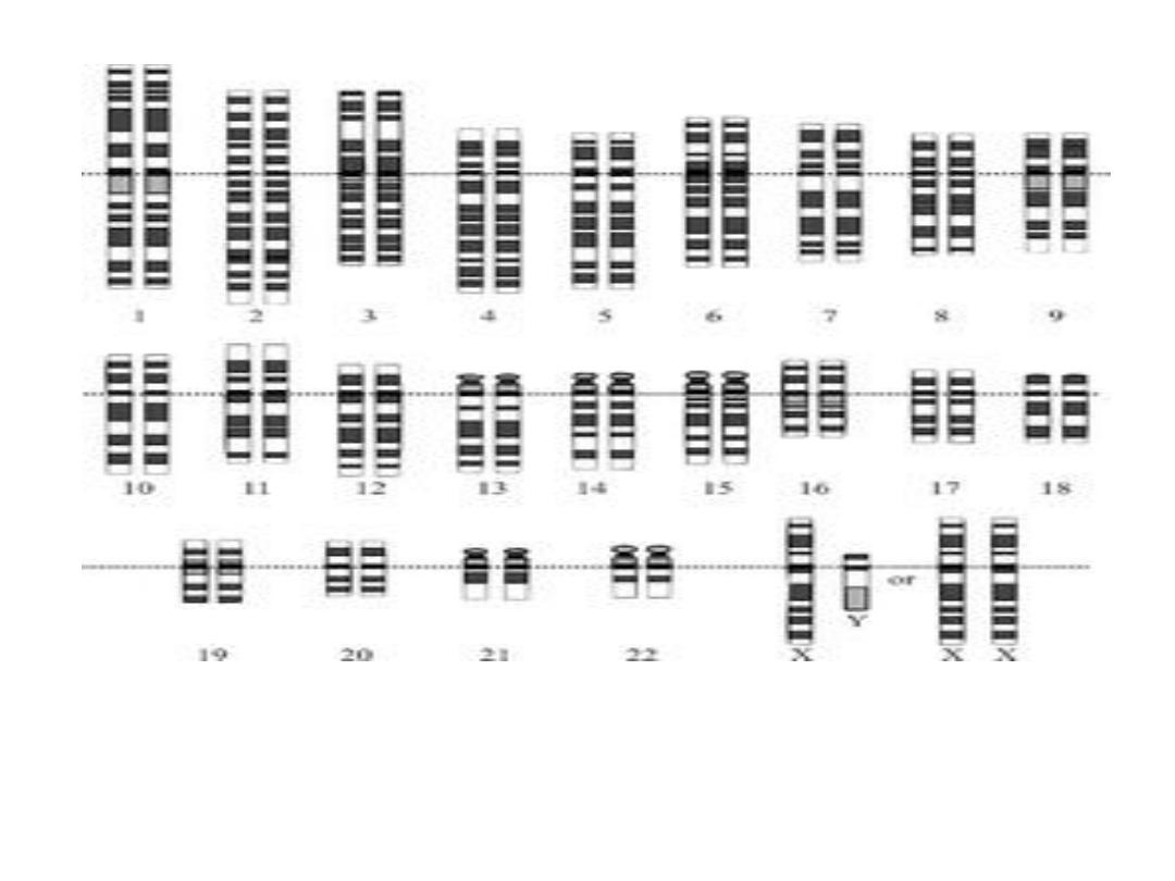
The human diploid
karyotype
, showing the organization of the
genome
into chromosomes. shows both the female (XX) and male (XY) versions of
the 23rd chromosome pair.

• Genome;
all genetic data of a single cell. The genes in the
nucleus, & mitochondrial DNA.
(23 identical chromosomal pairs + Mitoch DNA)
• Genes;
is a section of the genome which codes for one
protein. It consists of various codons.
•
Codon;
Every codon consists of three adjacent
nucleotides.
• 3 Bases
• in order of increasing scale, we have: base -
codon - gene - chromosome - genome

• A genome is all genetic data of a single cell. That
includes the genes in the nucleus, but also that of
mitochondrial DNA.
A gene is a section of the genome which codes for
one protein. It consists of various codons.
• A set of three bases on a single strand of DNA is
called a "codon". This is because it codes for a
particular amino acid when the codon is transferred
to RNA and then used to construct a protein
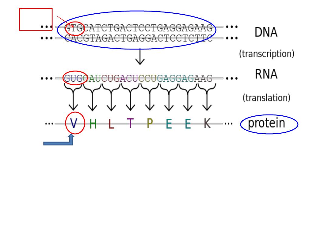
3 Bases - codon - gene - chromosome - genome.
Genes are expressed by being transcribed into RNA,
and this RNA then translated into protein.
codon
3
bases
gene
AA

Mitochondrial DNA
• All Mit DNA is derived from the mother as
sperm contribute no mitochondria to the
Zygote.

DISEASES
• GENETIC
• ENVIRONMENTAL
• BOTH

Hereditary (familial) disorders
Congenital diseases
Classes of genetic variants:
Mutations
:
genetic variations that cause diseases.
• germ cells
• somatic cells
Polymorphisms;
genetic variations that do not
cause disease
• single gene is insufficient to produce the disease.
• when several polymorphisms are present, disease
occurs, hence the term
multigenic or polygenic
.
Classes of Mutations
Nucleotide Substitution
• Synonymous
– resulting in a change in the codon
but no change in the AA and thus no phenotype
•
Missense
– change in the codon, resulting in an
A.A change in the protein
•
Nonsense
– introducing a premature stop codon,
resulting in truncation of the protein
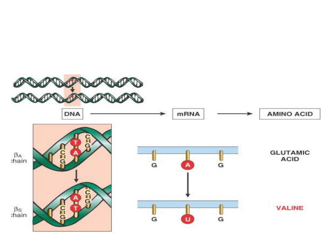
Point mutation - missense mutation
• Substitution of a
single nucleotide base
by a different base,
(
Sickle Cell anemia)
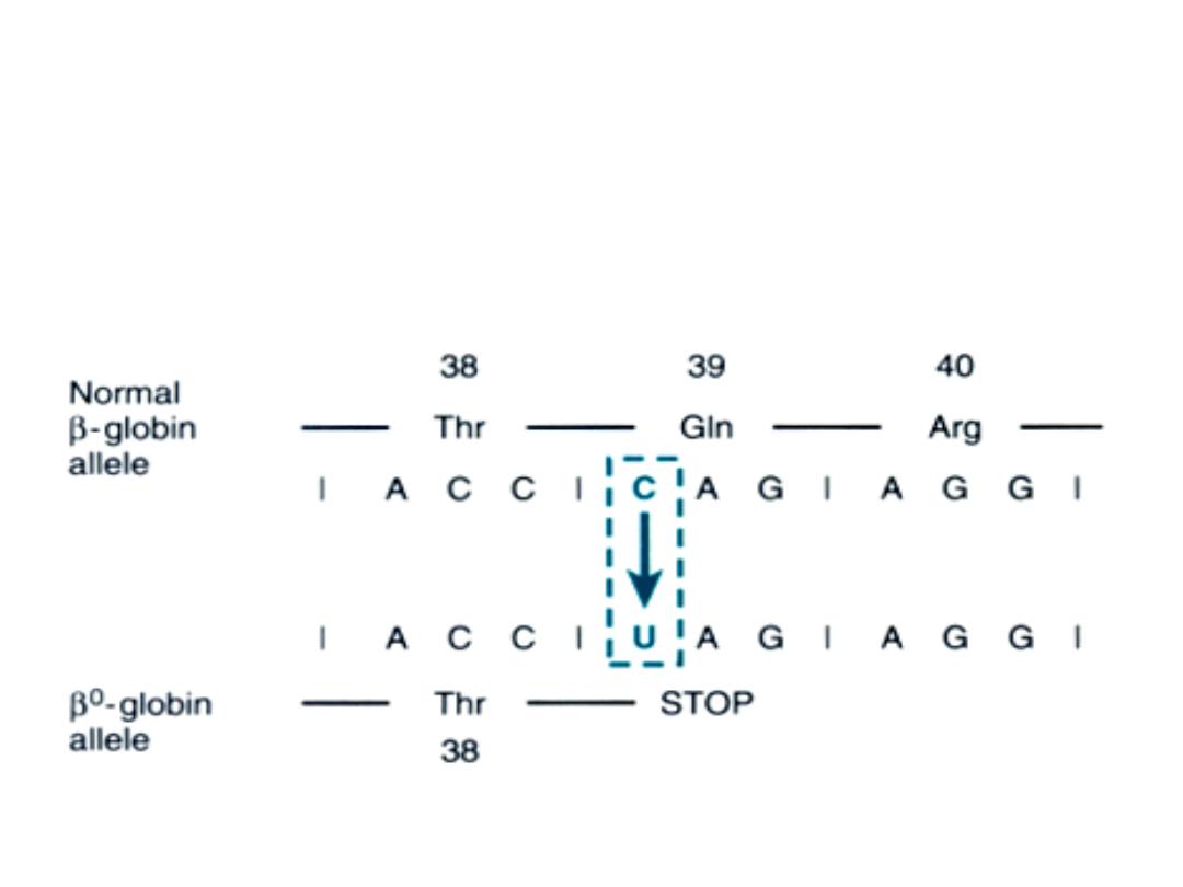
Point mutation - nonsense mutation
•
Change an
AA codon
to
a chain termination codon, or
stop codon
--- interrupt translation & the resultant protein
are rapidly degraded (
B Thalasemia)
Frameshift mutation
Insertion or deletion of 1 or 2 or > base pair alter the
reading frame of the DNA strand
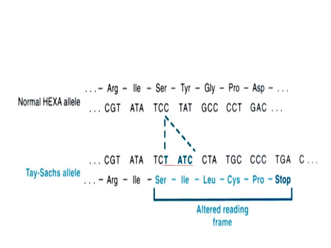
Four-base insertion

• Four-base insertion in the hexosaminidase A
gene, leading to a frameshift mutation. This
mutation is the major cause of Tay-Sachs
disease in Ashkenazi Jews.
Classes of Mutations
• Trinucleotide repeat mutations
A
mplification of sequence of 3 nucleotide
• e.g in fragile X syndrome, there are 250-4000 repeats of
the sequence CGG within the gene called FMR-1.
• In the normal population, the number of repeats is
small 29, so amplification of FMR-1 giving rise to mental
retardation
.
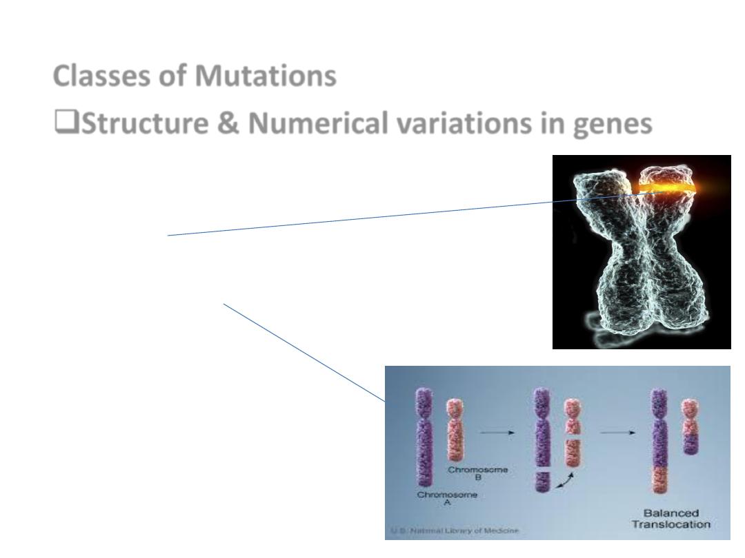
Classes of Mutations
Structure & Numerical variations in genes
• Germ line or Somatic cells.
• Amplification
• Deletion
• Insertion
• Translucation
Polymorphism or Mutation
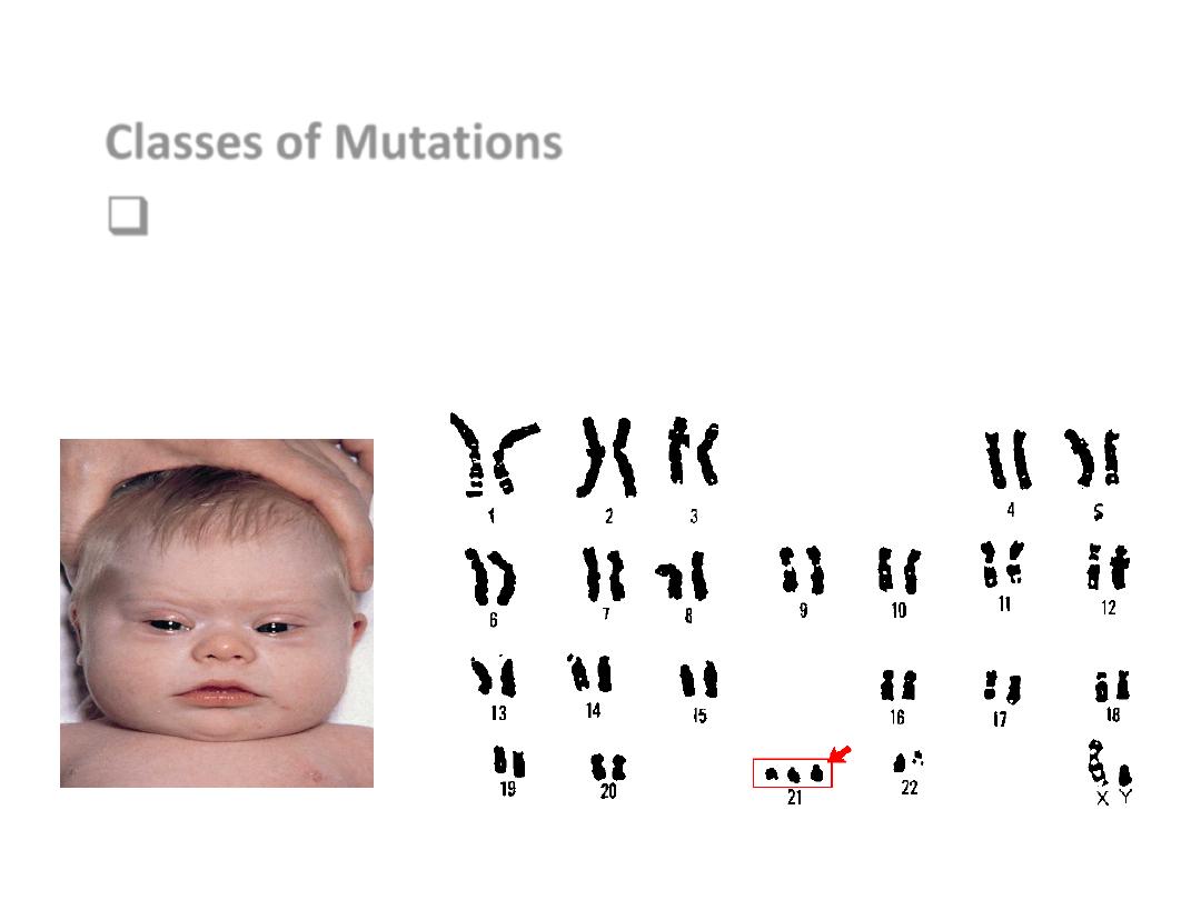
Classes of Mutations
Numerical chromosomal abnormality
e.g. Trisomy of Chromosome 21 (Downs
Syndrome).
Epigenetic Changes
• Modulation of genetic expression in absence of mutation
or structural changes.
• Don’t alter the sequence of the DNA code but have
biological effect in gene function.
• Histone Methylation
• Methylation, phosphorelation or acetylation.
• Affect on tumor suppressor gene
Cancer
.
• Cause progression of cancer.

Karyotype
Phenotype
Penetrance:
• Proportion of individual bearing a mutant allele
who develop the disease phenotype
Expression:
•
The degree of severity of dis expression
• E.g. NF 100% pentrance with variable expressiomn.
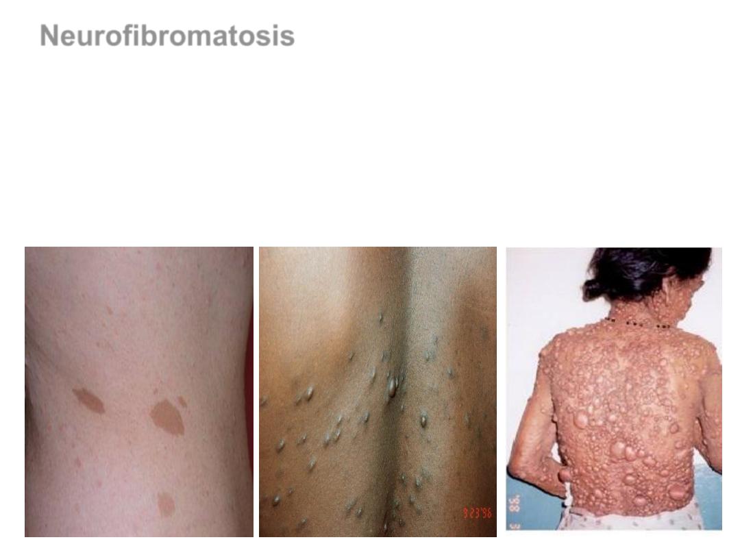
Neurofibromatosis
Fully (100%) penetrant
VARIABLE EXPRESSIVITY
•
The
Environmental factors
• and/or Variation in other genes
that act as modifiers of the
mutated gene’s
.

Types of genetic diseases
:
1-
Mutation in single gene;
Mendelian disorders. Single
gene mutations. highly penetrant,
2- Diseases arising from abnormality in No. or structure
of chromosome. high penetrance
3- Diseases with
multifactorial inheritance
(genetic &
environmental) .
4-
Heterogenous group
that is single gene but do not
follow Mendelian rules of inheritance.
e.g. Mutation in mitoch. DNA, Triple repeat mutation,
epigenetic (genomic imprinting).

Complex multifactorial disorders
• more common .
• interactions between multiple variant genes &
environmental factors.
• low penetrance.
• multifactorial disorders.
• Atheroscl., DM, hypertension, & autoimmune diseases.
• Even normal traits such as height and weight are governed
by polymorphisms in several genes.
• Multifactorial disorders = Multigenic (several polymorphic
genes) + Environmental factors

1-
Diseases caused by single gene defects
include:
• 1% of adult admission to hospital.
• 6 – 8 % of pediatric admission.
*
AD
* AR
* X-linked diseases
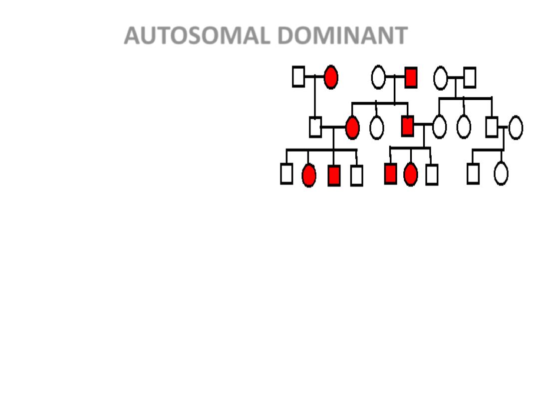
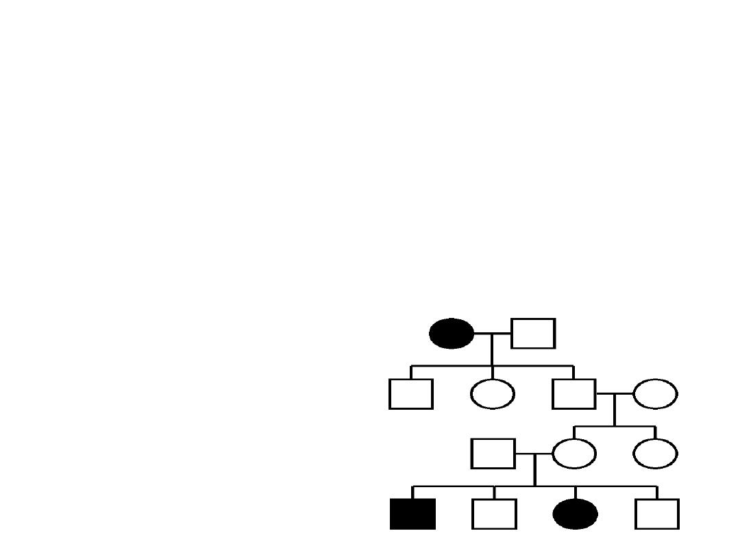
AUTOSOMAL DOMINANT
• Disease is in
Heterozygote
• Healthy parent
(New Mutation)
• PENETRANCE.
• VARIABLE EXPRESSIVITY,
(e.g. Neurofibromatosis)
The
environmental factors and/or variation in other
genes
that act as modifiers of the mutated gene’s function
????
.
-
BOTH SEXES INVOLVED
- GENERATIONS NOT SKIPPED
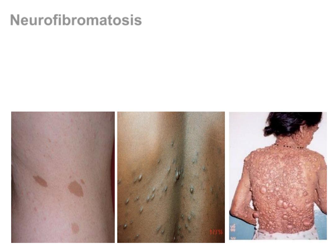
Neurofibromatosis
Fully (100%) penetrant
VARIABLE EXPRESSIVITY
The
environmental factors and/or variation in other genes
that act as
modifiers of the mutated gene’s function are mostly unknown.
Examples of AD disorders:
• Marfan syndrome
• hypercholesterolemia
• Polycystic kidney disease
• Hereditary spherocytosis
• Familial polyposis coli
AUTOSOMAL RECESSIVE
• Largest group of Mendelian disorders
• Disease in
HOMOZYGOTES
• Often
COMPLETE PENETRANCE
• Onset usually
EARLY in life
• Proteins show
LOSS of FUNCTION
• Include ALL
Inborn Errors of Metabolism
-
BOTH SEXES INVOLVED
- GENERATIONS SKIPPED
• Examples of AR disorders:
• Sickle cell anemia
• Thalassemias
• Cystic fibrosi
• Phenylketonuria
• Wilson disease
• Glycogen storage diseases
• Albinism
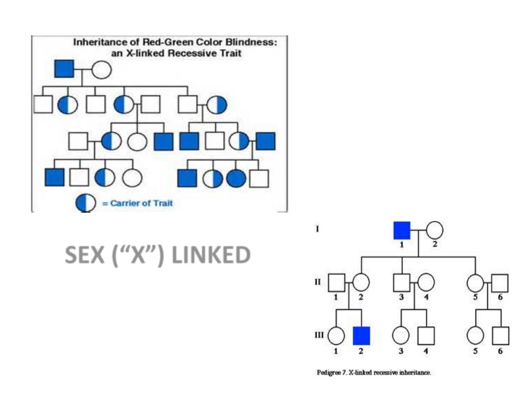
SEX (“X”) LINKED
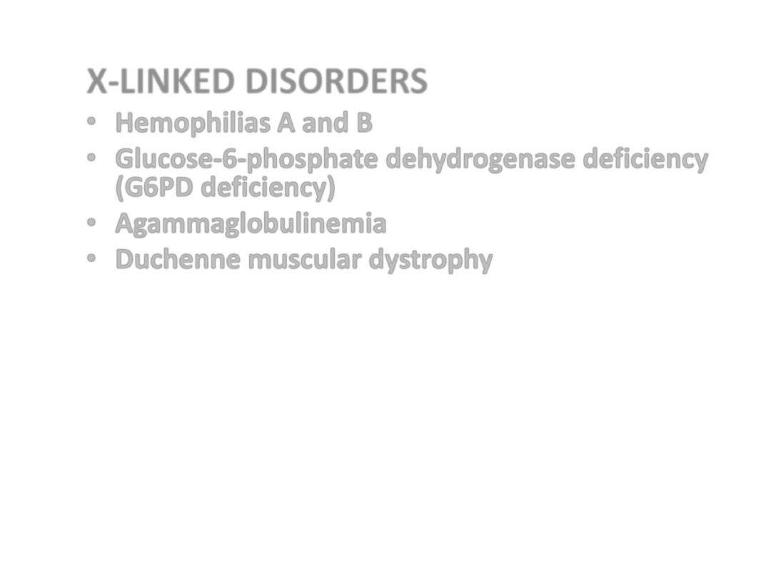
X-LINKED DISORDERS
• Hemophilias A and B
• Glucose-6-phosphate dehydrogenase deficiency
(G6PD deficiency)
• Agammaglobulinemia
• Duchenne muscular dystrophy
progressive weakness and degeneration of
skeletal muscles due to absence of
dystrophin
.
Mainly in boys, onset 3-5 yrs, by 12 years can’t
walk, and later needs respirator
.
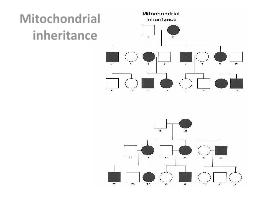
Mitochondrial
inheritance
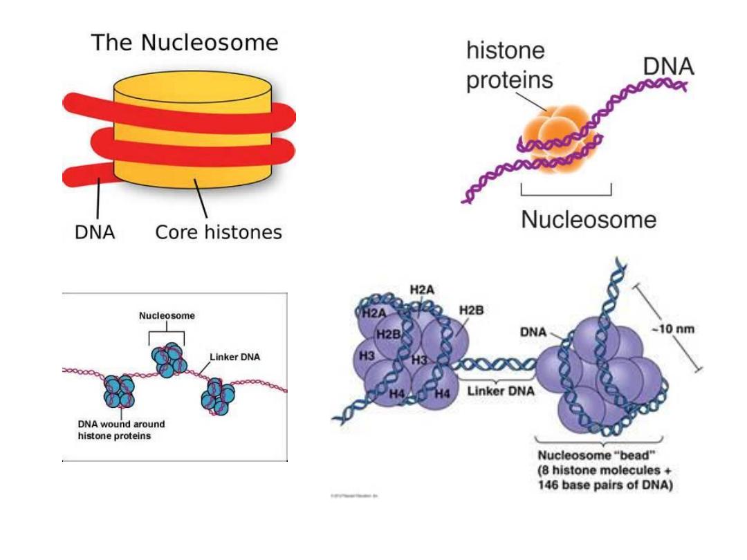
• Chromatin is a complex of macromolecules found in
cells, consisting of
,
. The
primary functions of chromatin are 1) to package
DNA into a smaller volume to fit in the cell, 2) to
reinforce the DNA macromolecule to allow
,
3) to prevent DNA damage, and 4) to control
and DNA replication. The primary protein
components of chromatin are
the DNA. Chromatin is only found in
(cells with defined nuclei)