
By
INTRODUCTION TO
MYCOLOGY
Dr. Mohammed H. Mushrif
Lecturer of Mycology

• What the mean mycology ?
• Aims and Objectives
After reading this lecture , you will be able of :
• Describe the basic physical characteristics of
fungi.
• Name the fungi of medical importance.
• Describe the diseases associated with fungi of
medical importance.
• Describe the basic methods used to diagnose
fungal infections.
4/30/16
2

• This term from the Greek is mean :-
• Myco = fungi - ology = science
• MYCOLOGY
is the study of fungi,
it is the branch of biology concerned with
the systematic study of fungi , including
their genetic and biochemical properties ,
their taxonomy, and their use to humans
as a source of medicine (e.g., penicillin),
food (e.g., beer, wine, cheese , edible
mushrooms), as well as their dangers,
such as poisoning or infection.
4/30/16
3
• Fungi are eukaryotic organisms.
• Their cell wall consists of chitin.
• Their cell membrane contains ergosterol.
Fungi differ from bacteria in the following points:-
4/30/16
General mycology4
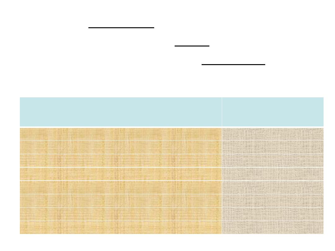
Eukaryotes
(Fungi)
Prokaryotes
(Bacteria)
4-15microns
Diameters 1 micron
Nuclear membrane
nuclear membrane No nuclear membrane
multiple
Chromosomes Single chromosome
Mitotic division
Division Binary fission
Organelles
cytoplasme No organelles
Chitin
Cell wall Peptidoglycan
Ergosterol
Cell membrane No ergosterol
80 S
Ribosome 70 S
Classification
4/30/16
General mycology5
4/30/16
General mycology6
Morphological
Clinical
Systematic

4/30/16
General mycology7
Fungal
morphology
Yeast
Mold
Dimorphic
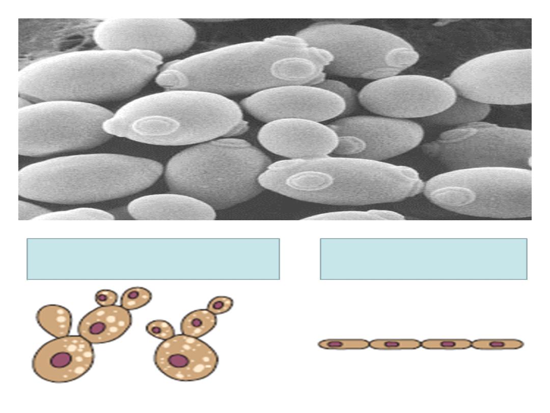
• Oval or round cells that reproduce by budding to form
blastospores.
• May form pseudohyphae (if blastospores remain
attached to each other).
• Examples: Candida, Cryptococcus.
4/30/16
General mycology8
Yeasts
4/30/16
General mycology9
Budding yeast cells
Pseudohyphae

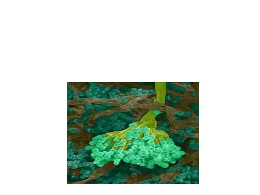
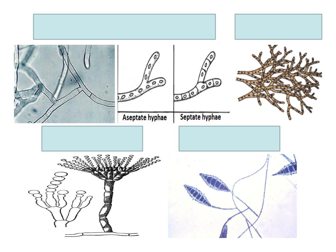
• Also called filamentous fungi or mycelial fungi.
• Formed of filaments called hyphae.
• Hyphae interlace to form mycelium.
• Hyphae on culture plate are two types: vegetative
hyphae for absorbing nutrients and aerial hyphae that
carry conidia.
• Hyphae may be septate or aseptate.
• Reproduce by formation of conidia.
• Conidia may be unicellular (microconidia) or
multicellular (macroconidia).
• Examples are: dermatophytes & aspergillus.
4/30/16
General mycology10
Molds
4/30/16
General mycology11
Hyphae
Microconidia
Macroconidia
Mycelium
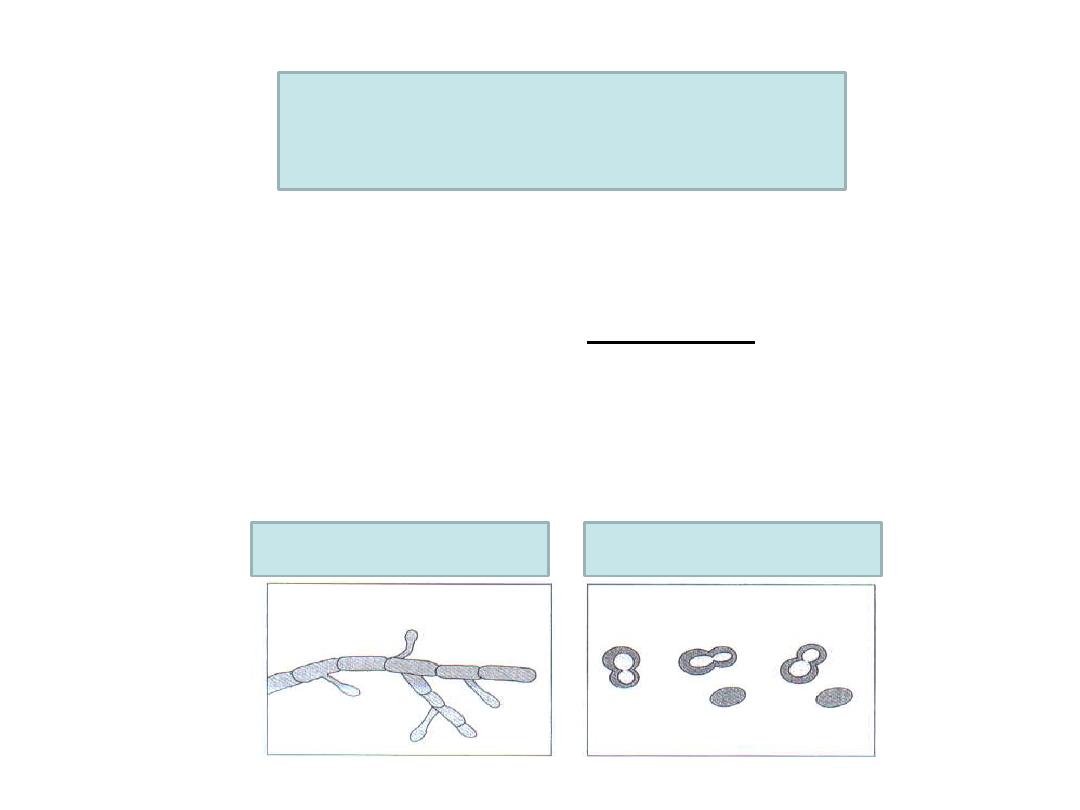
• These fungi occur in two forms:
v At the room temperature (25 degree), it appears as
mold.
v In the body (37 degree), it appears as yeast cells.
• Examples: Histoplasma & Blastomyces.
4/30/16
General mycology12
Dimorphic fungi
At 25 degree
At 37 degree
4/30/16
General mycology13
Clinical
classification
Superficial mycoses
Cutaneous mycoses
Subcutaneous
mycoses
Systemic mycoses
Opportunistic
mycoses
Allergy & mycetismus
& mycotoxicosis
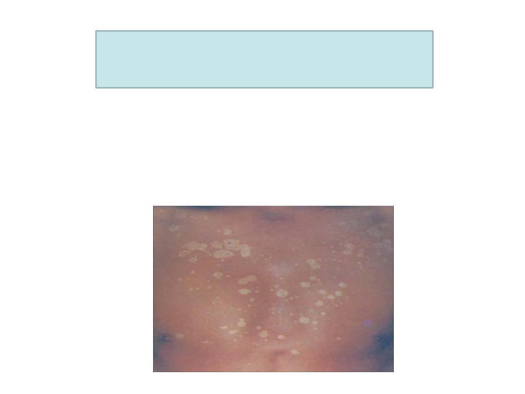
• Fungal infections confined to the stratum
corneum without tissue invasion.
• Example: Tinea versicolor caused by
Malassezia furfur.
4/30/16
General mycology14
Superficial mycoses
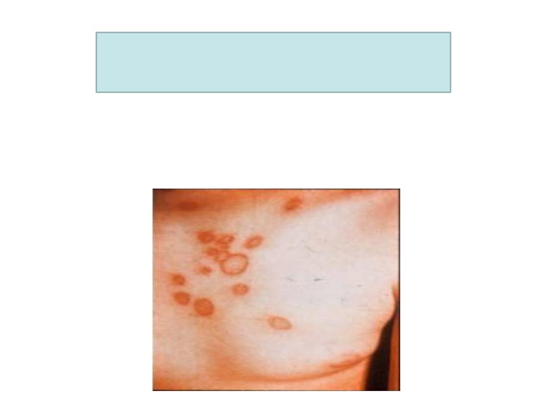
• Fungal infections that involve keratinized
tissues as skin, hair, nail.
• Example: Tinea caused by dermatophytes.
4/30/16
General mycology15
Cutaneous mycoses
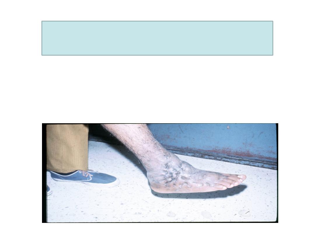
• Fungal infections that are confined to
subcutaneous tissues without dissemination
to distant sites.
• Example: mycetoma (madura foot).
4/30/16
General mycology16
Subcutaneous mycoses

• Also called endemic mycoses.
• Begin as primary pulmonary lesions that may
disseminate to any organ.
• Caused by dimorphic fungi.
4/30/16
General mycology17
Systemic mycoses

• Affect immunocompromised individuals
• Examples are:
1. Candidiasis caused by Candida albicans.
2. Cryptococcosis caused by Cryptococcus
neoformans.
3. Aspergillosis caused by aspergillus fungus.
4. Pneumocystis pneumonia caused by
pneumocystis jiroveci in AIDS patients.
4/30/16
General mycology18
Opportunistic mycoses

• Allergy occurs to fungal spores particularly those of
aspergillus fungus.
• Example: Seasonal allergies and asthma.
• The fungal flesh itself is toxic.
• Example: Amanita mushroom poisoning.
4/30/16
General mycology19
Allergy
Mycetismus

1. Aflatoxins produced by Aspergillus flavus
which infects grains and peanuts. This toxin
is hepatotoxic and cause tumors in animals
and suspected of causing hepatic carcinoma
in humans.
2. Ergotism which is caused by the mold
Claviceps purpura. This mold infects grains
and produce alkaloids (ergotamine ) that
cause neurological effects.
4/30/16
General mycology20
Mycotoxicosis

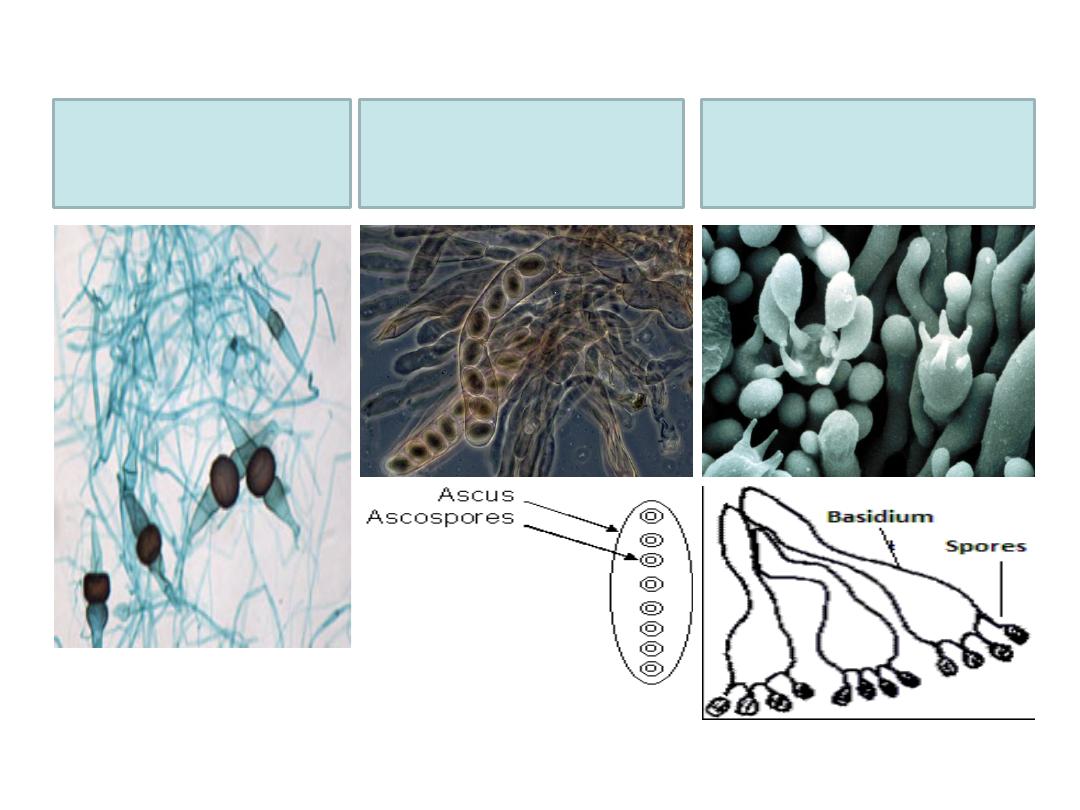
q It is based on the type of fungal spores:
" Sexual spores
" Asexual spores
4/30/16
General mycology21
Systematic
classification
q
Zygospores:
• Fungi forming zygospores are called zygomycetes.
q
Ascospores:
• Ascospores are carried in ascus.
• Fungi forming ascospores are called ascomycetes.
q
Basidiospores:
• Basidiospores are carried on basidium.
• Fungi forming basidiospores are called basidiomycetes.
4/30/16
General mycology22
Sexual
spores
Deuteromycetes are fungi whose sexual spores
are unknown. But, they produce asexual spores.
4/30/16
General mycology23
Zygospores
Ascospores
Basidiospores
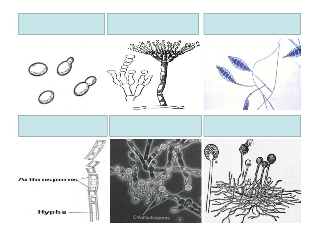
q
Blastospores:
• Produced by budding of the yeast cells.
q
Conidia:
• Produced by molds.
• May be microconidia or macroconidia
.
q
Arthrospores:
• Produced by fragmentation of hyphae.
q
Chlamydospores:
• Rounded thick walled spores produced by candida fungus.
q
Sporangiospores:
• Spores formed within a sac called sporangium. Formed by
zygomycetes.
4/30/16
General mycology 24
Asexual spores
4/30/16
General mycology25
Blastospores Microconidia
Macroconidia
Arthrospores
Chlamydospores Sporangiospores

4/30/16
26
Laboratory diagnosis of fungal
infections

v Specimen:
ü According to the site of infection.
ü For example, skin scales, nails, hair clippings
for dermatophyte examination.
v Microscopic examination of these specimens
using KOH 10%:
ü KOH dissolves keratin but does not affect
fungi. Branching hyphae are detected among
epithelial cells.
ü Fungal stains such as lactophenol cotton blue
could be used.
4/30/16
General mycology27
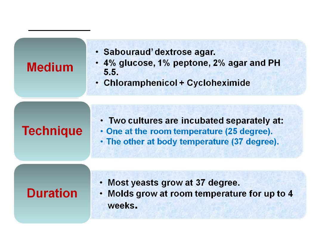
v Culture:
4/30/16
General mycology28
v Identification of the isolated fungus on culture
is done by:
ü
For molds: identification is done by :
4/30/16
General mycology29
Macroscopic
examination
Microscopic
examination
Colony
morpholog
y
+
color on
surface and
reverse
Slide
culture to
study
morpholog
y of conidia
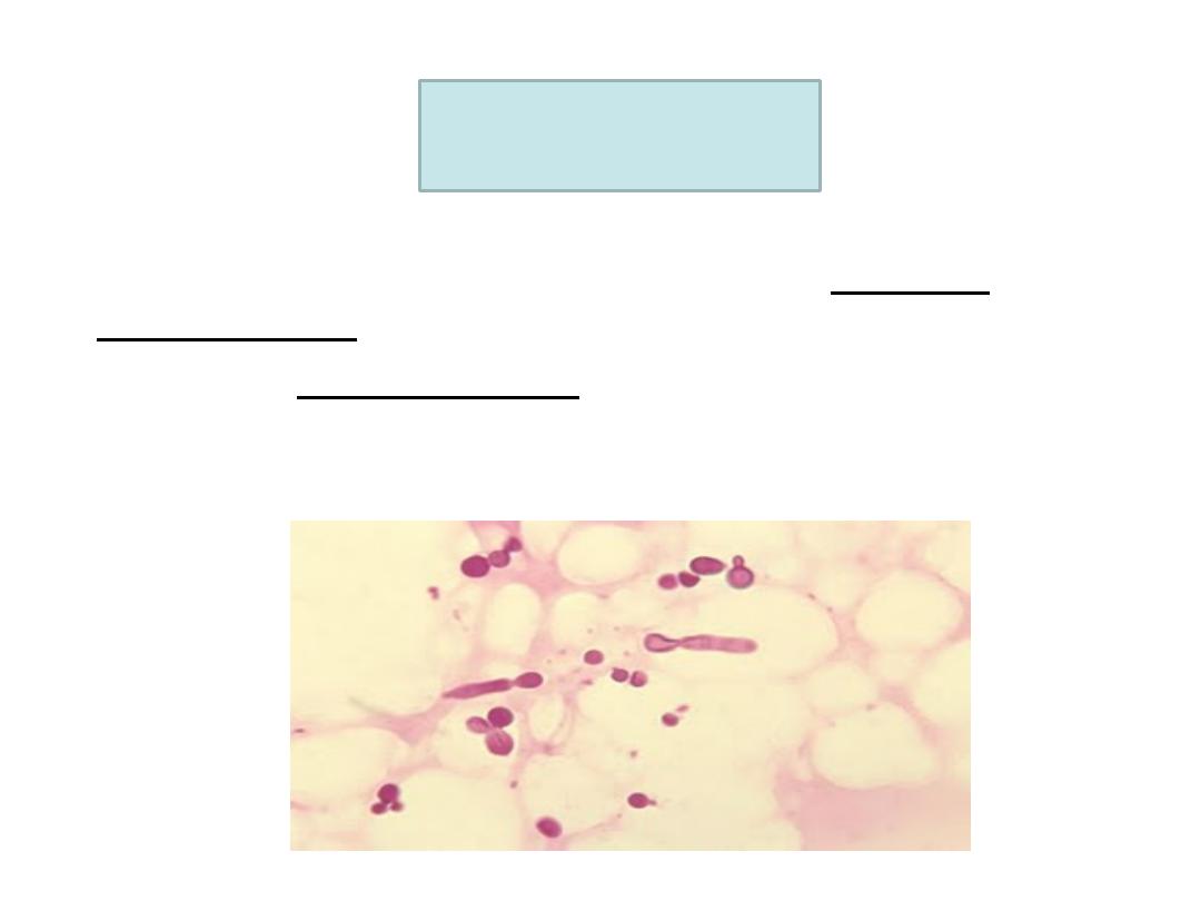
ü For yeasts: identification is done by :
4/30/16
General mycology30
Microscopic
examination
Biochemical
reactions
Oval
budding
Gram +Ve
yeast cells.